University of Pennsylvania
ScholarlyCommons
Publicly Accessible Penn Dissertations
Summer 8-13-2010
The Effect of T Cell Receptor Specificity on
CD4+CD25+ Regulatory T Cell Function in an
Autoimmune Setting
Soyoung Oh
University of Pennsylvania, [email protected]
Follow this and additional works at:http://repository.upenn.edu/edissertations
Part of theImmunology and Infectious Disease Commons, and theLaboratory and Basic Science Research Commons
Recommended Citation
Oh, Soyoung, "The Effect of T Cell Receptor Specificity on CD4+CD25+ Regulatory T Cell Function in an Autoimmune Setting"
(2010).Publicly Accessible Penn Dissertations. 205.
The Effect of T Cell Receptor Specificity on CD4+CD25+ Regulatory T
Cell Function in an Autoimmune Setting
Abstract
The studies presented in this dissertation examine how the T cell receptor (TCR) specificity of CD4+CD25+ regulatory T (Treg) cells affects their function in a mouse model of autoimmune arthritis. TS1xHACII mice co-express CD4+ T cells that express the transgenic 6.5 TCR, which is specific for the S1 determinant of influenza virus PR8 hemagglutinin (HA), and HA as a neo-self antigen under the MHC Class II I-Eα
promoter. The majority of TS1xHACII mice develop inflammatory arthritis that is driven by recognition of S1 peptide by 6.5+CD4+ T cells. Notably, arthritis develops despite the presence of CD4+CD25+Foxp3+ Treg cells, including a population that is specific for the disease target antigen S1 peptide. However, prophylactic administration of exogenous CD4+CD25+ Treg cells can prevent arthritis in TS1xHACII mice,
demonstrating that the disease is susceptible to Treg cell activity. Interestingly, we have found that the ability of CD4+CD25+ Treg cells to suppress arthritis is highly dependent on the TCR specificity(s) of the Treg cell population. Polyclonal CD4+CD25+ Treg cells, but not antigen-specific 6.5+CD4+CD25+ Treg cells can prevent arthritis development in TS1xHACII mice. Our data suggest that CD4+CD25+ Treg cells that are strongly reactive for a highly expressed target antigen can be detrimental in the context of certain autoimmune diseases, and that the balance of certain TCR specificities between CD4+CD25+ Treg and effector CD4+ T cells plays an important role in determining the maintenance of tolerance versus the development
autoimmunity.
Degree Type
Dissertation
Degree Name
Doctor of Philosophy (PhD)
Graduate Group
Immunology
First Advisor
Andrew J. Caton, Ph.D.
Keywords
CD4+CD25+ Treg cells, autoimmunity, arthritis
Subject Categories
THE EFFECT OF T CELL RECEPTOR SPECIFICITY ON CD4+CD25+
REGULATORY T CELL FUNCTION IN AN AUTOIMMUNE SETTING
Soyoung Oh
A DISSERTATION in
Immunology
Presented to the Faculties of the University of Pennsylvania in
Partial Fulfillment of the Requirements for the Degree of Doctor of Philosophy 2010
Andrew J. Caton, Ph.D., Supervisor of Dissertation
Steven Reiner, M.D., Graduate Group Chairperson
Dissertation Committee: Jan Erikson, Ph.D.
Christopher Hunter, Ph.D. Terri Laufer, M.D.
To Mom and Dad,
ACKNOWLEDGMENTS
There are many people whom I would like to sincerely thank for their support, help, and encouragement during my time as a graduate student. First, a heartfelt thank you to my thesis advisor, Andy Caton, for his invaluable
mentorship. I am a better scientist for having trained with him. I would also like to thank all current and former members of the Caton lab, particularly Andy Rankin, Malinda Aitken, Alissa Basehoar, Felipe Bedoya, Guang-Shing Cheng, Abby Liebow, Christina Mergenthaler, Olivia Perng, and Donnie Simons for providing thoughtful scientific discussions and encouragement throughout this project. In addition, I would like to thank Victoria Garcia, Lori Mroz, and Nardine Zakhary for their work in maintaining the countless transgenic mouse lineages that have gone into my dissertation work. I must also thank The Wistar Animal Facility, in particular Darryl Green who has taken care of our transgenic mouse lineages during my time in the Caton lab. Also a sincere thank you to the Wistar Flow Facility – Jeffrey Faust, Dave Ambrose, Dan Hussey, and Matt Farabaugh – for accommodating the numerous sorts that have gone into the studies presented here. I would also like to thank the members of my thesis committee: Drs. Jan Erikson, Chris Hunter, Terri Laufer, Larry Turka, and John Wherry for their guidance throughout this project.
ABSTRACT
THE EFFECT OF T CELL RECEPTOR SPECIFICITY ON CD4+CD25+ REGULATORY T CELL FUNCTION IN AN AUTOIMMUNE SETTING
Soyoung Oh
Andrew J. Caton, Ph.D.
The studies presented in this dissertation examine how the T cell receptor (TCR) specificity of CD4+CD25+ regulatory T (Treg) cells affects their function in
a mouse model of autoimmune arthritis (TS1xHACII mice). TS1xHACII mice co-express CD4+ T cells that express the transgenic 6.5 TCR, which is specific for the S1 determinant of influenza virus PR8 hemagglutinin (HA), and HA as a neo-self antigen under the MHC Class II I-Eα promoter. The majority of adult
TS1xHACII mice develop inflammatory arthritis that is driven by recognition of S1 peptide by 6.5+CD4+ T cells. Notably, arthritis develops despite the presence of CD4+CD25+Foxp3+ Treg cells, including a population of 6.5+ cells that are
specific for the disease target antigen S1 peptide. However, the administration of exogenous CD4+CD25+ Treg cells to pre-arthritic TS1xHACII mice can prevent
Interestingly, we have found that the ability of CD4+CD25+ Treg cells to
suppress arthritis in TS1xHACII mice is highly dependent on the TCR specificity(s) of the Treg cell population. Polyclonal CD4+CD25+ Treg cells (including cells that have not developed in the presence of the disease target antigen S1 peptide), but not CD4+CD25+ Treg cells that are enriched for S1-specific 6.5+ cells are able to prevent arthritis development in TS1xHACII mice.
Although polyclonal CD4+CD25+ Treg cells can suppress arthritis in TS1xHACII mice, our data suggest that successful CD4+CD25+ Treg cell mediated
suppression of arthritis is remarkably sensitive to the balance of Treg cells and pathogenic 6.5+CD4+ T cells. We also present data indicating that CD4+CD25+ Treg cells that are strongly reactive for a highly expressed disease target antigen can be detrimental in the context of certain autoimmune diseases. Collectively, these studies indicate that the antigen-specificity of CD4+CD25+ Treg cells is a critical determinant of their ability to suppress arthritis in TS1xHACII mice. Moreover the data presented here also suggest that the balance of certain TCR specificities between CD4+CD25+ Treg and effector CD4+ T cells plays an
Table of Contents
DEDICATION……….ii
ACKNOWLEDGMENTS………..iii
ABSTRACT………...……iv
Table of Contents……….…vi
List of Figures………..xii
List of Abbreviations………...xv
Chapter 1: Introduction and Overview...1
1.1 Introduction ...1
1.2 Rheumatoid Arthritis ...2
1.3 The role of CD4+ T cells in mouse models of arthritis ...3
1.4 The role of IL-17 in arthritis ...7
1.5 CD4+CD25+ regulatory T cells ...9
1.5.1 The role of TCR specificity in the thymic selection of CD4+CD25+ Treg cells...11
1.5.2 The role of TCR specificity in CD4+CD25+ Treg cell function...13
1.5.3 Mechanisms of CD4+CD25+ Treg cell function...17
1.5.4 Plasticity of CD4+CD25+ Treg cells...18
1.6 CD4+CD25+ Treg cells in human arthritis...25
1.7 CD4+CD25+ Treg cells in mouse models of arthritis...30
Chapter 2 : Materials and Methods ...37
2.1 Transgenic mice...37
2.3 Arthritis assessment in TS1xHACII mice ...41
2.4 Flow cytometry...41
2.5 Cell sorting...42
2.6 anti-IL-17 antibody treatment of TS1xHACII mice...43
2.7 MACS purification of CD4+ cells...43
2.8 CFSE labeling...44
2.9 Adoptive transfers...44
2.10 Analysis of in vivo CD4+CD25+ Treg cell proliferation...45
2.11 Intracellular cytokine staining...45
2.12 Unfractionated LN cultures ...46
2.13 In vitro CD4+CD25+ Treg cell suppression assays...46
2.14 ELISA...47
2.15 Luminex ...47
2.16 Statistical analyses ...48
2.17 Solutions ...48
Chapter 3 : Polyclonal CD4+CD25+ Treg cells can prevent arthritis development in TS1xHACII mice...50
3.1 Introduction ...50
3.2 Results...54
3.2.1 Arthritis development in TS1xHACII mice is accompanied by the accumulation of peripheral 6.5+CD4+ T cells ...54
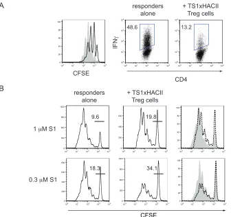
3.2.3 CD4+CD25+ cells from TS1xHACII mice possess in vitro suppressor
function...60 3.2.4 CD4+CD25+Foxp3+ cells are present in the joint-draining LNs of
TS1xHACII mice ...63 3.2.5 Polyclonal CD4+CD25+ Treg cells can prevent arthritis development in TS1xHACII mice ...65 3.2.6 Protective CD4+CD25+ Treg cells do not alter the accumulation or activation of 6.5+CD4+ T cells in TS1xHACII mice ...68 3.2.7 Preferential reduction of IL-17 in the popliteal LNs of TS1xHACII mice that received protective CD4+CD25+ Treg cells ...70 3.2.8 Protective CD4+CD25+ Treg cells suppress the systemic Th-17
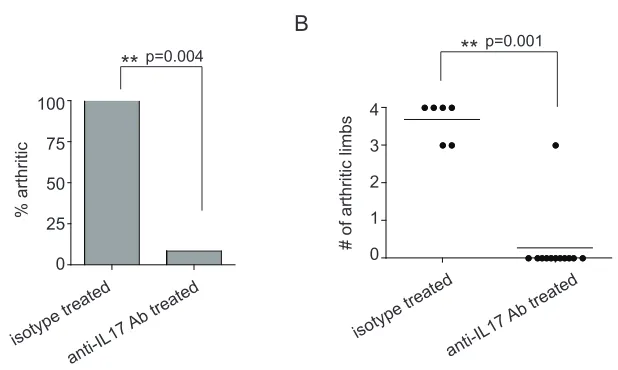
response in TS1xHACII mice...73 3.2.9 IL-17 is critical for the development of spontaneous autoimmune
arthritis in TS1xHACII mice...76 3.2.10 CD4+CD25+ cells from TS1xHACII mice can modify arthritis
development if 6.5+CD4+CD25+ cells are depleted...79
3.2.11 Suppressor function of CD4+CD25+ Treg cells is highly sensitive to balance of Treg and effector cells...84 3.2.12 Polyclonal CD4+CD25+ Treg cells that have not developed in the
3.3.1 TS1xHACII mice contain endogenous CD4+CD25+Foxp3+ cells,
including a population that expresses the 6.5 TCR and recognizes the
disease initiating target antigen ...91 3.3.2 Exogenous CD4+CD25+ Treg cells can inhibit the pathogenic Th-17
response in TS1xHACII mice...95 3.3.3 Specificity for the disease initiating target antigen is not required for CD4+CD25+ Treg cell suppression of arthritis in TS1xHACII mice...100 3.3.4 CD4+CD25+ Treg cell activity is highly sensitive to the balance of Treg and effector cells ...101 Chapter 4 : CD4+CD25+ Treg cells that are enriched in specificity for a disease target antigen fail to suppress arthritis in TS1xHACII mice ...106
4.1 Introduction ...106 4.2 Results...110 4.2.1 In vitro suppressor function of CD4+CD25+ cells from TS1xHA28 mice
can be activated by S1 peptide...110 4.2.2 CD4+CD25+ Treg cells enriched in specificity for a disease target
4.2.6 Strong antigenic stimulation coupled with lymphopenia can induce CD4+CD25+Foxp3+ Treg cells to produce IL-17...129 4.2.7 6.5+CD4+CD25+ Treg cells from TS1xHA28.Foxp3EGFP mice do not differentiate to produce IL-17 in TS1xHACII mice...133 4.2.8 Strong antigenic stimulation induces CD4+CD25+ Treg cell
downregulation of CD25 ...136 4.2.9 CD4+CD25+ Treg cells can maintain CD25 expression in the presence of low levels of their cognate antigen...140 4.2.10 S1 peptide can activate the in vitro suppressor function of CD4+CD25+
cells expressing a TCR with low reactivity for S1 ...143 4.2.11 CD4+CD25+ Treg cells expressing a TCR with low reactivity for S1
peptide can modulate arthritis development in TS1xHACII mice...147 4.2.12 CD4+CD25+ Treg cells enriched for the TS1(SW) TCR proliferate less and can maintain CD25 expression upon exposure to high levels of the mis-matched S1 peptide...151 4.3 Summary of Results and Discussion ...156
4.3.1 CD4+CD25+ Treg cells that are enriched in specificity for a disease target antigen fail to suppress arthritis development in TS1xHACII mice ...156 4.3.2 Interactions with high levels of cognate antigen can induce phenotypic changes in CD4+CD25+ Treg cells...158 4.3.3 CD4+CD25+ Treg cells enriched for cells with low reactivity for S1
4.3.4 CD4+CD25+ Treg cells enriched in specificity for S1 peptide may be
unable to suppress arthritis development in TS1xHACII mice...166
Chapter 5 : Discussion and Speculation ...169
5.1 Introduction ...169
5.2 The role of CD4+CD25+ Treg cells in arthritis development ...170
5.3 The effect of CD4+CD25+ Treg cell antigen-specificity in preventing autoimmunity ...183
5.4 Plasticity of CD4+CD25+ Treg cells: implications for suppression of autoimmunity ...193
List of Figures
Chapter 3: Polyclonal CD4+CD25+ Treg cells can prevent arthritis development in TS1xHACII mice
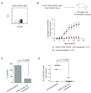
3-1. Key steps in the development of spontaneous inflammatory 52 arthritis in TS1xHACII mice
3-2. 6.5+CD4+ T cells accumulate in the periphery of arthritic 55
TS1xHACII mice
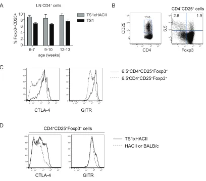
3-3. TS1xHACII mice contain S1-specific CD4+CD25+Foxp3+ 58 cells
3-4. CD4+CD25+ cells in TS1xHACII mice possess in vitro 61
suppressor function
3-5. 6.5+CD4+CD25+Foxp3+ cells are enriched in the popliteal 64
LNs of TS1xHACII mice
3-6. The adoptive transfer of CD4+CD25+ cells from HACII 67 mice can prevent arthritis development in TS1xHACII
mice
3-7. Protective HACII CD4+CD25+ Treg cells do not inhibit 69
the accumulation or activation of 6.5+CD4+ T cells in TS1xHACII mice
3-8. Protective HACII CD4+CD25+ Treg cells inhibit Th-17 72
cells in the popliteal LNs of TS1xHACII mice
3-9. Protective HACII CD4+CD25+ Treg cells inhibit systemic 74 IL-17 production in TS1xHACII mice
3-10. IL-17 is critical for the development of arthritis in 78 TS1xHACII mice
3-11. The adoptive transfer of 6.5-depleted TS1xHACII 81 CD4+CD25+ cells can modify arthritis in TS1xHACII
3-12. CD4+CD25+ Treg cell activity is sensitive to the balance 86
of Treg and effector cells
3-13. The adoptive transfer of CD4+CD25+ cells from BALB/c 89 mice can prevent arthritis in TS1xHACII mice
Chapter 4: CD4+CD25+ Treg cells that are enriched in specificity for a disease target antigen fail to suppress arthritis development in TS1xHACII mice
4-1. CD4+CD25+ cells from TS1xHA28 mice are enriched in 111 6.5+Foxp3+ cells
4-2. TS1xHA28 CD4+CD25+ cells do not suppress arthritis 113 development in TS1xHACII mice
4-3. TS1xHA28.Foxp3EGFP GFP+CD4+CD25+ cells do not 116 prevent arthritis in TS1xHACII mice
4-4. TS1xHA28.Foxp3EGFP GFP+CD4+CD25+ cells persist in 119 TS1xHACII mice
4-5. TS1xHA28 CD4+CD25+ cells do not suppress the Th-17 122 response in the popliteal LNs of TS1xHACII mice
4-6. TS1xHA28.Foxp3EGFP GFP+CD4+CD25+ cells do not 124 suppress the Th-17 response in the popliteal LNs of
TS1xHACII mice
4-7. TS1xHA28 CD4+CD25+ cells fail to suppress the 127 systemic Th-17 response in TS1xHACII mice
4-8. 6.5+CD4+CD25+Foxp3+ cells produce IL-17 and IFN-γ 131 upon exposure to high levels of cognate antigen in a
lymphopenic environment
4-9. TS1xHA28.Foxp3EGFP 6.5+CD4+CD25+Foxp3+ cells do 135
not produce IL-17 or IFN-γ in TS1xHACII mice
4-11. CD4+CD25+ Treg cells can maintain high levels of CD25 142 expression upon exposure to low, but not high, levels of
cognate antigen
4-12. The suppressor function of CD4+CD25+ cells expressing 145 the TS1(SW) TCR can be activated by non-cognate inter-
actions with S1 peptide
4-13. TS1(SW)xPevSW CD4+CD25+ cells modify arthritis 149
development in TS1xHACII mice
4-14. Foxp3 and CD25 profile of TS1(SW)xPevSW CD4+CD25+ 153 cells exposed to high levels of the mis-matched S1 peptide
Chapter 5: Discussion and Speculation
5-1. Working model: The balance of CD4+CD25+ Treg cells to 201
pathogenic 6.5+CD4+ T cells is a critical parameter for
List of Abbreviations
AOD autoimmune ovarian disease
APC antigen presenting cell
BDC2.5 transgenic mouse expressing a TCR that is specific for a pancreatic β cell islet antigen
BDC-6.9 transgenic mouse expressing a TCR that is specific for a pancreatic antigen present in NOD but not BALB/c mice
CFSE 5-(and-6)-carboxyfluroscein diacetate, succinimidyl ester
CIA collagen-induced arthritis
CNS central nervous system
CNS2 conserved noncoding sequence 2
GFP green fluorescent protein
GPI glucose-6-phosphoisomerase
GVHD graft-versus-host disease
EAE experimental autoimmune encephalitis
EAP experimental autoimmune prostatitis
HA hemagglutinin
HLA human leukocyte antigen
IPEX immunodysregulation polyendocrinopathy enteropathy X-linked syndrome
JIA juvenile idiopathic arthritis
K/BxN transgenic mouse strain that is a cross between KRN and BxN mice; develops spontaneous arthritis driven by a CD4+ T cell recognition of GPI and the production of GPI-specific antibodies by B cells
LN lymph node
NOD non-obese diabetic mouse strain
NOD.C6 non-obese diabetic mouse strain that does not express the peptide for the BDC-6.9 transgenic TCR
MBP myelin basic protein
MHC major histocompatibility complex
MOG myelin oligodendrocyte protein
MS multiple sclerosis
PLP myelin proteolipid protein
PLP1 pre-activated Ig-proteolipid protein 1
RA rheumatoid arthritis
SCID severe combined immunodeficiency
SKG mouse strain described by Sakaguchi and colleagues; develops spontaneous arthritis due to altered negative selection of
autoreactive thymocytes
TCR T cell receptor
TS1 transgenic mouse expressing a TCR specific for the S1 determinant of influenza virus PR8 HA
TS1(SW) transgenic mouse expressing a TCR specific for S1(SW) (an analog of S1 peptide) from the influenza virus SW HA
Chapter 1: Introduction and Overview
1.1 Introduction
The adaptive immune system is designed to be highly diverse, allowing for
the recognition of a wide range of pathogens. However, this diversity must be
balanced with the requirement of self-tolerance, and within tightly controlled
numbers of peripheral lymphocytes. During T cell development, gene
rearrangement mechanisms produce an enormous array of T cell receptors
(TCRs). This diverse pool of TCRs is subsequently screened for autoreactivity,
and highly self-reactive T cells are eliminated or functionally suppressed via
central and peripheral tolerance mechanisms (Palmer 2003; Hogquist et al. 2005;
Mueller et al. 2010), which include the activity of CD4+CD25+ regulatory T (Treg)
cells (Sakaguchi 2004). The combination of these processes allows the adaptive
immune system to achieve a diverse T cell repertoire while maintaining
self-tolerance. However, these tolerance mechanisms are not foolproof, and
approximately 3 to 5 percent of the population develops autoimmunity (Jacobson
et al. 1997).
How and why CD4+CD25+ Treg cells fail to prevent the development of
autoimmunity is poorly understood. The studies presented in this dissertation
inflammatory arthritis that is dependent on the autoreactive CD4+ T cell
response. We also address how the antigen-specificity of the CD4+CD25+ Treg
cells affects their in vivo suppressor function. This introductory chapter begins
with an overview of rheumatoid arthritis and mouse models of arthritis with a
particular focus on how CD4+ T cells can contribute to disease. The following
section presents a summary of relevant topics in CD4+CD25+ Treg cell biology.
The chapter concludes with an overview of the current understanding of
CD4+CD25+ Treg cell activity in human arthritis and mouse models of disease.
1.2 Rheumatoid Arthritis
Approximately 0.8 percent of the population develops rheumatoid arthritis
(RA) (Lipsky). Although the etiology of the disease remains unknown, a major
genetic risk factor for RA is the MHC Class II allele HLA-DR4 (DRβ1*0401).
Interestingly, other MHC Class II alleles (HLA-DR5, HLA-DR2, HLA-DR3, and
HLA-DR7) may be protective, as they are found at lower frequencies in RA
patients than in individuals who have not developed RA (Lipsky). However, the
mechanisms (e.g. deletion of autoreactive T cells, selection of Treg cells) by
which these particular MHC Class II alleles could confer protection are unknown.
tyrosine phosphatase (Lyp) that participates in TCR signal transduction, has
been identified as another genetic risk factor for RA (Lipsky; (Lundy et al. 2007).
Collectively, the association of genes that encode MHC Class II and TCR
signaling molecules with RA development strongly supports a role for CD4+ T
cells in disease. Indeed, studies in multiple mouse models of arthritis have
identified a critical role for CD4+ T cells in arthritis development. A summary of
these findings is presented in the following section.
1.3 The role of CD4+ T cells in mouse models of arthritis
Mouse models of inflammatory arthritis possess several characteristics
that resonate with human RA, and have been an instrumental tool in gaining
better understanding of this autoimmune disorder. Work in several different
arthritis models has clearly demonstrated a requirement for autoreactive CD4+ T
cells in arthritis pathogenesis. One of the most commonly used systems to study
disease is the inductive model of collagen-induced arthritis (CIA). In CIA,
disease development is initiated by immunizing mice with bovine type II collagen
in complete Freund’s adjuvant. The ensuing antigen-specific CD4+ T cell
response is required for B cell generation of collagen-specific antibodies that
ultimately dependent on the production of collagen-specific antibodies and
arthritis does not develop in B cell deficient mice (Svensson et al. 1998).
K/BxN mice develop spontaneous arthritis that also requires T and B cell
responses for disease development. Arthritis in K/BxN mice is initiated by KRN
transgenic TCR CD4+ T cell recognition of a glucose-6-phosphate isomerase
(GPI) peptide presented by the MHC Class II molecule Ag7, which activates
GPI-specific B cells, leading to the production of pathogenic autoantibodies that bind
to articular surfaces and initiate an inflammatory cascade that leads to joint
damage (Kouskoff et al. 1996; Matsumoto et al. 1999; Matsumoto et al. 2002).
Chimeric mice, generated by administration of KRN.RAG-/- bone marrow to
irradiated BxN.TCRα-/- mice, develop arthritis, demonstrating that CD4+ T cells of
other specificities are not required for the initiation of arthritis development in
K/BxN mice (Mangialaio et al. 1999). The transfer of serum from arthritic K/BxN
mice into BxN.RAG-/- mice induces arthritis, indicating that the presence of
autoantibodies alone is sufficient to mediate arthritis development, and that T
cells are not required for the effector phase of disease (Korganow et al. 1999).
However, although CD4+ T cells are not necessary for the development of serum
transfer induced arthritis, they can contribute to disease as co-administration of
CD4+ T cells with the serum transfers exacerbates arthritis by a mechanism that
While disease development in CIA and K/BxN arthritis models requires
both T and B cell responses, it has been also shown that autoreactive CD4+ T
cells alone can initiate arthritis. Sakaguchi and colleagues have described the
SKG mouse, which develops spontaneous arthritis that is attributed to a failure in
negative selection of autoreactive thymocytes due to a mutation in the ZAP-70
(ζ-associated protein of 70 kDa) gene (Sakaguchi et al. 2003). The adoptive
transfer of SKG CD4+ T cells into RAG-/- mice can induce arthritis, demonstrating
that B cell antibody production is not required for arthritis development, and that
CD4+ T cells alone can initiate disease (Hirota et al. 2007). However, while it is
evident that autoreactive CD4+ T cells drive arthritis development in SKG mice,
the nature of the peptide(s) that the cells recognize is unknown. Moreover,
arthritis development in SKG mice is also dependent on additional environmental
cues, as SKG mice housed in specific pathogen free conditions do not develop
disease (Yoshitomi et al. 2005). Our lab has recently described another mouse
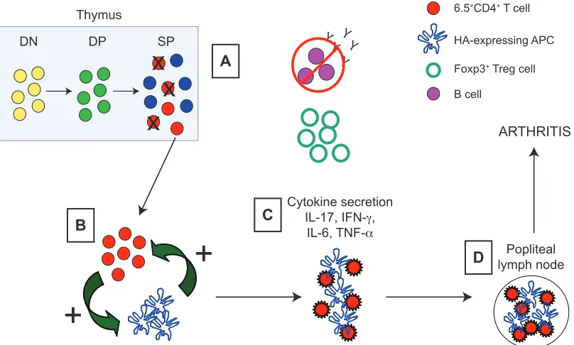
model of arthritis that is dependent on autoreactive CD4+ T cells. TS1xHACII
mice develop spontaneous inflammatory arthritis that is driven by CD4+ T cell
recognition of a single systemically presented peptide and does not require B
cells (Rankin et al. 2008). Specific aspects of arthritis development in
TS1xHACII mice will be discussed in greater detail in Chapter 3 of this
While CD4+ T cells clearly play a critical role in the initiation of the immune
response that leads to arthritis development, other aspects of the immune
response can also contribute arthritis pathogenesis. Dysregulation of
inflammatory pathways can be a key contributing factor to disease, as the
over-expression of cytokines such as TNF-α and IL-1α, or expression of a mutated
IL-6R gp130 that enhances gp130-mediated signal transducer and activator of
transcription (STAT3) activation also leads to arthritis development (Keffer et al.
1991; Niki et al. 2001; Sawa et al. 2006). Interestingly, the arthritis that develops
in the IL-6R gp130 mutant mice is attributed to CD4+ T cell activation resulting
from enhanced homeostatic proliferation, and disease can be ameliorated by
inhibiting the homeostatic proliferation of CD4+ T cells (Sawa et al. 2006). In
SKG mice, genetic deficiencies in IL-6, IL-1, and TNF-α resulted in either
complete protection from arthritis or delayed disease onset accompanied by
reduced incidence and disease severity (Hata et al. 2004), demonstrating that
the autoreactive CD4+ T cells are acting in concert with certain inflammatory
pathways. In addition to IL-6, IL-1, and TNF-α, there are reports that IL-17,
recently identified as defining a unique T helper cell lineage (Harrington et al.
2005; Park et al. 2005), is also involved in arthritis. In the next section we will
summarize the relevant findings on IL-17 in human arthritis and mouse models of
1.4 The role of IL-17 in arthritis
The cytokine IL-17 is thought to play a critical role in several autoimmune
diseases, including experimental autoimmune encephalitis (EAE), psoriasis, and
arthritis (Afzali et al. 2007; Korn et al. 2009). In human RA patients, IL-17 has
been detected in the peripheral blood, serum, synovial fluid, and/or synovium
(Chabaud et al. 1999; Kotake et al. 1999; Hirota et al. 2007; Sarkar et al. 2010).
Interestingly in the synovium, IL-17 is primarily found in T cell enriched areas,
and is mostly secreted by memory CD4+ T cells isolated from the peripheral
blood of RA patients (Sarkar et al. 2010), suggesting that CD4+ T cells are a
major source of IL-17 in RA. IL-17 has also been detected in the synovial fluid
and joints of arthritic mice (Hirota et al. 2007). IL-17 may contribute to arthritis
pathogenesis by a variety of mechanisms. IL-17 can induce the upregulation of
cytokines such as IL-1β, TNF-α, and IL-6 (Chabaud et al. 1998; Jovanovic et al.
1998; Sarkar et al. 2010), which not only contribute to arthritis pathogenesis but
can also promote additional differentiation of Th-17 cells (Weaver et al. 2007;
Korn et al. 2009). IL-17 can also recruit innate immune cells such as monocytes
or neutrophils, which are potential sources for the cytokines described above, to
sites of inflammation (Lundy et al. 2007; Shahrara et al. 2009; Pelletier et al.
2010). Moreover, the IL-17R is found on chondrocytes, synoviocytes, and
arthritis pathogenesis (Sarkar et al. 2010). Indeed, in vitro studies have
demonstrated that IL-17 can act upon osteoblasts to induce osteoclastogenesis
(Sato et al. 2006), and potentially contribute to bone resorption. Thus, IL-17 can
act upon cells in the joint and also activate and/or recruit other cell types to the
primary sites of inflammation and autoimmune pathology.
Studies in several mouse models of arthritis have demonstrated that IL-17
plays an important role in disease pathogenesis. Mice lacking the IL-1 receptor
antagonist (IL1-Ra-/-) develop inflammatory arthritis, and interestingly the CD4+ T
cells from these mice produce elevated amounts of IL-17 (Nakae et al. 2003).
When IL1-Ra-/- mice are bred onto an IL17-/- background, the mice are protected
from arthritis, demonstrating the critical role for IL-17 in mediating the disease
that develops in IL1-Ra-/- mice (Nakae et al. 2003). Moreover, treating arthritic
IL1-Ra-/- mice with anti-IL17 antibody can reduce disease severity (Koenders et
al. 2008). Work in the CIA model has shown that increasing systemic IL-17
production in mice by gene transfer with an adenoviral vector expressing IL-17
accelerates diseases onset and severity (Lubberts et al. 2001). Complementary
studies examining the effects of IL-17 or IL-17R blockade have demonstrated
that inhibiting the biological activity of IL-17 reduces the severity of CIA (Lubberts
et al. 2001; Lubberts et al. 2004; Egan et al. 2008). Furthermore, IL17-/- mice
mice (Nakae et al. 2003). Similarly, in an LPS induced model of arthritis, disease
incidence and severity are reduced in IL17-/- mice (Sato et al. 2006).
Interestingly, studies in K/BxN and SKG mice indicate that IL-17 production by
CD4+ T cells is important in arthritis development. In the K/BxN arthritis model,
the augmentation of serum transfer induced disease by KRN CD4+ T cells is
inhibited by treatment with anti-IL17 antibody, indicating that CD4+ T cell
production of IL-17 was responsible for the exacerbation autoantibody induced
arthritis (Jacobs et al. 2009). Additionally, Sakaguchi and colleagues have
shown that IL-17+/+CD4+, but not IL-17-/-CD4+ T cells from SKG mice, can transfer
arthritis upon transfer into RAG-/- mice, demonstrating that CD4+ T cell generated
IL-17 is critical for disease induction (Hirota et al. 2007).
1.5 CD4+CD25+ regulatory T cells
The concept of suppressor T cells was first suggested by experiments in
which thymectomized mice and adult rats developed autoimmune diseases that
could be prevented by the transfer of spleen cells from healthy donors (Nishizuka
and Sakakura 1969; Penhale et al. 1976). In the extensive work following those
initial observations, Treg cell populations have been more clearly defined, and
have been characterized, the best defined is the thymically generated
CD4+CD25+ Treg cell population that comprises approximately 5 to 10 percent of
the human and murine peripheral CD4+ T cell repertoires. This population was
first characterized by constitutive expression of the IL-2 receptor α chain (CD25)
(Sakaguchi et al. 1995), which is also upregulated by activated CD4+ T cells.
Subsequently the forkhead transcription factor Foxp3 was identified as a more
reliable marker for CD4+CD25+ Treg cells (Fontenot et al. 2003; Hori et al. 2003),
and Foxp3 was considered to be the “master regulator” of the CD4+CD25+ Treg
cell lineage.
However a recent set of studies has challenged the notion of Foxp3 as the
key lineage commitment factor of CD4+CD25+ Treg cells. Multiple groups have
reported that a subset of cells that have developed in the absence of Foxp3
express a genetic signature that is consistent with that of CD4+CD25+Foxp3+
cells, suggesting that CD4+CD25+ Treg cell lineage commitment occurs upstream
of Foxp3 expression (Gavin et al. 2007; Hill et al. 2007; Lin et al. 2007).
Moreover, Foxp3 expression can be transiently upregulated in activated human T
cells, which do not acquire regulatory function (Gavin et al. 2006; Mantel et al.
2006; Wang et al. 2007). Nonetheless, it is clear that Foxp3 expression is critical
for CD4+CD25+ Treg cell function, as mice lacking Foxp3 develop severe
humans, development of the autoimmune disorder IPEX has been attributed to
mutations in the FoxP3 gene (Bennett et al. 2001; Gambineri et al. 2003).
1.5.1 The role of TCR specificity in the thymic selection of CD4+CD25+ Treg cells
The initial day three thymectomy experiments indicated that the thymus
plays an important role in the generation of CD4+CD25+ Treg cells, however, the
exact nature of the signals (e.g. TCR engagement, cytokines, co-stimulation) that
guide the thymic development of CD4+CD25+ Treg cells remains unclear. While
the observation that there is overlap between the TCR repertoires of CD4+CD25+
Treg cells and conventional CD4+ T cells (Hsieh et al. 2004; Hsieh et al. 2006)
suggests that other signals (e.g. cytokines, co-stimulation) likely also play a role
in the thymic development of CD4+CD25+ Treg cells, a strong body of work
supports a significant role for TCR interactions with self-peptides in the thymic
selection of CD4+CD25+ Treg cells. The key findings in support of this concept
are presented below.
Experiments using transgenic TCRs provided the first evidence that
CD4+CD25+ Treg cells were selected via TCR interactions with self-peptide. It
was observed that mice containing CD4+ T cells expressing a transgenic TCR
background but not in intact mice (Olivares-Villagomez et al. 1998; Van de Keere
and Tonegawa 1998; Hori et al. 2002). Subsequent studies showed that mice on
an intact background did not develop disease because expression of a
secondary TCR, resulting from endogenous α chain rearrangements, allowed for
the selection of MBP-specific CD4+CD25+ Treg cells (Hori et al. 2002),
suggesting a role for TCR and self-peptide interactions in CD4+CD25+ Treg cell
selection. Indeed, studies in other transgenic systems have shown that
introducing the cognate antigen for the transgenic TCR onto a RAG-/- background
results in the development of CD4+CD25+ Treg cells expressing the transgenic
TCR (Apostolou et al. 2002; Kawahata et al. 2002; Walker et al. 2003).
Work from our laboratory has further demonstrated the importance of TCR
interactions with self peptides for the development of CD4+CD25+ Treg cells.
Studies in TS1xHA-transgenic mice have demonstrated that CD4+ T cells
expressing the 6.5 TCR (Kirberg et al. 1994), which confers specificity for the S1
determinant of PR8 HA, undergo efficient CD4+CD25+ Treg cell selection in
HA28 mice, which express PR8 HA driven by an SV40 promoter, demonstrating
that interactions with the agonist peptide can guide CD4+CD25+ Treg cell
development (Jordan et al. 2001). The requirement for peptide specificity for
CD4+CD25+ Treg cell selection appears to be quite stringent, as CD4+ T cells
CD4+CD25+ Treg cells (Jordan et al. 2001). Interestingly, while 6.5+CD4+CD25+
Treg cells can also be selected in other TS1xHA lineages, the numbers are lower
than what is generated in TS1xHA28 mice, and this appears to result from
enhanced deletion of 6.5+ thymocytes due to higher levels of HA expression in
the thymus (Picca et al. 2006; Simons et al. 2010), indicating that lower levels of
the cognate antigen favor more abundant CD4+CD25+ Treg cell selection.
1.5.2 The role of TCR specificity in CD4+CD25+ Treg cell function
TCR stimulation is required to activate CD4+CD25+ Treg cell suppressor
function (Takahashi et al. 1998; Thornton and Shevach 1998). However, in vitro
studies show that although CD4+CD25+ Treg cell activation is TCR specific,
CD4+CD25+ Treg cells are able to inhibit responder CD4+ T cells of different TCR
specificities (Takahashi et al. 1998; Thornton and Shevach 2000), indicating that
the suppression itself is not antigen-specific. Correspondingly, work in multiple
murine autoimmune diseases (e.g. CIA, colitis, EAE, diabetes, gastritis) have
shown that that the adoptive transfer of polyclonal CD4+CD25+ cells can prevent
or ameliorate disease (Olivares-Villagomez et al. 1998; Van de Keere and
Tonegawa 1998; Singh et al. 2001; Sarween et al. 2004; Morgan et al. 2005;
Zwar et al. 2006; Kelchtermans et al. 2009), indicating that CD4+CD25+ Treg
mediate suppression of an organ specific disease. However, there is also a
significant body of work suggesting that antigen-specific CD4+CD25+ Treg cells
are more effective than polyclonal CD4+CD25+ Treg cells at suppressing
organ-specific autoimmune diseases.
In the nonobese diabetes (NOD) model, the adoptive transfer of in vitro
expanded CD4+CD25+ cells expressing the BDC2.5 TCR, which recognizes a
pancreatic β cell islet antigen (Katz et al. 1993), into NOD.BDC2.5 mice (which
also express the transgenic TCR) can suppress diabetes (Tang et al. 2004;
Tarbell et al. 2004). Using a disease transfer model in which diabetes
development is induced by the administration of splenocytes from diabetic NOD
mice into NOD.scid mice, Steinman and colleagues also showed that lower
numbers of in vitro expanded antigen-specific CD4+CD25+ cells could suppress
disease, whereas greater numbers of in vitro expanded polyclonal CD4+CD25+ T
cells could not (Tarbell et al. 2004). Work with induced CD4+CD25+ Treg cells
also suggests that antigen-specific interactions are important for regulatory
function. TGF-β induced regulatory cells expressing the BDC-6.9 TCR can
suppress diabetes in NOD mice, which contain their cognate antigen, but not
NOD.C6 mice, which do not contain their cognate antigen (Tonkin et al. 2008). It
to poor trafficking, expansion, and/or survival at the primary site of autoimmune
pathology.
Studies in the EAE model also emphasize that CD4+CD25+ Treg cell
specificity for the target autoantigen is important for the prevention of
autoimmunity. As described previously, mice expressing a transgenic TCR
specific for the encephalotegenic MBP protein spontaneously develop EAE when
on a RAG-deficient but not wildtype background, and the difference in disease
development can be attributed to the absence of CD4+CD25+ Treg cells specific
for EAE associated peptides in the RAG-/- mice (Hori et al. 2002). In another
model of EAE where disease is driven by the immune response to the myelin
proteolipid protein (PLP), the difference between two strains of mice with differing
susceptibilities to disease appears to be the enhanced representation of
PLP-peptide specific CD4+CD25+ Treg cells in the more resistant strain (Reddy et al.
2004). Interestingly, CD4+CD25+ cells isolated from healthy individuals can
mediate better in vitro suppression of CD4+ T cell proliferation when stimulated
by MBP proteins than CD4+CD25+ cells isolated from multiple sclerosis (MS)
patients (Kumar et al. 2006), suggesting that in humans protection may also
correlate with the presence of CD4+CD25+ Treg cells that are specific for
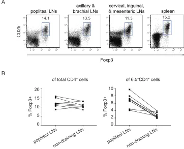
Studies in day 3 thymectomized mice that develop autoimmune ovarian
disease (AOD), show that CD4+CD25+ Treg cells isolated from the draining
lymph nodes are superior to CD4+CD25+ Treg cells isolated from non-draining
lymph nodes at suppressing AOD (Samy et al. 2005; Samy et al. 2008). This
apparent enrichment for disease-specific CD4+CD25+ Treg cells in the draining
lymph nodes was evident for other organs as well, as CD4+CD25+ Treg cells
isolated from the lacrimal or prostate draining lymph nodes were the most
effective at suppressing dacryoadenitis and experimental autoimmune prostatitis
(EAP) respectively (Samy et al. 2008; Wheeler et al. 2009). Collectively, these
observations suggest that draining lymph nodes, which have been shown to
uniquely present organ-specific peptides (Hoglund et al. 1999; Gagnerault et al.
2002; Scheinecker et al. 2002), are enriched for CD4+CD25+ Treg cells that are
specific for antigens derived from the organ. Recent work examining the
CD4+CD25+ Treg cell repertoires from various lymph nodes further corroborates
the concept that regional lymph nodes may be biased towards CD4+CD25+ Treg
cells that recognize organ-specific antigens, as Treg cells showed distinct
patterns of TCR usage in different LNs, similar to the skewing of TCR usage
among antigen-experienced conventional CD4+ T cells in different anatomical
1.5.3 Mechanisms of CD4+CD25+ Treg cell function
In spite of the clear observations that CD4+CD25+ Treg cells can suppress
immune responses and prevent autoimmune disease, how they exert their
regulatory function remains controversial. Indeed, a myriad of mechanisms by
which CD4+CD25+ Treg cells can mediate suppression have been reported. In
vitro studies indicate that direct cell contact between the regulatory and effector
populations is necessary, as suppression is prevented when the two cell
populations are separated in transwell experiments (Takahashi et al. 1998;
Thornton and Shevach 1998). Additional work indicates that CD4+CD25+ Treg
cell production of cytokines such as IL-10 or TGF-β is important for their
suppressor function (Huang et al. 2005). Alternatively CD4+CD25+ Treg cells
have been shown to act as a cytokine sink for cytokines such as IL-2, which can
promote apoptosis of effector CD4+ T cells (Pandiyan et al. 2007). It has also
been suggested that CD4+CD25+ Treg cells may suppress immune responses by
developing cytotoxic capabilities or modulating antigen presenting cells (APCs)
(Kryczek et al. 2006; Shevach et al. 2006; Vignali et al. 2008). Indeed,
Sakaguchi and colleagues have shown that a key contribution of CTLA-4 to
CD4+CD25+ Treg cell function may be through regulation of APCs (Wing et al.
2008). Interestingly, recent work has also demonstrated that CD4+CD25+ Treg
differentiation of the respective Th lineage cells (Chaudhry et al. 2009; Koch et
al. 2009; Zheng et al. 2009). It is possible that expression of transcription factors
specific for a certain Th lineage allows CD4+CD25+ Treg cells to acquire
properties (e.g expression of chemokine or cytokine receptors) that allow them to
traffic to the site of inflammation and/or compete for other soluble factors that
may potentiate the CD4+ Th cell response.
1.5.4 Plasticity of CD4+CD25+ Treg cells
CD4+CD25+Foxp3+ Treg cells can produce Th-1 and Th-17 associated cytokines
Recent studies have challenged the notion of CD4+CD25+ Treg cells as a
stable population of cells by demonstrating that under certain conditions
CD4+CD25+ Treg cells can differentiate to acquire properties that are normally
associated with effector CD4+ T cells. While multiple groups have documented
the loss of Foxp3 expression by CD4+CD25+ Treg cells (Hoffmann et al. 2006;
Duarte et al. 2009; Komatsu et al. 2009; Murai et al. 2009; Tsuji et al. 2009; Zhou
et al. 2009), there are also reports that CD4+CD25+ Treg cells that maintain
Foxp3 expression can develop phenotypic properties (e.g. cytokine production)
that are normally associated with effector CD4+ T cells. During Toxoplasma
response, a subset of CD4+CD25+Foxp3+ Treg cells differentiates to express the
transcription factor T-bet and to produce IFN-γ (Oldenhove et al. 2009). The
acquisition of Th-1 cell properties by CD4+CD25+ Treg cells is at least partially
dependent on a lack of IL-2 in the infectious setting, since treatment of T. gondii
infected mice with an IL-2 α-IL-2 antibody complex that has been shown to
enhance the biological activity of IL-2 (Boyman et al. 2006) reduces the presence
of IFN-γ+CD4+CD25+Foxp3+ cells (Oldenhove et al. 2009).
In light of the complicated and interconnected relationship of CD4+CD25+
Treg cells and Th-17 cells (Bettelli et al. 2006; Veldhoen et al. 2006; Mucida et al.
2007; Zhang et al. 2008; Zhou et al. 2008), there has been much interest in
determining whether CD4+CD25+Foxp3+ Treg cells can differentiate to produce
IL-17. Several groups have shown that IL-17+CD4+CD25+Foxp3+ cells can be
isolated from human peripheral blood (Ayyoub et al. 2009; Beriou et al. 2009;
Voo et al. 2009). The IL-17 producing CD4+CD25+ Treg cells also express CCR6
(Ayyoub et al. 2009; Beriou et al. 2009; Voo et al. 2009), a chemokine receptor
that is associated with Th-17 cells (Acosta-Rodriguez et al. 2007; Annunziato et
al. 2007). It appears that while IL-1β and IL-6 promote IL-17 production by
human CD4+CD25+Foxp3+ Treg cells (Beriou et al. 2009; Voo et al. 2009), the
presence of TGF-β in in vitro cultures is inhibitory to this differentiation process
stimulation can favor IL-17 production, as a high but not low dose of α-CD3
induced IL-17 production by CD4+CD25+Foxp3+ cells (Voo et al. 2009).
However, despite producing IL-17, CD4+CD25+ Treg cells can inhibit responder
CD4+ T cells in in vitro suppression assays (Beriou et al. 2009; Voo et al. 2009).
Murine CD4+CD25+ Treg cells can also be differentiated in vitro to generate a
population of IL-17+CD4+CD25+Foxp3+ cells (Xu et al. 2007; Osorio et al. 2008;
Yang et al. 2008). CD4+CD25+Foxp3+ cells expressing RORγt, the transcription
factor that governs Th-17 differentiation (Ivanov et al. 2006; Yang et al. 2008),
can be found in mice in vivo, and while also present in the spleen are primarily
enriched in the lamina propria and mesenteric lymph nodes (Lochner et al.
2008).
CD4+CD25+ Treg cells can lose Foxp3 expression
Recent studies have demonstrated that the expression of Foxp3, which is
critical for maintenance of the CD4+CD25+ Treg cell phenotype (Wan and Flavell
2007; Williams and Rudensky 2007), can be unstable in CD4+CD25+ Treg cells.
Using Foxp3EGFP reporter mice, Hori and colleagues showed that a subset of
CD4+EGFP+ cells transferred into RAG-/- mice or lympho-replete mice lost Foxp3
expression, and this loss is exacerbated in the RAG-/- hosts (Komatsu et al.
expression, as CD25+ cells were able to better maintain Foxp3 expression than
CD25- cells isolated from the same mice. This observation is consistent with
previous reports demonstrating that signaling through the IL-2R can potentiate
Foxp3 expression (Fontenot et al. 2005; Zorn et al. 2006). Additionally, Foxp3
loss was most prevalent in the CD4+CD25+ Treg cells that had undergone
extensive proliferation (Komatsu et al. 2009). This instability of Foxp3 can also
be seen in human CD4+CD25+ Treg cells, as prolonged in vitro stimulation and
expansion of CD4+CD25+ Treg cells isolated from peripheral blood mononuclear
cells results in the loss of Foxp3 expression in a subset of cells (Hoffmann et al.
2006; Hoffmann et al. 2009). While exposure to lymphopenia can induce loss of
Foxp3 expression in CD4+CD25+ Treg cells, exposure to inflammation during
autoimmune disease also contributes to the instability of Foxp3 expression
(Murai et al. 2009; Zhou et al. 2009). Interestingly, TCR engagement appears to
enhance this process, as a greater percentage of CD4+CD25+ Treg cells
expressing the BDC2.5 TCR, which is specific for a pancreatic islet antigen, lost
Foxp3 than polyclonal CD4+CD25+ Treg cells in NOD mice (Zhou et al. 2009).
The loss of Foxp3 in CD4+CD25+ Treg cells can be prevented or reduced
by the activity of cytokines such as TGF-β (Komatsu et al. 2009) or IL-10 (Murai
et al. 2009), although the mechanisms by which these cytokines are acting
cell Foxp3 expression only becomes apparent in conditions of inflammation.
Kronenberg and colleagues showed that during colitis, wildtype CD4+CD25+ Treg
cells transferred into an IL10-/-RAG-/- host, or IL10Rβ-/- CD4+CD25+ Treg cells
transferred into a RAG-/- host fail to suppress disease, which is associated with
loss of Foxp3 expression in the CD4+CD25+ Treg cells (Murai et al. 2009).
However, when IL10Rβ-/- CD4+CD25+ Treg cells were co-transferred with
wildtype CD4+CD25+ Treg cells so that that RAG-/- mice did not develop colitis,
the IL10Rβ-/- CD4+CD25+ Treg cells were able to maintain Foxp3 expression,
indicating that IL-10 is important for the maintenance of Foxp3 expression only
during conditions of inflammation (Murai et al. 2009).
Recent studies in both human and murine CD4+CD25+ Treg cells indicate
that the methylation status of the Foxp3 promoter and the conserved noncoding
sequence 2 (CNS2) is an important determinant of Foxp3 expression (Kim and
Leonard 2007; Janson et al. 2008). Whereas natural CD4+CD25+ Treg cells
could be characterized by demethylation at the Foxp3 promoter and CNS2, the
DNA sequences remained methylated in induced CD4+CD25+ Treg cells or
conventional CD4+ T cells that had transiently upregulated Foxp3 (Baron et al.
2007; Floess et al. 2007; Polansky et al. 2008). Moreover, CD4+CD25+ Treg
cells in which the CNS2 region of the Foxp3 locus has been knocked out show
CD4+CD25+ Treg cells (Zheng et al. 2010). Whereas wildtype CD4+CD25+ Treg
cells could maintain Foxp3 expression after multiple cell divisions,
CNS2-deficient CD4+CD25+ Treg cells exhibited dramatic loss of Foxp3 expression after
three to four cell divisions (Zheng et al. 2010). Interestingly, work with human
CD4+CD25+ Treg cells shows that the cells that lose Foxp3 upon repeated in
vitro stimulation have methylated the critical regions in the Foxp3 promoter
(Hoffmann et al. 2009).
CD4+CD25+ Treg cells lose Foxp3 expression can acquire effector properties
CD4+CD25+ Treg cells that lose Foxp3 can differentiate to acquire effector
functions. Strober and colleagues first showed that CD4+GFP+ cells purified from
Foxp3EGFP reporter mice could differentiate to generate both Foxp3+ and Foxp3
-IL-17 producing cells when cultured in vitro in the presence of IL-6 (Xu et al.
2007). Subsequently other groups have also demonstrated that CD4+CD25+
Treg cells can lose Foxp3 and acquire the ability to produce IL-17, and suggest
that the cytokines IL-6, IL-1, and IL-23 play important roles in this process
(Osorio et al. 2008; Yang et al. 2008). The dichotomy between CD4+CD25+ Treg
and Th-17 cells appears to be explained by antagonistic interactions between the
lineage specific transcription factors Foxp3 and RORγt (Yang et al. 2008; Zhang
the conversion of CD4+CD25+ Treg cells to Th-17 cells, it has also been shown
that CD4+CD25+ Treg cells that lose Foxp3 expression can acquire the ability to
produce other cytokines such as IL-2 and IFN-γ (Duarte et al. 2009; Hoffman et
al. 2009; Komatsu et al 2009; Murai et al 2009; Zhou et al 2009).
Upon transfer into T cell deficient mice, CD4+CD25+ Treg cells can lose
Foxp3 and differentiate into T follicular helper (TFH) cells, which promote germinal
center formation and IgA secretion by B cells, in the Peyer’s patches (Tsuji et al.
2009). Differentiation into TFH cells, but not Foxp3 downregulation, required B
cell interactions (Tsuji et al. 2009), indicating that environmental signals that do
not contribute to Foxp3 downregulation can influence the differentiation of
CD4+CD25+ Treg cells that have lost Foxp3 expression. The de-differentiation of
CD4+CD25+ Treg cells can also have overtly negative physiological
consequences. In the transfer colitis model, loss of Foxp3 by CD4+CD25+ Treg
cells results in a failure to prevent disease (Murai et al. 2009). Work in the NOD
mouse model of diabetes has demonstrated that the loss of Foxp3 expression in
CD4+CD25+ Treg cells can directly contribute to disease pathogenesis, as the
adoptive transfer of ex-Treg cells (CD4+CD25+Foxp3+ cells that no longer
express Foxp3) into NOD.RAG-/- mice can induce diabetes development (Zhou et
1.6 CD4+CD25+ Treg cells in human arthritis
In light of the compelling evidence that CD4+CD25+Foxp3+ Treg cells play
an active role in preventing the spontaneous development of systemic
autoimmunity, many recent studies have aimed at determining whether some
deficits in CD4+CD25+ Treg cell activity might contribute to the development of
autoimmune diseases such as RA. Interestingly, many of these studies have
reached the seemingly paradoxical conclusion that autoimmune arthritis can
develop despite the presence of CD4+CD25+ Treg cells. For example, it appears
that CD4+CD25+ Treg cells can be enriched in arthritic patients, since increased
frequencies of CD4+CD25+ T cells have been found in synovial fluid (i.e. the
primary disease site) (de Kleer et al. 2004; Mottonen et al. 2005; Ruprecht et al.
2005; Cao et al. 2006; Lawson et al. 2006) and in some cases also systemically
in the peripheral blood of arthritic patients (Han et al. 2008). Indeed, an
enhanced representation of CD4+CD25+ Treg cells in the joints and synovial fluid
of affected individuals has been observed in patients with RA, with juvenile
idiopathic arthritis (JIA), and with other rheumatic diseases in which arthritis is a
secondary manifestation of disease (de Kleer et al. 2004; Mottonen et al. 2005;
Ruprecht et al. 2005; Cao et al. 2006; Lawson et al. 2006; Han et al. 2008).
Identifying CD4+CD25+ Treg cells based only on CD25 expression is limiting,
supported the conclusion that the CD4+CD25bright population isolated from RA
patients was indeed enriched for Treg cells (de Kleer et al 2004; Ruprecht et al.
2005; Cao et al. 2006). A potential explanation for the enrichment of CD4+CD25+
Treg cells in arthritic joints is that the expression of specific patterns of
chemokine receptors leads to preferential trafficking of CD4+CD25+ Treg cells to
the disease site(s). Studies of human peripheral blood CD4+CD25+ Treg cells
have shown that they express certain chemokine receptors, such as CCR4, and
studies of mouse CD4+CD25+ Treg cells indicated that there are many different
subsets of chemokine receptor expression on Treg cells, that could promote
trafficking to specific locations (Iellem et al. 2001; Wei et al. 2006). A comparison
of CD4+CD25+ T cells from the synovial fluid and peripheral blood of patients with
active RA showed a significant enrichment in the synovial fluid of CD4+CD25+
Treg cells expressing the chemokine receptors CCR4, CCR5, and CXCR4 which
are associated with migration to sites of inflammation (Jiao et al. 2007).
Additionally, comparison of the chemokine profiles of dendritic cells and synovial
tissue from RA patients and healthy individuals indicated that certain chemokines
are enriched during RA, potentially resulting in the preferential recruitment of a
variety of immune system cells, including CD4+CD25+ Treg cells (Radstake et al.
2005; Wei et al. 2006). Thus in RA patients, disease develops not only despite
the presence of CD4+CD25+ Treg cells, but in spite of an enrichment of the
These observations raise the question of whether the CD4+CD25+ Treg
cells that are present in arthritic patients are perhaps dysfunctional, or are
functional and are either unable to prevent disease, or are modifying it in some
manner. There is evidence for both effective and dysfunctional CD4+CD25+ Treg
cell activity in disease settings. Support for the beneficial effects of CD4+CD25+
Treg cells that localize in arthritic joints arose in studies of patients with JIA,
where greater numbers of CD4+CD25+ T cells were found in patients with
persistent-oligoarticular JIA (which is a relatively mild form of the disease) than in
patients with the more severe extended-oligoarticular JIA (de Kleer et al. 2004).
Additionally, CD4+CD25+ Treg cells isolated from patients with the milder form of
JIA expressed higher levels of Foxp3 mRNA, which have been correlated with
better suppressor function, than did CD4+CD25+ Treg cells from patients with
more severe disease (de Kleer et al. 2004). Even within individual JIA patients,
there appeared to be a divergence of CD4+CD25+ Treg cell function based on the
location from which the CD4+CD25+ Treg cells were isolated. Results of in vitro
suppression assays indicated that CD4+CD25+ T cells from the synovial fluid of
JIA patients were more effective suppressors than those isolated from the
peripheral blood, suggesting that the CD4+CD25+ Treg cells at the primary
disease site possessed more potent regulatory function (de Kleer et al. 2004;
the duration of remission following corticosteroid treatment in JIA patients
showed a positive correlation with the number of CD4+CD25+ Treg cells present
in the synovial fluid (de Kleer et al. 2004). Thus, in JIA patients there seemed to
be a correlation between an increased frequency of CD4+CD25+ Treg cells and a
reduction in disease severity, with the possibility that more effective Treg cells
localize to the joints and synovial fluid.
The alternative concept of dysfunctional CD4+CD25+ Treg cells in RA has
been supported by findings that Treg cells isolated from RA patients exhibit
reduced suppressor function (Ehrenstein et al. 2004; Valencia et al. 2006). Much
of this work has examined the possible effects of the inflammatory environment
in RA on CD4+CD25+ Treg cell function. Several groups have shown that
CD4+CD25+ Treg cells isolated from RA patients post-Infliximab (anti-TNF-α)
treatment show improved regulatory activity in in vitro suppression assays
(Ehrenstein et al. 2004; Valencia et al. 2006; Nadkarni et al. 2007). CD4+CD25+
T cells isolated from patients with active RA, pre-Infliximab treatment, were able
to suppress the in vitro proliferation, but not cytokine production, of responder
CD4+ T cells. However, after Infliximab treatment, CD4+CD25+ Treg cells
originating from RA patients acquired the ability to suppress responder cytokine
production (Ehrenstein et al. 2004). The improved suppressive activity of the
correspondingly, it has been shown that treatment of healthy donor Treg cells
with TNF-α leads to a reduction in Foxp3 expression and loss of suppressor
function (Valencia et al. 2006). Other in vitro work has shown that addition of
cytokines such as IL-2, IL-7, and IL-15 to suppression assays can abrogate
CD4+CD25+ Treg cell function, suggesting that multiple cytokines that may be
elevated in RA patients can negatively affect Treg cell function (Ruprecht et al.
2005; Valencia et al. 2006; van Amelsfort et al. 2007).
There is also work suggesting that anti-TNF-α treatment may lead to the
induction of peripheral CD4+CD25+ Treg cells rather than an improvement in the
function of pre-existing Treg cells (Nadkarni et al. 2007). After Infliximab
treatment, an increased percentage of CD4+Foxp3+ cells was observed in the
peripheral blood of active RA patients. Corresponding in vitro studies showed
that upon culture with Infliximab, a subset of CD4+CD25- T cells from RA patients
expressed Foxp3, which could be prevented by TGF-β blockade. Interestingly,
this increase in Foxp3 expressing cells was not observed when CD4+CD25- T
cells from healthy donors were cultured with Infliximab (Nadkarni et al. 2007).
The lack of Foxp3 induction in conventional CD4+ T cells from healthy individuals
upon Infliximab treatment suggests that not only CD4+CD25+ Treg cells, but also
effector CD4+ T cells from RA patients exhibit phenotypic changes in response to
conventional CD4+ T cells isolated from the synovial fluid of RA patients are
refractory to suppression by CD4+CD25+ Treg cells (de Kleer et al. 2004; van
Amelsfort et al. 2007). While these studies of CD4+CD25+ T cells in RA have
predominantly focused on the possibility of detrimental effects of the
inflammatory environment on CD4+CD25+ Treg cell function, more recent work
has shown that Treg cells from RA patients can exhibit deficiencies in CTLA-4
regulation that may also affect their suppressor capabilities (Flores-Borja et al.
2008). It has also been shown that higher percentages of CD4+CD25+Foxp3+ T
cells and monocytes from RA patients express GITR and GITR-L respectively
than in healthy donors (Han et al. 2008). Ligation of GITR has been linked to
abrogration of CD4+CD25+ Treg cell function (Shimizu et al. 2002; Ji et al. 2004),
suggesting another possible mechanism by which Treg cells might be rendered
dysfunctional in RA patients.
1.7 CD4+CD25+ Treg cells in mouse models of arthritis
Studies in multiple mouse models of inflammatory arthritis have indicated
that CD4+CD25+ Treg cells are capable of modifying disease, and the role of
Treg cells has been most extensively studied in the collagen-induced and K/BxN
arthritis models. As seen in human arthritis, CD4+CD25+ Treg cells can be found
al. 2005; Gonzalez-Rey et al. 2007; Nguyen et al. 2007). CD4+CD25+ T cells
isolated from arthritic mice are capable of exerting suppressor function in in vitro
assays (Kang et al. 2008; Monte et al. 2008), although in some situations they
have been found to be less functional than their counterparts in healthy mice
(Manoury-Schwartz et al. 1997). CD4+CD25+ T cells from IFN-γ receptor
knockout mice, which develop accelerated and more severe CIA
(Manoury-Schwartz et al. 1997; Vermeire et al. 1997), exhibit less potent suppressive
activity in vitro and express lower levels of Foxp3 mRNA, akin to the effects of
TNF-α on Treg cells seen in RA patients (Kelchtermans et al. 2005). Additional
work suggested that in the absence of IFN-γ, other cytokines such as IL-17 are
unchecked and contribute to exacerbated disease (Chu et al. 2007), suggesting
that the altered and perhaps more severe inflammatory environment in the IFN-γ
receptor knockout mice is affecting the phenotype and function of CD4+CD25+
Treg cells.
Work examining the impact of CD4+CD25+ Treg cell deficiency on arthritis
development, achieved by genetic means or antibody depletion, has provided
evidence that CD4+CD25+ Treg cells indeed modulate the autoimmune response
in inflammatory arthritis. K/BxN mice, which develop spontaneous inflammatory
arthritis initiated by a CD4+ T cell response to a GPI peptide, have been crossed
CD4+CD25+Foxp3+ Treg cells affects disease development. K/BxN.Foxp3-sf
mice were found to develop an accelerated and more severe disease than K/BxN
mice containing CD4+CD25+ Treg cells, suggesting that while the Treg cells do
not ultimately prevent arthritis, their activity is affecting disease pathogenesis and
severity (Nguyen et al. 2007). These mice did not suffer from the multi-organ
autoimmunity associated with Foxp3-sf mice, presumably because the TCR
repertoire was restricted by expression of the transgenic TCR. In contrast to the
effects of a genetic deficiency in CD4+CD25+ Treg cells, CD4+CD25+ T cell
depletion by antibody treatment did not appear to affect arthritis development in
K/BxN mice, as neither disease onset nor severity were affected (Kang et al.
2008). However, CD4+CD25+ T cell depleted K/BxN mice exhibited more
extensive lymphocyte infiltration into other organs and also an increase in serum
anti-dsDNA antibody levels, indicating that the endogenous CD4+CD25+ Treg
cells are able to modulate other aspects of an autoimmune response even as
arthritis develops (Kang et al. 2008). In the CIA model, depletion of CD4+CD25+
T cells by antibody treatment did lead to increased disease severity (Morgan et
al. 2003; Kelchtermans et al. 2005). The effect on arthritis development could be
reversed by transferring CD4+CD25+ T cells into Treg cell depleted mice,
suggesting that while the Treg cells are not able to prevent disease development,
Complementary studies involving the transfer of exogenous CD4+CD25+ T
cells into pre-arthritic mice also suggest that Treg cells are capable of modifying
arthritis development. Transferring pre-activated CD4+CD25+ T cells from
healthy mice or Foxp3-transduced CD4+ T cells ameliorated disease in the CIA
model (Morgan et al. 2005; Ohata et al. 2007; Kelchtermans et al. 2009).
However, there appeared to be a limited time frame in which transferred Treg
cells could modulate arthritis severity. CD4+Foxp3+ T cells were best at
ameliorating CIA when they were transferred prior to the primary immunization
with collagen. At later time points, such as after a secondary immunization, a
greater number of transferred Treg cells was required to achieve a comparable
reduction in disease severity, and if transferred after the booster immunization
the Treg cells had no effect on arthritis development (Ohata et al. 2007). It has
been shown that as soon as one day post transfer, CD4+CD25+ T cells that have
been injected systemically can be found in the draining lymph node, synovial
fluid, and synovial tissue, indicating that the cells are trafficking to the disease
site (Morgan et al. 2005). Interestingly CD4+CD25+ T cell transfers were more
effective at modifying disease severity when the cells were injected systemically
rather than directly into the joints, raising questions about how and where the
Treg cells are acting to modulate arthritis (Ohata et al. 2007). Together, the
results of depletion and addition of Treg cells in multiple mouse models of