Vein of Galen Aneurysmal Malformation Associated
With an Endoglin Gene Mutation
abstract
A child with vein of Galen aneurysmal malformation (VGAM) presented with cardiac failure in the neonatal period. The family history revealed his mother to have hereditary hemorrhagic telangiectasia. The child underwent an endoglin genetic analysis after the newborn period, which eventually demonstrated an endoglin mutation. The pathogene-sis of VGAM is currently unknown. The findings of this case suggest that an endoglin mutation might be linked with VGAM.Pediatrics2011;128: e1307–e1310
AUTHORS:Yoshiyuki Tsutsumi, MD,aRika Kosaki, MD,
PhD,bYushi Itoh, MD,cKeiko Tsukamoto, MD,cRumiko
Matsuoka, MD, PhD,d,eMasaki Shintani, PhD,d,eShunsuke
Nosaka, MD, PhD,aHidekazu Masaki, MD, PhD,aand Yuo
Iizuka, MD, PhDf
Departments ofaRadiology,bClinical Genetics and Molecular Medicine, andcNeonatology, National Center for Child Health and Development, Tokyo, Japan;dInternational Research and Educational Institute for Integrated Medical, Science (IREIIMS), andeDepartment of Pediatric Cardiology, Tokyo Women’s Medical University, Tokyo, Japan; andfDepartment of Radiology, Toho University Ohashi Medical Center, Tokyo, Japan
KEY WORDS
vein of Galen, aneurysmal malformations, endoglin
ABBREVIATIONS
HHT—hereditary hemorrhagic telangiectasia VGAM—vein of Galen aneurismal malformation
www.pediatrics.org/cgi/doi/10.1542/peds.2010-0961
doi:10.1542/peds.2010-0961
Accepted for publication Jun 24, 2011
Address correspondence to Yoshiyuki Tsutsumi, MD, National Center for Child Health and Development, 2-10-1 Okura Setagaya, Tokyo 157-8535, Japan. E-mail: [email protected]
PEDIATRICS (ISSN Numbers: Print, 0031-4005; Online, 1098-4275).
Copyright © 2011 by the American Academy of Pediatrics
FINANCIAL DISCLOSURE:The authors have indicated they have no financial relationships relevant to this article to disclose.
Boynton and Morgan1reported compli-cating cerebrovascular malformation in an infant with a family history of he-reditary hemorrhagic telangiectasia (HHT) in 1973. The imaging and autopsy findings noted in that report sug-gested that the patient may have had vein of Galen aneurysmal malforma-tion (VGAM).2
HHT is a disease of autosomal-dominant inheritance; thus, there is a 50% probability that the affected infant was a carrier of the disease. An au-topsy of the patient revealed no ex-tracranial vascular lesions, and no means of genetically diagnosing HHT had been discovered at that time. Con-sequently, it is unclear whether the pa-tient had an HHT gene anomaly.
In this report we describe a case of VGAM in a patient with a family history of HHT. The patient underwent genetic testing for HHT. To our knowledge, this is the first report of a case of VGAM in a patient with a confirmed anomaly in a gene responsible for HHT.
CASE REPORT
The patient was a male who was deliv-ered vaginally by a 36-year-old mother in cephalic presentation at a fetal age of 384⁄7 weeks. His birth weight was 2880 g, his length was 49.0 cm, his head circumference was 32.5 cm, and his Apgar scores at 1 and 5 minutes were 9 and 9, respectively. This was the mother’s first pregnancy and child-birth, and there were no remarkable events in the mother’s pregnancy and parturition history. There was a family history of lip telangiectasia and epi-staxis in the patient’s maternal grand-mother, which was diagnosed as HHT on the basis of clinical symptoms. Lip telangiectasia was also seen in the mother, and she had experienced re-peated epistaxis since she was in her teens. A definitive diagnosis of HHT was made on the basis of the diagnostic criteria.3
There was no skin telangiectasia or other remarkable findings in the child at birth. A cardiac bruit was noticed 12 hours after delivery, and plain chest radiography revealed cardiomegaly (cardiothoracic ratio: 70%). Echocardi-ography revealed no intracardiac mal-formation, but there was tricuspid and mitral valve regurgitation. VGAM was suspected on the basis of head sonog-raphy performed when the child was 3 days of age, and the patient was trans-ferred to our facility (National Center for Child Health and Development, To-kyo, Japan).
This patient was admitted with a tem-perature of 36.6°C, and his blood pres-sure was 64/30 mm Hg. There were no abnormal skin findings. His anterior fontanel was flat. Respiratory sounds were clear. A systolic bruit was de-tected (Levine 2/6), and the liver was palpated 4 cm below the right subcos-tal margin. Laboratory testing re-vealed a hemoglobin level of 15.4 g/dL, a white cell count of 6.6 ⫻ 109/L, a platelet count of 118⫻109/L, an acti-vated partial thromboplastin time of 56.5 seconds, a prothrombin
time-international normalized ratio of 1.29, an aspartate aminotransferase level of 30 IU/L, an alanine aminotransfer-ase level of 9 IU/L, a lactate
dehydroge-nase level of 465 IU/L, a total protein level of 5.0 g/dL, and a C-reactive pro-tein level of⬍0.2 mg/dL. Head sonog-raphy revealed a dilated midline vessel
corresponding to the dilated median vein of the prosencephalon.
MRI and magnetic resonance angiog-raphy revealed dilated vessels that were thought to be arteries that emp-tied into the dilated median vein of the prosencephalon at 3 days of age. The
draining pathway was thought to in-volve the parietal superior sagittal si-nus via the falcine sulcus (Fig 1). There were areas of high signal intensity on
T1-weighted images of the left parietal lobe subcortical white matter, and a region of pronounced low signal inten-sity on T2-weighted images of the same area was also noted. This was seen
as an isodense area on computed-tomography imaging performed at 4 days of age and was thought to indi-cate a prenatal hemorrhagic change.
FIGURE 1
T2-weighted sagittal MRI. The dilated galenic vein, namely the median vein of prosencephalon (thick arrow), located midline in the cistern of velum interpositum, drains into the parietal superior sagittal sinus (thin arrow) via the persistent primitive falcine sinus (arrowhead).
e1308 TSUTSUMI et al
at Viet Nam:AAP Sponsored on August 28, 2020
www.aappublications.org/news
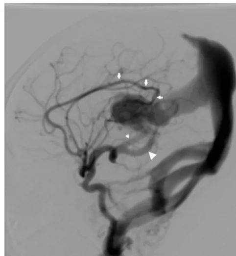
The patient’s cardiorespiratory failure worsened despite catecholamine, di-uretic, and human atrial natriuretic peptide administration. Therefore, the patient was intubated and placed on mechanical ventilation at 4 days of age. Initial angiography was performed at 11 days of age. Digital subtraction angiography revealed that multiple shunts had formed in the median vein of the prosencephalon. The arterial supply, including the left pericallosal, right lenticulostriate, right anterior choroidal, and right posterior choroi-dal arteries, indicated a diagnosis of choroidal-type VGAM. The galenic vein drained into the parietal superior sag-ittal sinus via the persistent primitive falcine sinus. Stenosis was observed at the tentorium cerebelli in the drain-age pathway. Further draindrain-age path-ways included the transverse, acces-sory transverse, sigmoid, occipital, and marginal sinuses (Figs 2 and 3).
A microcatheter was guided into a shunt vessel from the right medial posterior choroidal artery, which was the vessel with the largest caliber, and emboliza-tion was performed by using the mixture of n-butyl cyanoacrylate (n-BCA), ethio-dized oil, and tantalum powder. The em-bolization procedure remarkably im-proved the patient’s respiratory status, and the patient was extubated at 22 days of age. Additional embolization of the 2 shunt vessels of the right anterior and left posterior choroidal arteries was per-formed when the patient was 1 year of age. Spinal cord MRI and thoracoab-dominal contrast-enhanced computed tomography at 1 year of age revealed nothing that was clearly suggestive of ar-teriovenous fistula in the lungs, liver, or spinal cord. MRI and magnetic reso-nance angiography performed when the patient was 2 years 6 months of age re-vealed a slight residual shunt lesion and mild dilation of the vein of Galen, but the patient’s development was otherwise normal.
FIGURE 2
Lateral view of the right internal carotid angiogram. The arterial supply including the left anterior pericallosal artery (arrows), right medial posterior choroidal arteries (small arrowhead), and right lateral posterior choroidal arteries (large arrowhead) are shown.
FIGURE 3
Lateral view of the left internal carotid angiogram. Multiple feeding arteries from the left medial posterior choroidal arteries (small arrowhead) and left lateral posterior choroidal arteries (large arrowhead) demonstrate shunting at the inferior-caudal portion of the dilated galenic vein (arrow). These features are characteristic of typical choroidal-type VGAM.
Genetic testing of both the patient and his mother was performed. The genomic DNA used in the testing was isolated from the peripheral blood. The regions that encode the proteins of the affected genes in HHT, the endoglin and activin receptor-like kinase (ALK1) genes, were directly sequenced. A de-letion of 13 bases was seen in exon 11 of the endoglin gene, and a frame-shift mutation in the stop codon (c.1672_1684delGGGTCTCAAGACC p. Gln558fsX568) was seen. The mother carried the same mutation. No anoma-lies were seen in theALK1gene.
DISCUSSION
The pathogenesis of VGAM is unknown. One article concerning the genetics of VGAM reported an anomaly of the
RASA1 gene.4 The RASA1 gene is the causative gene in capillary malformation-arteriovenous malfor-mation (CM-AVM) and is seen in the proteins of transforming growth fac-tor in vascular endothelial cells. CM-AVM has autosomal dominant inheri-tance. A diagnostic sign of this disease
is multiple capillary malformations, which are highly expressive of its occurrence.
HHT, or Rendu-Osler-Weber disease, is a rare disorder with an incidence of 1 to 2 in 100 000 people5; however, this incidence may be an underestimation.6 Epistaxis is the most common presen-tation. More than 90% of cases mani-fest by the age of 21 years.7Skin and mucosa telangiectasia and arterio-venous malformations in the lungs, brain, liver, and spinal cord are com-mon manifestations. The phenotypes of HHT are diverse, and, as in the pres-ent case, there are often no symptoms such as epistaxis or skin/lip telangiec-tasia during the neonatal period.
Cerebral arteriovenous malformation is a common manifestation in patients with HHT. Maher et al8 reported the cases of 12 patients with a history of cerebral vascular malformations among 321 patients with HHT. Ten patients had arteriovenous malforma-tions, 1 had a dural arteriovenous fistula, and 1 had a cavernous malformation.
There have been few reports concern-ing VGAM in patients with HHT. Al-though there has been a report of vein
of Galen–like vascular malformations in familial HHT, it preceded the era in which genetic testing was performed.1
Several culprit genes have been
identi-fied for HHT. The endoglin (HHT type 1) gene has been identified on 9q34.1,9 and the ALK1 (HHT type 2) gene has been identified on 12q11-q14.10 The gene responsible for HHT type 3 has been identified on 5q31.3-q32.11In addi-tion, a genetic anomaly on 7p14 has been reported for HHT type 4.12
The anomaly of the endoglin gene seen in our patient was reported to be asso-ciated with intracranial arteriovenous malformation.13
CONCLUSIONS
We have described here a case of VGAM that occurred in combination with HHT. The accumulation of further
genetic investigations in other pa-tients may help to elucidate the patho-physiology of VGAM.
REFERENCES
1. Boynton RC, Morgan BC. Cerebral arterio-venous fistula with possible hereditary tel-angiectasia.Am J Dis Child. 1973;125(1): 99 –101
2. Lasjaunias P, Beugge KG, Berenstein A. Sur-gical Neuroangiography 3: Clinical and In-terventional Aspects in Children.2nd ed. Berlin, Germany: Springer-Verlag; 2006
3. Shovlin CL, Guttmacher AE, Buscarini E, et al. Di-agnostic criteria for hereditary hemorrhagic tel-angiectasia (Rendu-Osler-Weber syndrome).Am J Med Genet. 2000;91(1):66 – 67
4. Revencu N, Boon LM, Mulliken JB, et al. Parkes Weber syndrome, vein of Galen an-eurysmal malformation, and other fast-flow vascular anomalies are caused byRASA1 mutations.Hum Mutat. 2008;29(7):959 –965 5. Bianchi DW. Telangiectasia, Osler hemor-rhagic. In: Buyse ML, ed.Birth Defects
Ency-clopedia. Cambridge, United Kingdom: Wiley-Blackwell; 1990:1654 –1655
6. Guttmacher AW, McKinnon WC, Upton MD. Hereditary hemorrhagic telangiectasia: a disorder in search of the genetics commu-nity.Am J Med Genet. 1994;52(2):252–253
7. AAssar OS, Friedman CM, White RI Jr. The natural history of epistaxis in hereditary hemorrhagic telangiectasia.Laryngoscope. 1991;101(9):977–980
8. Maher CO, Piepgras DG, Brown RD Jr, Friedman JA, Pollock BE. Cerebrovascular manifestations in 321 cases of hereditary hemorrhagic telangi-ectasia.Stroke. 2001;32(4):877– 882
9. McAllister Mc, Grogg KM, Jonson DW, et al. Endoglin, a TGF-beta binding protein of en-dothelial cells, is the gene for hereditary haemorrhagic telangiectasia type 1. Nat Genet. 1994;8(4):345–351
10. Johnson DW, Berg JN, Baldwin MA, et al. Mu-tations in the activin receptor-like kinase 1 gene in hereditary haemorrhagic telangiec-tasia type 2.Nat Genet. 1996;13(2):189 –195
11. Cole SG, Begbie ME, Wallace GM, Shovlin CL. A new locus for hereditary haemorrhagic telangiectasia (HHT3) maps to chromosome 5.J Med Genet. 2005;42(7):577–582
12. Bayrak-Toydemir P, McDonald J, Akarsu N, et al. A fourth locus for hereditary hemor-rhagic telangiectasia maps to chromosome 7. Am J Med Genet A. 2006;140(20): 2155–2162
13. Paquet ME, Pece-Barbara N, Vera S, et al. Analysis of several endoglin mutants re-veals no endogenous mature or secreted protein capable of interfering with normal endoglin function. Hum Mol Genet. 2001; 10(13):1347–1357
e1310 TSUTSUMI et al
at Viet Nam:AAP Sponsored on August 28, 2020
www.aappublications.org/news
DOI: 10.1542/peds.2010-0961 originally published online October 10, 2011;
2011;128;e1307
Pediatrics
Masaki Shintani, Shunsuke Nosaka, Hidekazu Masaki and Yuo Iizuka
Yoshiyuki Tsutsumi, Rika Kosaki, Yushi Itoh, Keiko Tsukamoto, Rumiko Matsuoka,
Mutation
Vein of Galen Aneurysmal Malformation Associated With an Endoglin Gene
Services
Updated Information &
http://pediatrics.aappublications.org/content/128/5/e1307
including high resolution figures, can be found at:
References
http://pediatrics.aappublications.org/content/128/5/e1307#BIBL
This article cites 11 articles, 2 of which you can access for free at:
Subspecialty Collections
http://www.aappublications.org/cgi/collection/genetics_sub
Genetics
following collection(s):
This article, along with others on similar topics, appears in the
Permissions & Licensing
http://www.aappublications.org/site/misc/Permissions.xhtml
in its entirety can be found online at:
Information about reproducing this article in parts (figures, tables) or
Reprints
http://www.aappublications.org/site/misc/reprints.xhtml
DOI: 10.1542/peds.2010-0961 originally published online October 10, 2011;
2011;128;e1307
Pediatrics
Masaki Shintani, Shunsuke Nosaka, Hidekazu Masaki and Yuo Iizuka
Yoshiyuki Tsutsumi, Rika Kosaki, Yushi Itoh, Keiko Tsukamoto, Rumiko Matsuoka,
Mutation
Vein of Galen Aneurysmal Malformation Associated With an Endoglin Gene
http://pediatrics.aappublications.org/content/128/5/e1307
located on the World Wide Web at:
The online version of this article, along with updated information and services, is
by the American Academy of Pediatrics. All rights reserved. Print ISSN: 1073-0397.
the American Academy of Pediatrics, 345 Park Avenue, Itasca, Illinois, 60143. Copyright © 2011 has been published continuously since 1948. Pediatrics is owned, published, and trademarked by Pediatrics is the official journal of the American Academy of Pediatrics. A monthly publication, it
at Viet Nam:AAP Sponsored on August 28, 2020
www.aappublications.org/news