© 2018 IJSRST | Volume 4 | Issue 2 | Print ISSN: 2395-6011 | Online ISSN: 2395-602X Themed Section: Science and Technology
Detection of Cholera Toxin Using Lactose-Decorated Silver
Nanoparticles
Ratnadeep R. Koyale, Imran L. Patel, Samradni D. Pingale
*Department of Biotechnology, Sinhgad College of Science, Ambegaon Bk, Pune, Maharashtra, Pune, Maharastra, India
ABSTRACT
A novel method for biosensor preparation is mechanized, which uses silver nanoparticles as sensing molecules. These nanoparticles were prepared using banana leaf extract-mediated synthesis, which is decorated using PEG to stabilize their surface. The silver nanospheres were synthesized with size 100 to 120 nm enveloped with PEG coating with the stability of -16 mV. They were characterized using UV-spectroscopy, particle size analyzer and SEM imaging. These particles are coated with lactose decorated silver nanoparticles against food pathogen such as cholera toxin. The food samples were exposed to these plates and the surface plasmon absorbance was recorded at 419 nm. Changes in surface Plasmon absorbance showed biosignals that specify the presence of toxin producing bacteria in the sample.
Keywords: Biosensor, Cholera toxin, Surface Plasmon Absorbance, Zeta Potential.
I.
INTRODUCTION
Nanoparticles can be synthesized by different methods like physical, chemical and biological methods [1]. Due to several rising food borne diseases nowadays scientists are doing research on use nanoparticles for the detecting purpose i.e. Preparation of biosensors using nanoparticles for the detection of different food pathogens from contaminated food or canned food [2,3]. Nanoparticles are synthesized from clusters of atoms which are nanoscopic in nature [4]. These particles are not visible to naked eyes, hence require large magnification instruments for observation. Nanoparticles are fine entities having size ranging from 100 to 2500 nanometers [5]. The branch of science which deals with studies and the application of these particles is called nanotechnology.
Silver nanoparticles and gold nanoparticles are highly studied and applied in the field of medical sciences in comparison to others [6]. Silver nanoparticles in hydrosols are one of the most attractive inorganic
materials due to their tremendous applications in photography [7], catalysis [8], biosensor [9], Biomolecular detection [10], diagnostics [11], and particularly antimicrobial activity [12,13,14].
Different methods have been used in the past for the synthesis of silver nanoparticles, which include reduction in solution [15], radiation assisted [16], chemical and photoreduction in reverse micelles [17], thermal decomposition of silver compound [18] and recently by beginners or green synthesis route [19,20,21]. Biological synthesis of silver nanoparticles using different leaf extracts is investigated and dispersed particle size has been reported [22,23,24,].
[26].Nanobiosensor is a measurement system that uses nanoparticles to detect analyte such as toxins produced by bacteria.Food pathogens like cholera toxin has the ability to produce the toxin that causes
watery diarrhea in humans which may be lethal. The increased applications of nanoparticles have
resulted in raising hopes of employing nanoparticles as alternative detecting agent. This study involves biosynthesis of silver nanoparticles using Musa paradisiaca leaf extract (Banana- Green extract), its surface modification using PEG and its mechanism for detection of cholera toxin.
II.
METHODS AND MATERIAL
2.1.Reagents:
Analytical grade silver nitrate (AgNO3), polyethylene
glycol (PEG), phosphate buffered saline (PBS), NaCl, Polyvinylpyrrolidone (PVP), lactose sugar, rectangular glass chips.
2.2.Biosynthesis & PEGlyation of silver nanoparticles: Silver nanoparticles were synthesized using the reduction method induced by polyphenols from the leaves of Musa paradisiaca [. The leaves of Musa
paradisiaca were obtained from Pune, India and were
identified by consulting botanical experts in Sinhgad College of Science, Pune. The leaf materials were washed to remove excess dirt and contaminants, chopped into pieces and oven dried at 150 oC until the
dry weight was uniform. Then dried leaves were crushed into powder form. Then 5 g of powder leaves was boiled into 250ml glass beaker containing 100 ml deionized water for 5 min. The aqueous extract was filtered using Whatman no. 1 filter paper to remove larger plant material clumps. The extract was stored at 4oCto be used for the biosynthesis of silver
nanoparticles from silver nitrate solution. An aqueous solution of AgNO3 (1 mm) was prepared in 250 ml
Erlenmeyer flask and 0.3% PEG was added to induce PEGlyation of nanoparticles and increase stability. The aqueous extract of Musa paradisiaca was added to the above solution to initiate reduction of Silver ions. The reaction (1 ml aqueous extract + 99 ml 1mM AgNO3
with 0.3% PEG) was carried out in light at room temperature. The reaction mixture was observed for
color changes from slight yellowish to red and finally into brown color, which indicated the synthesis of silver nanoparticles. Further the AgNPs were filtered, centrifuged and freeze dried.
2.3.Characterization of AgNP‟s:
This is an important aspect of nanoparticles research to achieve better interpretation of results with primitive understanding. The color changes were recorded along with periodic sampling and scanning using UV-Vis spectrophotometry (Systronics type 108-double beam) ranging from 200 to 680 NM for a maximum time period upto 120 min. Further reaction mixture was centrifuged at 10000 rpm for 10 mins at 4oC, dissolved in distilled water and washed by
centrifugation to remove plant impurities.Particle size analysis (Nanophox NX0088, SympatecGmbh) and zeta potential measurement (DelsaTm Nano, Beckman Coulter) were carried to analyse the size and stability of AgNPs. An FTIR spectrum (Bruker) of the AgNPs was measured in the transmittable mode in the range 4000 to 500 cm-1 in KBr pellets. Further
the FESEM analysis (Nova Nano SEM 450) i.e. Field emission scanning electron microscopy was performed to confirm the size and morphology of the AgNPs.
2.4. Preparation of Nano chip:
The stabilized AgNp‟s were tagged on the surface on glass chip with the help of PVP polymer. PVP polymer has the glass property which can tag nanoparticles on the surface of glass chip. The layer of lactose sugar was given on the surface of those nanoparticles to induce protein-ligand interaction.
III.
RESULTS AND DISCUSSION
3.1.Biogenic synthesis and characterization of AgNPs:
ethylene glycol) mediated surface coating was performed. In addition, of aqueous plant extract to silver nitrate, the reaction mixture turned transparent to light yellow, further into reddish brown. This was a clear indication for AgNPs synthesis using polyphenol-reduction method. In this study, it is suggested that PEG coating creates thiol group on the surface of AgNPs, increasing the stability.
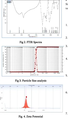
UV-Vis spectroscopy plays an important role in the characterization of silver nanoparticles synthesis. A characteristic surface plasmon peak was observed at 418nm with a sharp peak for treating PEG-AgNPs. It is well understood that the color of AgNPs influences the size of particles due to which a characteristic surface plasmon peak is observed in the solution. An FTIR spectrum confirms the presence of glycolated (PEGlyated) thiol groups due to peak observed at 2865 and 3157 cm-1. This spectra explains the presence of
free AgNP‟s coated with PEG i.e functional hydroxyl groups.2865 cm-1 peak represents CH stretching
alkanes while 3157 cm-1 represents Alcohol/Phenol –
OH stretch which indicates presence of AgNPs coated with glycol group.
The diameter and zeta potential of PEG-AgNPs were detected using Nanophox and Beckman coulter Delsa Tm. It was observed that uniform AgNPs were formed with average size of 131±18 nm(Fig). The negative Zeta potential of AgNPs was observed to be at -15.8 mv which confirms the higher stability of particles. Greater zeta potential indicates higher negative charge which creates stronger repulsive forces. This avoids agglomeration and increase the stability. The characterization of nanoparticles by FESEM revealed that PEG-AgNPs had a spherical shape observed with uniformity and monodispersity. This confirms that PEG-AgNPs had narrow particle size distribution due to PEG coating as compared to other plant mediated synthesis.
3.2. Preparation of Nano chip:
The importance of this lactose sugar is that the cholera toxin has the highest affinity towards the lactose sugar, it can bind to the lactose sugar easily and the interaction between lactose sugar and cholera toxin is like protein-ligand interaction. After the preparation of nanochip, it was dried at temperature 34 for 2 hours. After the proper incubation period the cholera toxin was exposed to the nanochip on the surface of lactose. The results were observed.
Silver Nanochip
Fig 2. FTIR Spectra
Fig 3. Particle Size analysis
Fig. 4. Zeta Potential
IV.
CONCLUSION
The color of surface of nanochip was previously red due to the absorption of light by nanoparticles. After the exposure of cholera toxin the toxins were binding on the surface of lactose sugar because of that the surface area of nanoparticles was indirectly covered by toxin and hence the color of nanochip changed to the faint purple. Red color changes to faint purple color on Bionanochip. Stabilized Ag-Np‟s were synthesized using PEG as a stabilizer and embedded in
a PVP matrix to prepare nanochip. Lactose-decorated Silver Nanoparticles can be used for the detection of toxin in a food sample.
V.
REFERENCES
1. Iravani, S., et al. "Synthesis of silver nanoparticles: chemical, physical and biological methods “(2014).
2. .Nuria S. etal. “Nanoparticles based biosensors for detection of pathogenic bacteria. “ (2009). 3. Claire L etal. “Glyconanoparticles
forcolourimetric detection of Cholera toxin. ”
Analytical chemistry (2007).
4. Kunwar, Puskal, et al. "Sub-micron scale patterning of fluorescent silver nanoclusters using low-power laser." Scientific reports 6 (2016).
5. Ruzer, Lev S. "Exposure and Dose: Health Effect Studies Associated with Nanometer Aerosols."
Journal of Nanomedicine & Nanotechnology
(2011).
6. Söderstjerna, Erika, et al. "Silver and gold nanoparticles exposure to in vitro cultured retina–studies on nanoparticle internalization, apoptosis, oxidative stress, glial-and microglial activity." PloS one 9.8 (2014): e105359.
7. Colvin, V. L., M. C. Schlamp, and A. Paul Alivisatos. "Light-emitting-diodes made from cadmium selenide nanocrystals and a semiconducting polymer." Nature 370.6488 (1994): 354-357.
8. Wang, Ying, and N__ Herron. "Nanometer-sized semiconductor clusters: materials synthesis, quantum size effects, and photophysical properties." The Journal of Physical Chemistry
95.2 (1991): 525-532.
9. Schmid, Guenter. "Large clusters and colloids. Metals in the embryonic state." Chemical
Reviews 92.8 (1992): 1709-1727.
10. Hoffman, A. J., et al. "Q-sized cadmium sulfide: synthesis, characterization, and efficiency of
0 10 20 30 40 50 60 70 80 90 100
cum
ul
at
ive
d
is
tri
bu
ti
on
Q
(x
) /
%
0 1 2 3 4 5 6 7 8
de
ns
it
y
di
st
ri
bu
ti
on
q
*(
x)
0.5 1.0 5 10 50 100 500 1000 5000 10000
photoinitiation of polymerization of several vinylic monomers." The Journal of Physical
Chemistry 96.13 (1992): 5546-5552.
11. Hamilton, J. F., and R. C. Baetzold. "Catalysis by small metal clusters." Science 205.4412 (1979): 1213-1220.
12. Mansur, Herman S., et al. "Photoelectrochemical properties of „Q-state‟CdS particles in arachidic acid Langmuir–Blodgett films." Journal of the
Chemical Society, Faraday Transactions 91.4
(1995): 665-672.
13. Mukherjee, Priyabrata, et al. "Fungus-mediated synthesis of silver nanoparticles and their immobilization in the mycelial matrix: a novel biological approach to nanoparticle synthesis."
Nano Letters 1.10 (2001): 515-519.
14. Klaus-Joerger, Tanja, et al. "Bacteria as workers in the living factory: metal-accumulating bacteria and their potential for materials science." TRENDS in Biotechnology 19.1 (2001): 15-20.
15. Sylvestre, Jean-Philippe, et al. "Stabilization and size control of gold nanoparticles during laser ablation in aqueous cyclodextrins." Journal of
the American Chemical Society 126.23 (2004):
7176-7177.
16. Dolgaev, S. I., et al. "Nanoparticles produced by laser ablation of solids in liquid environment."
Applied surface science 186.1 (2002): 546-551.
17. Kim, Sungil, et al. "Catalytic effect of laser ablated Ni nanoparticles in the oxidative addition reaction for a coupling reagent of benzylchloride and bromoacetonitrile." Journal
of Molecular Catalysis A: Chemical 226.2 (2005):
231-234.
18. Link, Stephan, et al. "Laser-induced shape changes of colloidal gold nanorods using femtosecond and nanosecond laser pulses." The
Journal of Physical Chemistry B 104.26 (2000):
6152-6163.
19. Iravani, S., et al. "Synthesis of silver nanoparticles: chemical, physical and biological
methods." Research in pharmaceutical sciences
9.6 (2014): 385.
20. Mafuné, Fumitaka, et al. "Formation of gold nanoparticles by laser ablation in aqueous solution of surfactant." The Journal of Physical
Chemistry B 105.22 (2001): 5114-5120.
21. Tarasenko, N. V., et al. "Synthesis of nanosized particles during laser ablation of gold in water."
Applied surface science 252.13 (2006):
4439-4444.
22. Vilchis-Nestor, Alfredo R., et al. "Solventless synthesis and optical properties of Au and Ag nanoparticles using Camellia sinensis extract."
Materials Letters 62.17 (2008): 3103-3105.
23. Begum, Naznin Ara, et al. "Biogenic synthesis of Au and Ag nanoparticles using aqueous solutions of Black Tea leaf extracts." Colloids
and surfaces B: Biointerfaces 71.1 (2009):
113-118.
24. Kesharwani, Jayendra, et al. "Phytofabrication of silver nanoparticles by leaf extract of Datura metel: hypothetical mechanism involved in synthesis." Journal of Bionanoscience 3.1 (2009): 39-44.
25. Cooper, Dustin L., and Sam Harirforoosh. "Effect of formulation variables on preparation of celecoxib loaded polylactide-co-glycolide nanoparticles." PloS one 9.12 (2014): e113558. 26. Chang, Wei-Chang, et al. "Surface PEGylation
of Silver Nanoparticles: Kinetics of Simultaneous Surface Dissolution and Molecular Desorption."