Correlation between peripapillary retinal nerve fiber layer
thickness and visual function changes in patients receiving
ethambutol
ABSTRAK
Latar belakang: Terdapat beberapa studi yang telah menunjukkan adanya penurunan jumlah sel ganglion retina pada hewan yang diberikan etambutol. Studi ini bertujuan mengevaluasi efek etambutol pada ketebalan serabut saraf retina peripapil manusia. Selain itu studi ini juga ingin melihat korelasi antara perubahan ketebalan retina tersebut dengan perubahan beberapa parameter fungsi penglihatan.
Metode: Studi kohort pada 29 subyek penderita tuberkulosis yang berobat di salah satu pusat pengobatan tuberkulosis di Jakarta. Seluruh subyek menjalani pemeriksaan ketebalan retina menggunakan optical coherence tomography
(OCT) dan beberapa parameter fungsi penglihatan (tajam penglihatan, sensitivitas kontras, sensitivitas warna dan lapang pandangan) sebelum dan dua bulan setelah mengkonsumsi etambutol. Perbedaan parameter tersebut sebelum dan setelah konsumsi etambutol dianalisis menggunakan uji T-berpasangan ataupun uji Wilcoxon. Hubungan antara perubahan ketebalan retina dan perubahan fungsi penglihatan dinilai dengan uji Spearman.
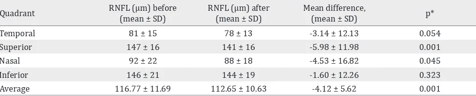
Hasil: Sebelum dan setelah pengobatan dengan etambutol terdapat perubahan bermakna pada ketebalan retina di
kuadran superior (147 dan 141 μm, p = 0,001), nasal (92 dan 88 μm, p = 0,045), dan rerata seluruh kuadran (116,77 dan 112,65 μm, p = 0,001). Tidak terdapat hubungan antara perubahan
ketebalan retina ini dengan perubahan fungsi penglihatan.
Kesimpulan: Konsumsi etambutol selama dua bulan dalam dosis yang dianjurkan dianggap masih aman terhadap retina manusia dan terhadap fungsi penglihatan.
ABSTRACT
Background: Recent animal studies revealed the decreased amount of retinal ganglion cells after treatment with ethambutol. The aim of this study was to evaluate the changes of peripapillary retinal nerve fiber layer (RNFL) thickness in patients receiving ethambutol therapy, as well as to know the correlation of RNFL thickness changes with the changes of visual function.
Methods: This was a cohort study on 29 subjects at one tuberculosis treatment center, Jakarta. Subjects underwent RNFL thickness measurement using optical coherence tomography (OCT) and several visual function parameters (visual acuity, contrast sensitivity, color sensitivity and visual field) before and two-months following ethambutol therapy. Statistical comparison between pre- and post- ethambutol measurements were done using either paired T-test or Wilcoxon test. Correlations between anatomical and functional changes were assessed with Spearman correlation test.
Results: There were significant changes of peripapillary RNFL thickness in superior (147 and 141 μm, p = 0.001), nasal (92 and 88 μm, p = 0.045) quadrants and average RNFL thickness (116.77 and 112.65 μm, p = 0.001). No significant correlation between RNFL thickness changes and the changes of visual function parameters (p > 0.05).
Conclusion: Ethambutol consumption for two months in recommended dose is still considered safe to human retina and visual status.
Keywords: ethambutol, optical coherence tomography, optic neuropathy, retinal nerve
pISSN: 0853-1773 • eISSN: 2252-8083 • http://dx.doi.org/10.13181/mji.v24i1.1065 • Med J Indones. 2015;24:19-23 • Received 10 Sept 2014 • Accepted 20 Feb 2015
Correspondence author: Dialika, [email protected] Clinical Research
Copyright @ 2015 Authors. This is an open access article distributed under the terms of the Creative Commons Attribution-NonCommercial-ShareAlike 4.0 International License (http://creativecommons.org/licenses/by-nc-sa/4.0/), which permits unrestricted non-commercial use, distribution, and reproduction in any medium, provided the original author and source are properly cited.
Dialika,1 Muhammad Sidik,2 Syntia Nusanti,2 Aria Kekalih3
1 Department of Ophthalmology, Faculty of Medicine, Universitas Indonesia, Jakarta, Indonesia
2 Department of Ophthalmology, Faculty of Medicine, Universitas Indonesia, Cipto Mangunkusumo Hospital, Jakarta, Indonesia
3 Department of Community Medicine, Faculty of Medicine, Universitas Indonesia, Cipto Mangunkusumo Hospital, Jakarta,
Ethambutol is one of the first line drugs used in treating tuberculosis (TB) together
with isoniazid, rifampicin, pyrazinamide and streptomycin.1 According to WHO report of
2011, Indonesia is in the fourth-highest number of TB infection in 2010, followed with India, China and South Africa2 One of the serious side effects caused by ethambutol consumption is
ocular side effect known as ethambutol optic neuropathy (EON).
The incidence of EON varies from 1.5-10% of all
patients consuming ethambutol, depending on the dosage and duration of drug consumption.3,4
Leibold5 found an incidence of 11% in patients
consuming 35 mg/kgBW/day of ethambutol, and only 2% in patients consuming less than 30 mg/ kgBW/day.
The clinical manifestation of EON involves central fibers damage leading to loss of visual acuity, color vision, contrast sensitivity and field defect (mainly central scotoma).6-8 This ocular
toxicity has variable prognosis from reversible
to permanent disturbance.9,10 Early detection is
generally considered to improve prognosis.3
Various studies performed in animal have shown
decreased amount of retinal ganglion cells in animals treated with ethambutol.11,12 So that nowadays retinal ganglion cell damage is more
widely believed to cause EON.
Optical coherence tomography (OCT) is a non-contact examination technique, which shows cross-sectional images of the retina. Assessing retinal thickness quantitatively, either in the
macula or peripapillary region, has been made possible with this device. Evaluating peripapillary
retinal nerve fiber layer (RNFL) is popular in optic
neuropathy cases especially glaucoma.13
There have been some cross-sectional studies evaluating peripapillary RNFL in patients with EON. Those studies revealed RNFL thinning in patients diagnosed with EON.10,14,15 This condition
is assumed to happen due to damage in retinal
ganglion cells in the disease. Cross-sectional data of RNFL thickness is considered not sufficient in making conclusion of RNFL thinning among patients with EON, due to the highly variable baseline RNFL thickness. This study aimed to evaluate the course of peripapillary RNFL
thinning after patients consume ethambutol and also to evaluate the correlation with some visual
function parameters. In the future it is expected that OCT scanning of peripapillary RNFL can
be used as an alternative diagnostic procedure
among patients with possible EON.
METHODS
This was a cohort study on 30 individuals newly diagnosed with TB infection, conveniently
recruited from PPTI (perkumpulan pemberantasan
tuberkulosis Indonesia) clinic, DKI Jakarta region
beginning from March - July 2013 (including two-month follow-up). Anti-tuberculosis regimens were all given in the form of a fixed dose combination (FDC) consisting of ethambutol, isoniazid,
rifampicin and pyrazinamide, in accordance to
WHO guideline for TB treatment. Inclusion criteria were patient age 18-60 year-old, agree to not
consuming alcohol during the course of study, and
agree to sign the study consent. While patients with visual acuity less than 6/6, defect in color sensitivity, intra-occular pressure (IOP) > 21 mmHg, presence
of retinal defect, optic nerve abnormality or any
opacity in the visual axis which may contribute to poor OCT results, were excluded.
All subjects underwent complete ophthalmological and OCT examination before and two months after consuming ethambutol. Each examination
included an assessment of visual acuity using the early treatment diabetic retinopathy study
(ETDRS) Chart, color sensitivity using the hardy-rand-rittler (HRR, Richmond products, US) pseudoisochromatic plates, and Pelli-Robson
chart was used for contrast sensitivity test.
Humphrey 24-2 standard automated perimetry using the Swedish Interactive Test Algorithm strategy (Carl Zeiss Meditec, Dublin, California, USA) was performed at each visit. OCT Stratus (Carl Zeiss Meditec, Inc, Dublin, CA) was used to examine peripapillary RNFL on both eyes of each patient using the retinal nerve fiber
layer analysis protocol, as has been described elsewhere. Patients who were not able to attend
the scheduled follow-up time, were considered
lost to follow up.
differences between pre- and post-ethambutol
consumption were assessed using the paired
T-test or Wilcoxon test. Correlations between
anatomical and functional changes were assessed
with Spearman correlation test. P value less than 0.05 is considered significant. The study protocol has been approved by the Ethics Committee of the Faculty of Medicine, Universitas Indonesia (No. 48/H2.F1/ETIK/2013).
RESULTS
There were initially 30 subjects enrolled in the
study, one subject dropped out before the second
examination owing to stopped continuing TB
treatment that only 29 subjects included in the study analysis. Patient’s body weight was also recorded to get a more precise dosage received
according to body weight. There were 21 (72.4%) male subjects and 8 (27.6%) females subject enrolled in the study, with age ranging from 18 to 55 years old (median 37 years old). The mean
calculated dosage of ethambutol received per body
weight in the study was 16.44 mg/kgBW/day.
Before ethambutol
median (min-max) median (min-max)After ethambutol p* Visual acuity† 0 (0–0.14) 0 (0-0.12) 0.022
Color
sensitivity 19 (13–20) 19 (14–20) 0.001 Contrast
sensitivity 1,95 (1,35–1,95) 1,95 (1.5-1.95) 0.737 Mean
deviation -4 (-25-0) -2.29 (-23.73-0.2) 0.001 Table 1. Visual function changes associated with two months ethambutol consumption
* Wilcoxon test, p < 0,05 significant
† logMar
Quadrant RNFL (μm)(mean ± SD)before RNFL (μm) after(mean ± SD) Mean difference,(mean ± SD) p*
Temporal 81 ± 15 78 ± 13 -3.14 ± 12.13 0.054
Superior 147 ± 16 141 ± 16 -5.98 ± 11.98 0.001
Nasal 92 ± 22 88 ± 18 -4.53 ± 16.82 0.045
Inferior 146 ± 21 144 ± 19 -1.60 ± 12.26 0.323
Average 116.77 ± 11.69 112.65 ± 10.63 -4.12 ± 5.62 0.001
Table 2. Changes in peripapillary retinal nerve fiber layer thickness before and two months after receiving ethambutol
RNFL: retinal nerve fiber layer, SD: standard deviation *Dependent T-test, before vs after ethambutol
Visual acuity
(logMar) sensitivityColor sensitivityContrast Visual field (MD)
p value 0.142 0.744 0.084 0.515
r value* -0.195 -0.044 -0.229 -0.087
Tabel 3. Correlation between visual function parameters and changes of mean RNFL thickness
Effects of ethambutol consumption here were divided into 2, functional and anatomical effects, as seen in table 1 and table 2. There are
statistically significant changes of visual acuity, color sensitivity and visual field after consuming ethambutol (p < 0.05) (Table 2). Two out of 58 eyes examined (from 2 different subjects) were revealed suitable for the EON diagnostic criteria. One has decreased contrast sensitivity and the other has decreased color sensitivity. On both
subjects, ethambutol consumption has been
discontinued after the second examination. There are no statistically significant correlation between mean peripapillary RNFL thickness change and all visual function parameters changes as expressed
in table 3.
DISCUSSION
Optic neuropathy due to mitochondrial
dysfunction in retinal ganglion cells can happen
either acquired or inherited. These diseases are
similar in signs and symptoms one with another.16
Some of the most common cause for acquired
mitochondrial optic neuropathies are vitamin
deficiency, nutritional deficiency, toxic substance exposure and drug exposures. Some drugs that are well known to cause optic neuropathies
include ethambutol, chloramphenicol, linezolid, erythromycin and streptomycin.16,17 Ethambutol
as one of first line anti-TB drug, is well known to
be able to cause ethambutol optic neuropathy, which was postulated to be related to damage on mitochondria of retinal ganglion cells.18
Demographic data shows that there are more
male subjects with male to female ratio of
2.62.19 This unequal sex distribution happens in almost all parts of the world, due to several
possible reasons. This male-female ratio is also in accordance with WHO report 2011 regarding sex distribution of TB patients in South East Asia.2
Mean age of subjects in this study (35 year-old) is also in accordance with WHO report 2011.2
Anti-TB drugs given to all subjects in this study were in the form of fixed dose combination (FDC), which contain 275 mg of ethambutol and was
prescribed according to patient’s body weight.
In this study, subjects received ethambutol with average dose of 16.4 mg/kgBW/day. This dose is
in the acceptable range of dose as recommended
by WHO, that is between 15-20 mg/kgBW/day,
and considered to cause very minimal side effects.
During the study, no patient complained of any visual disturbance. This can be explained since EON (if any) is mostly asymptomatic at the beginning. Decrease of color or contrast sensitivity might
happen without any complaint from the patient.7,8
This study found statistically significant decrease of RNFL thickness in superior and nasal quadrants.
This is similar with earlier studies in animals, that found decreased retinal ganglion cell density among animals fed by ethambutol.11,12 However, the reduction is still in the standard deviation range, and is still less than the value considered
significant set at the beginning of the study (10 μm). Recalculation of samples size revealed that the power was less than 80%, so the sample size of this study is considered insufficient. Chung, et
al20 and Tang, et al21 in their study also did not find
any significant RNFL difference two months after
ethambutol consumption.
Sadun, et al17 have previously explained a theory
that in mitochondrial optic neuropathy cases, there is a time period where visual function
deficit happens without any reduction of ganglion cell axons. This is made possible, due to an earlier compensatory phase in the axons,
which is mitochondrial congregation, right before
apoptosis start taking place. This compensatory
phase is considered to already be able to cause visual function defects.
Some studies have reported that EON can occur
as early as two months after starting ethambutol consumption.3,9,18 This duration is different
among different studies, with other report
mentioning a mean duration 3-5 months of drug consumption and one descriptive study in Korea even reporting that EON cases occured after nine
months of drug ethambutol consumption.3 This study only evaluated patients who consumed the drug for two months, so it is also possible that it is
too early to expect some effect in RNFL thickness.
Apart from RNFL thickness, this study found statistically significant reduction of mean acuity, color sensitivity and visual field (expressed with mean deviation). However, the changes of each
visual function parameter are clinically considered
insignificant. These results are different when
compared to earlier study by Menon, et al22 which
is able to detect visual function defect in 20% of
their subjects.
Some earlier studies (especially in glaucoma field) have found good correlation between RNFL thickness and severity of glaucoma. However, we
found no correlation between visual function
changes and average RNFL thickness changes.
This is thought to be due to the fact that most subjects only suffer a minimum reduction of
RNFL thickness (less than 10 μm), thus, no visual
function changes happened yet.
This study has some limitations, in which consecutive sampling method was used and
the samples only came from one center. Other limitation is that the HFA result in one-third of all subjects did not meet the required criteria as an accurate result (false positive, false negative and fixation losses index). This has made sensitivity changes difficult to interpret. The short
follow-up duration was also considered as a limitation,
as it is well known that occurrence of EON is influenced by the duration of drug consumption.
In conclusion, this preliminary study found
a significant decrease of RNFL thickness in
some patients after two months of ethambutol consumption without changes in visual function
parameters. In other words, ethambutol
dose is still considered safe to human retina and their visual status.
Conflicts of Interest
The authors affirm no conflict of interest in this
study.
REFERENCES
1. Perhimpunan Dokter Paru Indonesia. Tuberkulosis: pedoman diagnosis dan penatalaksanaan di Indonesia. Jakarta: Perhimpunan Dokter Paru Indonesia; 2011. p. 20-30. Indonesian.
2. World Health Organization [Internet]. Global tuberculosis control: WHO report 2011 [cited 2012 Sept 13] Available from: http://apps.who.int/iris/ bitstream/
3. Lee EJ, Kim SJ, Choung HK, Kim JH, Yu YS. Incidence and clinical features of ethambutol-induced optic neuropathy in Korea. J Neuroophthalmol. 2008;28(4):269-77. 4. Goyal JL, De Sarmi, Singh NP, Bhatia A. Evaluation of
visual functions in patients on ethambutol therapy for tuberculosis: a prospective study. J Commun Dis. 2003;35(4):230-43.
5. Liebold JE. The ocular toxicity of ethambutol and its relation to dose. Ann NY Acad Sci. 1966;135:904-9. 6. Bartlett JD. Clinical ocular pharmacology. 5th ed.
Bartlett JD, Jaanus SD, editors. Missouri: Butterworth Heinemann; 2008. p. 736-7.
7. Junita TP, Sidik M, Nusanti S. Characteristic of color perception and contrast sensitivity in patients treated with ethambutol at Cipto Mangunkusumo Hospital Jakarta. Ophthalmologica Indonesiana. 2011;38(1):39-46.
8. Salmon JF, Carmichael TR, Welsh NH. Use of contrast sensitivity measurement in the detection of subclinical ethambutol toxic optic neuropathy. Br J Ophthalmol. 1987;71(3):192-6.
9. Fraunfelder FW, Sadun AA, Wood T. Update on ethambutol optic neuropathy. Expert Opin Drug Saf. 2006;5(5):615-8.
10. Chai SJ, Foroozan R. Decreased retinal nerve fibre layer thickness detected by optical coherence tomography in patients with ethambutol-induced optic neuropathy. Br J Ophthalmol. 2007;91(7):895–7.
11. Yudapratiwi N. Perbandingan densitas sel ganglion retina dengan dan tanpa suplementasi zinc pada tikus yang diberi etambutol [theses]. Bandung: Universitas Padjajaran; 2011. Indonesian.
12. Kinoshita J, Iwata N, Maejima T, Kimotsuki T, Yasuda M. Retinal function and morphology in monkeys with ethambutol-induced optic neuropathy. Invest Ophthalmol Vis Sci. 2012;53(11):7052-62
13. Sung KR, Kim DY, Park SB, Kook MS. Comparison of retinal nerve fiber layer thickness measured by cirrus HD and stratus optical coherence tomography. Ophthalmology. 2009;116(7):1264-70.
14. Zoumalan CI, Agarwal M, Sadun AA. Optical coherence tomography can measure axonal loss in patients with ethambutol-induced optic neuropathy. Graefes Arch Clin Exp Ophthalmol. 2005;243(5):410-6.
15. Kim YK, Hwang JM. Serial retinal nerve fiber layer changes in patients with toxic optic neuropathy associated with antituberculosis pharmacotherapy. J Ocul Pharmacol Ther. 2009;25(6):531-5.
16. Wang MY, Sadun AA. Drug-related mitochondrial optic neuropathies. J Neuroophthalmol. 2013;33(2):172-8. 17. Sadun AA, La Morgia C, Carelli V. Mitochondrial optic
neuropathies: our travels from bench to bedside and back again. Clin Experiment Ophthalmol. 2013;41(7):702-12. 18. Vistamehr S, Walsh TJ, Adelman RA. Ethambutol
neuroretinopathy. Semin Ophthalmol. 2007;22(3):141-6. 19. Neyrolles O, Quintana-Murci L. Sexual inequality in
tuberculosis. PLoS Med. 2009;6(12): e1000199. 20. Chung JK, Park YB, Park SP. Visual function test for
early detection of ethambutol-induced ocular toxicity. J Korean Ophthalmol Soc. 2012;53(5):694-9.
21. Tang WW, Lai JS, Tham CC, Chan KK, Chan KS. Scanning laser polarimetry in pulmonary tuberculosis patients on chemotherapy. Ann Acad Med Singapore. 2006;35(6):395-9.