SHORT COMMUNICATION
THE RESIN-EMBEDDED CORNEA PREPARED VIA RAPID
PROCESSING PROTOCOL : A GOOD HISTOMORPHOMETRIC
TARGET FOR CLINICAL INVESTIGATION IN OPHTHALMOLOGY
AND OPTOMETRY
Pike See Cheah**, Norhani Mohidin*, Bariah Mohd Ali*, Myint Maung& Azian Abdul Latif*.
Department of Anatomy, Faculty of Medicine;
*Department of Optometry, Faculty of Allied Health Sciences,
Universiti Kebangsaan Malaysia, Jalan Raja Muda Abdul Aziz, 50300, Kuala Lumpur, Malaysia. **Department of Human Anatomy, Faculty of Medicine & Health Sciences, Universiti Putra Malaysia,
43400 UPM Serdang, Selangor, Malaysia.
This study illustrates and quantifies the changes on corneal tissue between the paraffin-embedded and resin-embedded blocks and thus, selects a better target in investigational ophthalmology and optometry via light microscopy. Corneas of two cynomolgus monkeys (Macaca fascicularis) were used in this study. The formalin-fixed cornea was prepared in paraffin block via the conventional tissue processing protocol (4-day protocol) and stained with haematoxylin and eosin. The glutaraldehyde-fixed cornea was prepared in resin block via the rapid and modified tissue processing procedure (1.2-day protocol) and stained with toluidine blue. The paraffin-embedded sample exhibits various undesired tissue damage and artifact such as thinner epithelium (due to the substantial volumic extraction from the tissue), thicker stroma layer (due to the separation of lamellae and the presence of voids) and the distorted endothelium. In contrast, the resin-embedded corneal tissue has demonstrated satisfactory corneal ultrastructural preservation. The rapid and modified tissue processing method for preparing the resin-embedded is particularly beneficial to accelerate the microscopic evaluation in ophthalmology and optometry.
Key words :primate, cornea, paraffin, resin
Introduction
The cornea is a specialized and transparent connective tissue. It is frequently prepared for ultrastructural and morphological assessments in research of ophthalmology and optometry. Few studies have utilized the formalin-fixed and paraffin-embedded corneal sample for quantitative histological investigation on primate(1), cat(2) and rabbit(3) corneas via light microscopy. From the previous experience, we find that the semithin sections (300 – 500 nm in thickness) of the resin-embedded sample actually afford greater cellular definition and also suitable to be a target in histological quantitative studies via the light microscopy besides paraffin-embedded sample. In
this study, the presence of artifact was observed and the micro-alteration was quantified from the tissue sections of the paraffin-embedded and resin-embedded corneas.
Materials and Methods
The ethical approval was obtained from the Animal Ethics Committee, Universiti Kebangsaan Malaysia. Two young cynomolgous monkeys
(Macaca fascicularis) (aged 3 - 4 years) weighing
Pike See Cheah, Norhani Mohidin et. al
rim was excised from the intact eyeball and was bisected into two halves. One halve of the cornea was immersed immediately in 10% formalin with pH 7.4 and was subjected to overnight fixation at 4ºC. Then, it was processed and embedded in paraffin via the 4-day conventional protocol. The cornea was serially sectioned at 5 to 7 µm at room temperature and stained with haematoxylin and eosin for light microscopic evaluation.
Another halve of the cornea was immersed in 2.5% phosphate-buffered glutaraldehyde with pH 7.4. Four rectangular sections measured 1 x 1.5 mm were cut from the central 3 mm corneal regions and were subjected to overnight fixation at 4ºC. The corneal samples were processed and embedded in resin via the rapid protocol. The samples were added with two drops of 10% Bovine Albumin (Dominion Biological Limited, Nova Scotia, Canada) and were centrifuged at 12000 RPM for 10 minutes. Excess albumins were removed from the samples and were post-fixed in 2% osmium tetroxide for 20 minutes at room temperature. The samples were subjected to en bloc staining with 2% uranyl acetate for 10 minutes. At 5 minutes intervals, the samples were dehydrated by using 50:50, 25:75, 10:90 mixtures of distilled water and ethanol (99.5%). Again, at 5 minutes intervals, the samples were processed with 99.5% ethanol and pure propylene oxide for 2 changes respectively. Then, at 15 minutes intervals, the samples were infiltrated by using 50:50 mix of propylene oxide to resin followed by a 25:75 mix of propylene oxide to resin. Eventually, samples were embedded in undiluted epoxy resin and were cured at 75ºC for 45 minutes followed by another 45 minutes at 95ºC. The whole tissue processing
procedure requires 1.2 days to obtain the resin block. The samples were cooled at room temperature and were sectioned serially at 300 to 500 nm in thickness. The sections were stained with toluidine blue for light microscopic investigation.
Total corneal thickness and stromal thickness were measured directly from the representative sections. The epithelial thickness was calculated by subtraction (total – stromal thickness). Fifty readings were collected from 10 corneal sections with 5 readings from each section and subjected to statistical analysis (paired samples t-test). All data were reported as mean ± standard deviation (SD) with P-value 0.05 was considered significant. Gross examination was made from the representative sections via conventional light microscopy (Olympus BX 51).
Results
Table 1 shows that the average value for the corneal thickness in cynomolgous monkey was 444 ± 15 µm and 423 ± 32 µm in paraffin-embedded and resin-embedded corneal sections respectively. The stromal layer was significantly different between the paraffin-embedded and resin-embedded corneas with the average values of 422 ± 15 µm and 383 ± 28 µm respectively (P < 0.001, paired-samples t-test). The central epithelial thickness of the paraffin-embedded corneas (22.8 ± 3.7 µm) was also differed statistically from the resin-embedded corneas (39.9 ± 5.3 µm) (P < 0.001, paired-samples t-test).
Both corneal sections comprised of similar number of cell layers (6 - 8 layers) in constituting the epithelium. However, paraffin-embedded cornea
Table 1 Means and standard deviations of the central total corneal, stromal and epithelial
thickness of cynomolgous monkeys.
Source of sample
M1
M2
M1 & M2
Mean SD Mean SD Mean SD Mean SD Mean SD Mean SD 431 14 453 9 444 15 393 9 451 7 423 32 410 14 429 10 422 15 357 11 410 7 383 28 20.6 3.4 24.3 3.0 22.8 3.7 35.9 4.0 44.1 2.5 39.9 5.3 M1 M2
M1 & M2
TotalCorneal thickness parameters (μm)Stromal Epithelial
Paraffin- embedded corneas
demonstrated more histological artifacts compared to the resin-embedded corneal section (Figure 1, 2 & 3).
All measurements on corneal sections were significantly different (at the 0.05 confidence level; paired samples t-test) between the paraffin-embedded and resin-paraffin-embedded corneas.
Discussion
The present study showed that the paraffin-embedded corneal tissue experienced various morphological distortions and demonstrated more undesirable artifacts compared to the resin-embedded sample.
The corneal epithelium from the paraffin-embedded section is 42.5% thinner compared to the
resin-embedded cornea. As both the tissue processing protocols preserved the same total cell layers (6 - 8 layers) in the epithelium, the substantial difference in the epithelial thickness between the two groups is solely due to the volumic change (lost of cytoplasmic contents) in the cells with changes in membrane permeability. Part of this change could be associated with cross-linking action of the fixative that led to tissue contraction as proposed by Doughty et al.(4). However, it is also suggested that the evaporative artifacts could have contributed to the corneal epithelial thinning(5). These factors could be the source of serious errors in morphometry.
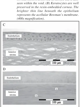
Tissue distortion was also revealed in the endothelium. Most of the paraffin-embedded sections composed of detached monolayer endothelium. Conversely, the endothelium remained intact in all resin-embedded sections due to the presence of albumin as supporting medium to the sample. Albumin solution is commonly employed
as an encapsulation medium to microorganisms(6). Santhana et al.(7) had integrated the albumin which functionally, helps to maintain the intactness of the resin-embedded sample and morphologically, allows complete visualization on the sample. Albumin has successfully preserved the overall structural integrity of the cornea particularly the vulnerable monolayer endothelium which is weak and easily tears off from the posterior cornea(7).
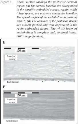
The presence of the swollen stromal region also makes the paraffin-embedded section less suitable for histomorphometrical studies. Its corneal stroma was approximately 10.2% thicker (+ 39 µm) as compared to the resin-embedded section. This difference is caused by the presence of numerous voids (empty spaces) among the lamellae layers. The fibroblasts of the stroma, termed keratocytes also encountered serious distortion as the lamellae separated from each other. The phenomenon of stromal swelling and the presence of voids among
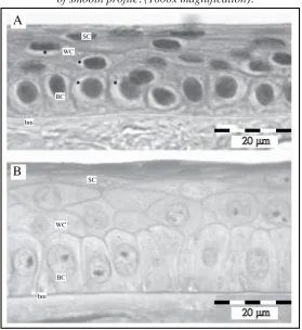
Figure 2 : Cross-section through the anterior corneal region. (A) Numerous voids (clear spaces) are seen clearly in the anterior stroma of the paraffin-embedded corneal section. Distorted keratocyte (K) is also seen within the void. (B) Keratocytes are well preserved in the resin-embedded cornea. The brighter thin line beneath the epithelium represents the acellular Bowman’s membrane. (400x magnification).
stromal lamellae in the paraffin-embedded corneal sample may probably be also due to the lengthy dehydration process. The dehydration time should be as brief as possible to minimize the risk of extracting cellular components.
From this study, perhaps, we proved that the resin-embedded corneal sample has better preservation on corneal ultrastructures with less histological artifacts as compared to the paraffin-embedded corneal sample. Thus, its corneal section is a suitable target for clinical investigation in ophthalmology and optometry.
Acknowledgements
This project was supported by the Intensification of Research in Priority Areas (IRPA) Grant by the Ministry of Science, Technology and Innovation, Malaysia. The authors wish to express
their appreciation to the Department of Wildlife and National Parks, Malaysia for their kindness in providing the experimental subjects. We are grateful to Mr King-Hwa Ling of The Walter and Eliza Hall Institute of Medical Research (WEHI), Australia, for his valuable advice and discussion.
Corresponding Author :
Dr. Pike-See Cheah BSc. (UKM), Ph.D (UKM) Department of Human Anatomy,
Faculty of Medicine & Health Sciences,
Universiti Putra Malaysia, 43400 UPM Serdang, Selangor, Malaysia.
Tel: + +60165680480 Fax: + 6097663353
Email : [email protected]; [email protected]
References
1. Metha AB, Crane AM, Rylander III HG, Thomsen SL and Albrecht DG. Maintaining the cornea and the general physiological environment in visual neurophysiology experiments. J Neurosci Metho 2001;
109: 153-166.
2. Choo JD, Caroline PJ, Harlin DD and Meyers W. Morphologic changes in cat epithelium following overnight lens wear with the Paragon CRT lens for corneal reshaping. Invest Ophthalmol Vis Sci 2004; 44:
E-1552.
3. Matsubara M, Kamei Y, Takeda S, Mukai K, Ishii Y and Ito S. Histologic and histochemical changes in rabbit cornea produced by an orthokeratology lens. Eye Contact Lens 2004; 30: 198-204.
4. Doughty MJ, Bergmanson JPG and Blocker Y. Shrinkage and distortion of the rabbit corneal endothelial cell mosaic caused by a high osmolarity glutaraldehyde-formaldehyde fixative compared to glutaraldehyde. Tissue Cell 1997; 29: 533-547.
5. Doughty MJ. Evidence for heterogeneity in a small squamous cell type (light cells in the rabbit corneal epithelium - a scanning electron microscope study. Doc Ophthalmol 1996; 92: 117-136.
6. Ingris TJJ, Rigby P, Robertson TA, Dutton NS, Henderson M and Chang J. Interaction between Burkholderia pseudomallei and Acanthaamoeba species results in coiling phagocytosis, endamebic bacterial survival, and escape. Infect Immun 2000; 68:
1681-1686.
7. Santhana RL, Cheah PS, Nor Asiah CP, Teh Hamidah Z, Mohidin N, Mohd-Ali B, Myint M and Azian AL. Rapid technique: modified protocol for transmission electron microscope visualization of cornea. Ann Microsc 2006; 6: 36-41