2014 | VOLUME 10 | 23 © ARCHIVES OF BUDO | SCIENCE OF MARTIAL ARTS
Postural patterns and adaptations in judo athletes
Wagner Castropil
ADE, Carla Arnoni
BCDInstituto Vita, Sao Paulo, Brazil
Source of support:
Departmental sources
Received:
31 October 2013;
Accepted:
30 January 2014;
Published online:
07 February 2014
ICID:
1090966
Abstract
Background
Regular practice results in physiological and postural adaptations that may correlate with a higher incidence of injuries in some sports. The postural adaptations in judo are not well understood. The aim of this study was the common posture patterns and misalignments of high-level judo athletes.Material & Methods:
The postures of 50 judo athletes (23 men and 27 women) were evaluated using a posture grid and plumb line. Full analysis was performed on 37 subjects (19 women and 18 men). This posture analysis method was cho-sen because it is accessible and can be easily applied by health professionals supporting athletes in the field.Results:
Only one athlete did not have any misalignment. The most common postural patterns observed were flat foot (80%), shoulder lateral asymmetry (70%), winged scapula (54%), and forward head posture (58%), all of which are likely to be related to the repetitive movements and sustained positions that are specific to judo.Conclusion:
This study provides the first complete description of postural patterns in judo athletes. Understanding the unique postural patterns and adaptations in judo will help improve training protocols to enhance performance while preventing injuries.Keywords: injuries • injury prevention • martial arts
Author’s address: Wagner Castropil, Instituto Vita, Rua Mato Grosso, 306 - 1o andar, Sao Paulo, 01239-020, Brasil; e-mail: [email protected]
& Study Aim:
I
ntroductIonThe regular and intense practice of certain sports is known to result in physiological and postural adapta-tions in an athlete’s body. In long-term studies, some of these adaptations have been reported to correlate with a higher incidence of injuries [1-3]. For instance, the scapula of the dominant shoulder in baseball play-ers is more internally rotated and anteriorly inclined compared to that of the non-dominant shoulder [4] Such scapular positioning can, in turn, lead to sub-acromial impingement syndrome, which is worsened by concomitant thoracic hyperkyphosis and cervical hyperlordosis [5].
Judo is an ancient Japanese martial art that became an Olympic sport during the 1964 Tokyo Olympic Games. As with many contact sports, judo has a high incidence of injuries [6-8]. There are no reports in the
literature that describe postural patterns and adap-tations that are specific to judo or that address their possible relationships with the most common judo injuries. Based on findings reported for other activi-ties [3,6,7], it is important to understand the postural patterns and adaptations that are unique to judo to design training protocols that emphasize the preven-tion of chronic and insidious injuries that may pre-vent athletes from competing.
Because frequent evaluations are necessary to assess the effectiveness of training or therapeutic interven-tion protocols, it is essential that the method used for posture evaluation be simple and accessible. We opted to use the posture grid, which meets these require-ments and has been validated in the literature [9,10]. This method also allows for the collection of quali-tative rather than quantiquali-tative posture data, which Judo – Japanese meaning “gentle
way” is a modern martial art created in Japan in 1882 by Jigoro Kano.
Uke – the receiver of the technique applied by the opponent.
Tori – an individual who applied a technique in judô training.
Postural pattern – shape or form of a stance.
Authors’ Contribution: A Study Design
B Data Collection
C Statistical Analysis
D Manuscript Preparation
E Funds Collection
24 | VOLUME 10 | 2014 www.archbudo.com
0 20 40 60 80 100
Flat HArch NL
%
o
f A
th
le
te
s
Women (n=27) Men (n=23) are appropriate for this descriptive groundwork that
aims to lay the foundation for future injurypreven-tion studies.
The aim of this study was the common posture pat-terns and misalignments of high level judo athletes and their implications for injury prevention.
M
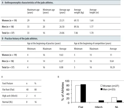
aterIalandMethodsFifty high-level judo athletes from the Brazilian national team were evaluated for this study. We included 23 males and 27 females between 16 and 33 years of age from different categories and with a minimum of five years of experience. Sixty-six percent of these athletes were international-level competi-tors. Although we assessed the posture of 50 athletes, anthropomorphic data and practice history were only collected from 37 athletes (19 females and 18 males) (Tab. 1). None of the selected athletes had trained for another sport.
Approval from the Ethic Committee was obtained before the commencement of this paper and all sub-jects gave written informed consent.
The athletes were asked to complete a questionnaire to obtain pertinent anthropomorphic data and infor-mation regarding their judo practice histories.
The athletes’ postures were evaluated using a symme-trographic posture grid (Sanny, Brazil) and a plumb line. This grid consists of a 2.0 X 1.0 m acrylic board divided into 10.0 X 10.0 cm squares [9,10]. The ath-letes were evaluated in their swimwear while standing on a 20-cm high step positioned in front of the grid. They were then photographed using a digital camera (Canon G12, USA) mounted on a 70-cm high tripod and positioned 2.5 m from the posture grid. Images of the anterior and lateral views of subjects in the ortho-static position were used to assess the alignment of certain body parts in relation to the gridlines. Angles were calculated using the Dartfish software (Dartfish,
A - Anthropomorphic characteristics of the judo athletes.
Maximum age (years) Minimum age (years) Average age (years) Average weight (kg) Average height (m)
Women (n = 19) 29 16 23.31 69.15 1.64
Men (n = 18) 33 20 26.50 89.56 1.77
Total (n = 37) 33 16 24.86 7.06 1.70
B - Practice history of the judo athletes.
Age at the beginning of practice (years) Age at the beginning of competition (years)
Minimum Maximum Average Minimum Maximum Average
Women (n = 19) 4 16 9.63 5 15 11
Men (n = 18) 4 14 6.27 5 16 9.64
Total (n = 37) 4 16 8.00 5 16 10.29
Foot Posture n %
Flat foot (Flat) 40 80
High arch (HArch) 2 4
Normal (NL) 8 16
Figure 1. Postural pattern of the foot (total and gender differences).
A B
Table 1. Anthropomorphic characteristics and practice histories of the judo athletes.
0 20 40 60 80 100
FH NL
%
o
f
A
th
le
te
s Women (n=27)
Men (n=23)
0 20 40 60 80 100
ShA FSh N-Sh WG N-Sc
%
o
f A
th
le
te
s
Women (n=27)Men (n=23)
0 20 40 60 80 100
Flat HArch NL
%
o
f A
th
le
te
s
Women (n=27) Men (n=23)
USA). Relevant anatomical landmarks were marked with surgical tape to facilitate the image identifica-tion; these landmarks included the spinous process of C7, bilateral coracoid processes, bilateral antero-superior portion of the iliac crests, bilateral center of the patellae, and bilateral malleoli [11-13]. Each ath-lete’s posture was assessed for each body part and clas-sified as follows.
Cervical: Normal or Forward Head Posture. The cervi-cal angle was obtained from the lateral view photographs by drawing a horizontal line through C7 and a line from the superior aspect of its spinous process to the tragus of the ear. The normal angle is between 122.6° and 133° [11,14]. Forward head posture was assigned to the ath-letes with cervical angles that were less than 33.0°.
Shoulder: Normal, Lateral Asymmetry (one side higher than the other), and Forward Shoulder Posture. To assess lateral symmetry based on the anterior view photograph, we calculated the distance between each coracoid process and the nearest horizontal line of the grid. Lateral asymmetry was assigned to those athletes with different distances on the right and left sides. To determine forward shoulder posture, we used the lateral view photograph and drew a line connecting the lat-eral aspect of each shoulder to C7. The angle between this line and the horizontal line running through the scapula was obtained. Angles were considered normal when between 33.8° and 59.6° [14]. Forward shoulder posture was assigned when angles were less than 33.8°.
Scapula: Normal or Winged. The scapula was con-sidered to be winged when any of its borders (supe-rior, medial, or inferior) was prominent or lifted away from the ribcage [15].
Knee: Normal, Valgus, or Varus. Knee alignment was determined based on the Q angle. Normal ranges are between 6.4 and 16.2° for men (valgus Q angles > 16.2°; varus Q angles < 6.4°) and between 7.8 and 18.8° for women (valgus Q angle > 18.8°; varus < 7.8°) [16].
Ankle and Foot: Normal, Flat, or High Arch. The plantar arch was evaluated subjectively by an orthope-dic surgeon who specialized in foot and ankle surgery.
The knees, feet and scapulae were considered to be asymmetrical when at least one side was misaligned.
r
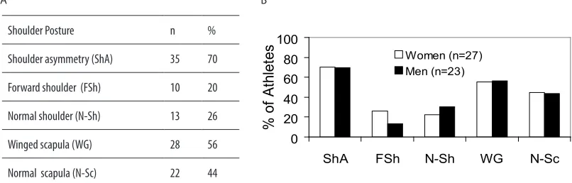
esultsOnly one of the 50 athletes who were evaluated for posture did not present with any form of mis-alignment. The most frequent postural changes affected the feet and ankles, shoulders, and cervi-cal region. The most common change was flat foot, which affected 80% of the athletes (Fig. 1), followed by shoulder lateral asymmetry (70%) and winged scapula (56%) (Fig. 2A). Forward shoulder posture was more frequent in the female athletes (Fig. 2B). Forward head posture was noted in more than half of all athletes (Fig. 3A) and seemed to affect women
A
A
B
B
Shoulder Posture n %
Shoulder asymmetry (ShA) 35 70
Forward shoulder (FSh) 10 20
Normal shoulder (N-Sh) 13 26
Winged scapula (WG) 28 56
Normal scapula (N-Sc) 22 44
Figure 2. Postural pattern of the shoulder and scapula (total and gender differences).
Cervical Posture n %
Forward head posture (FH) 29 58
Normal (NL) 11 22
26 | VOLUME 10 | 2014 www.archbudo.com more than men (Fig. 3B). The majority of all athletes
had varus knees (Fig. 4A); however, most female athletes had valgus knees (Fig. 4B).
d
IscussIonPhysiological and cardiovascular adaptations to exercise are well understood, and their implications are usually considered when implementing physical training pro-grams for athletes. However, biomechanical and pos-tural adaptations are still poorly understood for most sports. Normal posture ranges are rarely reported in the literature, and little is known about the postural pat-terns that are unique to each activity. In this study, we evaluated a population of high-level judo athletes with at least five years of experience and who competed in international events. The most prevalent postural changes found in judo athletes, regardless of gender, were flat foot (80%), lateral shoulder asymmetry (70%), forward head posture (58%), and winged scapula (54%).
Note that the main objective of the sport is to throw or takedown the opponent by moving his or her center of gravity away from the base, causing a loss of balance [17]. To efficiently subdue the opponent, the athlete must maintain his or her balance throughout the com-petition. The movement efficiency increases with the number of contact points between the bare foot and the tatami and with the firmness of the base (obtained by standing with a wide stance).
Sogabe analized the changes in plantar pressure of the eight directions of Kuzushi for top-ranked judo players and reported that uke attempt to maintain stability by counteracting external force by pronating the foot and concentrating weight on the ball of his foot [19].These unique characteristics of the judo stance may help to explain the flat foot posture that was observed in most of the athletes.
The normal plantar arch is supported by ligamentous structures that, if positioned repetitively under forced pronation, may stretch excessively and result in arch
collapse and flat foot [20]. Collapse of the plantar arch has been associated with tibial posterior muscle weak-ness and altered foot function, including prolonged calcaneal eversion, increased tibial internal rotation, increased forefoot abduction, reduced efficiency of gait, and reduced shock absorption. As a result, greater stress might be applied to those structures involved in con-trolling these movements during the stance phase of the gait. Additionally, abnormal foot motion may increase stress on more proximal structures, such as the knee joint, predisposing both the foot and knee to lesions [20,21].
Scapular and shoulder asymmetries, such as having winged scapula and one shoulder that is higher than the other, were also frequent findings in our study popula-tion. These findings may be explained by the fact that the shoulder region is subject to frequent and direct trauma in judo. To protect themselves, athletes will often con-tract the pectoral muscles, closing the chest and bring-ing the shoulders up towards the ears. Judo athletes also use their shoulders to perform complex movements and transfer energy from the trunk to the upper limbs, while grappling, choking, and taking down the opponent. It is important to minimize shoulder asymmetry because it may alter the biomechanical characteristics of the scap-ular region and compromise the transfer of energy from the trunk to the upper limbs. The inefficiency of this local kinetic chain leads to shoulder strain and increased risk for subacromial impingement syndrome [22,23]. Forward head posture, which was present in more than half of the judo athletes we studied, may also impact the incidence of shoulder lesions. This type of cervical misalignment leads to anterior inclination of the scap-ula and decreases its ability to rotate upward, which is a common problem in patients with shoulder impinge-ment [5,24]. Although there are no reports in the liter-ature correlating judo practice with the development of forward head posture, frequent practice may favor this type of head positioning in defensive stances.
We observed a marked gender difference in knee joint positioning among the judo athletes, with the majority of women having valgus knees (associated with larger Q
0 20 40 60 80 100
Val Var NL
%
o
f A
th
le
te
s Women (n=27)
Men (n=23)
Knee Posture n %
Valgus (Val) 17 34
Varus (Var) 27 54
Normal (NL) 6 12
Figure 4. Postural pattern of the knee (total and gender differences).
A B
angles) and most men having varus knees. However, this gender difference has been well described in the litera-ture, regardless of sport practiced [25]. While it is agreed that female athletes have more frequent knee lesions than male athletes [26], there is disagreement as to the direct role of the Q angle in the mechanism of knee lesions. Larger Q-angles, which are associated with valgus knees, are speculated to increase the lateral pull of the quadriceps muscles on the patella, thereby leading to pressure on the lateral facet of the patella and pain at the anterior knee [16]. However, other authors have not found a correla-tion between static knee misalignment (as evidenced by the Q angle) and knee lesion incidence; they believe that dynamic valgus alignment is more relevant [27]. Gender difference in the dynamic alignment of the lower limbs and knees during some activities, such as jumps and squats, is well studied and is particularly important in high-impact sports, such as volleyball, basketball, and floor exercises in gymnastics. Dynamic valgus, which occurs much more fre-quently in women, is exacerbated during landing, predis-posing the athlete to lesions. However, a direct role for the Q angle in the development of dynamic valgus has not been demonstrated [25], as women also present wider adduction angles for hips, wider abduction angles for knees, and weaker neuromuscular control, all of which character-ize a dynamic valgus alignment [28,29]. The knee joint is the most frequent site of injury in judo, most likely because of weight-bearing, closed-chain torsion [30,31]. However, the role of the Q angle in closed-chain torsions remains unknown and deserves further study.
Our results suggest that preventive training protocols for judo should focus on strengthening the serratus
and trapezius and stretching the pectoralis, which are responsible for stabilizing the scapula and support-ing cervical alignment and correct head positionsupport-ing. We also recommend strengthening the posterior tib-ial muscle, which plays an essenttib-ial role in support-ing a healthy plantar arch [24,32].
In summary, this report is the first to detail postural patterns and adaptations that are unique to judo, as determined by a simple and accessible evalua-tion method based on a posture grid. This subjec-tive posture evaluation study lays the groundwork for posture analysis in judo, but further objec-tive posture analysis of the cervical, shoulder, and plantar regions in judo athletes is warranted. Understanding how these posture patterns influ-ence lesion incidinflu-ence is essential for designing and evaluating the impact of lesion prevention proto-cols in this martial art.
a
cknowledgMentsWe thank the Brazilian judo team, their trainers and therapists for participating in this study. We also thank Suzana Sturlini Couto for translation and reviews, as well as the professionals at American Journal Experts for peer review and editing.
c
oMpetIngI
nterestsAuthors declare that we do not have any financial or personal relationships with other people or organisa-tions that could inappropriately influence our paper.
Knee Posture n %
Valgus (Val) 17 34
Varus (Var) 27 54
Normal (NL) 6 12
Figure 4. Postural pattern of the knee (total and gender differences).
r
eferences1. Laudner KG, Moline MT, Meister K. The relation-ship between forward scapular posture and poste-rior shoulder tightness among baseball players. Am J Sports Med 2010; 38(10): 2106-2112
2. Shultz SJ, Nguyen AD, Levine BJ. The Relationship Between Lower Extremity Alignment Characteristics and Anterior Knee Joint Laxity. Sports Health 2009; 1(1): 54-60
3. Santos SG, Detanico D, Graup S et al. Relation between posture changes, prevalence of injuries and impact magnitude on lower limbs as regards the handball athletes. Fit Perf 2007; 6(6): 388-393 4. Kobayashi T, Takeda N, Atsuta Y et al. Flattening
of sagittal spinal curvature as a predictor of vertebral fracture. Osteoporos Int 2008; 19(1): 65-69 5. Weon JH, Oh JS, Cynn HS, et al. Influence of forward
head posture on scapular upward rotators during isometric shoulder flexion. J Body Mov Ther 2010; 14(4): 367-374 6. Kujala UM, Taimela S, Antti-Poika I, et al. Acute
injuries in soccer, ice hockey, volleyball, basketball, judo, and karate: analysis of national registry data. BMJ 1995; 311: 1465-1468
7. Carazzato JG, Cabrita H, Castropil W. Repercussão no aparelho locomotor da prática do judô de alto nível. Rev Bras Ortop 1996; 31(12): 957-968
8. Pocecco E, Ruedl G, Stankovic N et al. Injuries in judo: a systematic literature review including sug-gestions for prevention. Br J Sports Med 2013; 47:1139–1143
9. Gadotti IC, Biasotto-Gonzalez DA. Sensitivity of clinical assessments of sagittal head posture. J Eval Clin Pract 2010; 16(1): 141-144
10. Juskeliene V, Magnus P, Bakketeig LS et al. Prevalence and risk factors for asymmetric posture in preschool children aged 6-7 years. Int J Epidemiol 1996; 25(5): 1053-1059
11. Hackney J, Bade D, Clawson A. Relationship between forward head posture and diagnosed inter-nal derangement of the temporomandibular joint. J Orofac Pain 1993; 7(4): 386-390
12. Guimarães MMB, Sacco ICN, João SMA. Caracterização Postural da Jovem Praticante de Ginástica Olímpica. Rev Bras Fisioter 2007; 11(3): 213-219 13. Sacco ICN, Alibert S, Queiroz BWC et al.
Confiabilidade da Fotogrametria em Relação a Goniometria para Avaliação Postural de Membros Inferiores. Rev Bras Fisioter 2007; 11(5): 411-417 14. Raine S, Twomey LT. Head and shoulder posture
variations in 160 asymptomatic women and men. Arch Phys Med Rehabil 1997; 78(11): 1215-1223
15. Kibler WB, Uhl TL, Maddux JW et al. Qualitative clinical evaluation of scapular dysfunction: a reli-ability study. J Shoulder Elbow Surg 2002; 11(6): 550-556
16. del Mar Carrion MM, Santiago FR, Calvo RP et al. Patellofemoral morphometry in patients with idio-pathic patellofemoral pain syndrome. Eur J Radiol 2010; 75(1): e64-67
17. Franchini E. Judô: Desempenho Competitivo. 1st ed Manole; 2001
18. Lehmkuhl LD, Smith LK. Cinesiologia Clínica de Brunnstrom. 4rd ed Manole; 1987
19. Sogabe A. Analysis of patterns of response to kuzushi in eight drections based on plantar pressure and reac-tion movement. Arch Budo 2008; 4: 70-77 20. Kaufman KR, Brodine SK, Shaffer RA et al. The effect
of foot structure and range of motion on musculoskeletal overuse injuries. Am J Sports Med 1999; 27(5): 585-93 21. Kamiya T, Uchiyama E, Watanabe K et al. Dynamic effect of the tibialis posterior muscle on the arch of the foot during cyclic axial loading. Clin Biomech (Bristol, Avon) 2012; 27(9): 962-966
22. Kibler WB, McMullen J. Scapular dyskinesis and its relation to shoulder pain. J Am Acad Orthop Surg 2003; 11(2): 142-151
28 | VOLUME 10 | 2014 www.archbudo.com
23. Borich MR, Bright JM, Lorello DJ et al. Scapular angular positioning at end range internal rotation in cases of glenohumeral internal rotation deficit. J Orthop Sports Phys Ther 2006; 36(12): 926-934 24. Lynch SS, Thigpen CA, Mihalik JP et al. The effects
of an exercise intervention on forward head and rounded shoulder postures in elite swimmers. Br J Sports Med 2010; 44(5): 376-381
25. Pantano KJ, White SC, Gilchrist LA et al. Differences in peak knee valgus angles between individuals with high and low Q-angles during a single limb squat. Clin Biomech (Bristol, Avon) 2005; 20(9): 966-972 26. Ford KR, Myer GD, Hewett TE. Valgus knee
motion during landing in high school female and
male basketball players. Med Sci Sports Exerc 2003; 35(10): 1745-50
27. Park SK, Stefanyshyn D. Greater Q angle may not be a risk factor of Patellofemoral Pain Syndrome. Clin Biomech (Bristol, Avon) 2011; 26(4): 392-396 28. Ford KR, Shapiro R, Myer GD et al. Longitudinal
sex differences during landing in knee abduction in young athletes. Med Sci Sports Exerc 2010; 42(10): 1923-1931
29. Zeller BL, McCrory JL, Kibler WB et al. Differences in kinematics and electromyographic activity between men and women during the single-legged squat. Am J Sports Med 2003; 31(3): 449-456
30. Koshida S, Deguchi T, Miyashita K et al. The com-mon mechanisms of anterior cruciate ligament inju-ries in judo: a retrospective analysis. Br J Sports Med 2010; 44(12): 856-861
31. Barsottini D, Guimarães AE, Morais PR. Relação entre técnicas e lesões em praticantes de judô. Rev Bras Med Esporte 2006; 12(1): 56-60
32. Imhauser CW, Siegler S, Abidi NA et al. The effect of posterior tibialis tendon dysfunction on the plan-tar pressure characteristics and the kinematics of the arch and the hindfoot. Clin Biomech (Bristol, Avon) 2004; 19(2): 161-9
Cite this article as: Castropil W, Arnoni C. Postural patterns and adaptations in judo athletes. Arch Budo 2014; 10: 23-28