INTRODUCTION
Several members in nod-like receptor (NLR) family are related to inflammasomes [1]. By far, NLR protein 3 (NLRP3) inflammasome is the most extensively studied inflammasome, consisting of NLRP3, adaptor protein apoptosis associated speck-like protein (ASC) and serine protease caspase-1 pre-cursor (procaspase-1) [2]. A variety of microbial pathogens can activate NLPR3 inflammasome, including fungi, bacte-ria and viruses, probably by their products, such as toxins, RNAs and DNAs [3,4]. On NLRP3 inflammasome activa-tion, procaspase-1 precursor is turned into active caspase-1 by auto-cleavage. Subsequently, caspase-1 cleaves pro-IL-1β to form active IL-1β.
Chronic alcohol consumption is a common cause of liver injury. One of the most important underlying mech-anisms is the gut-liver axis [5]. Studies have demonstrated that alcohol can lead to intestinal bacterial outgrowth and
enteric dysbiosis [6]. More importantly, alcohol increases gut permeability, causing microbial translocation [7]. It has been revealed that both ethanol and its metabolic prod-uct acetaldehyde can disrupt epithelial tight junctions [7]. In addition, dysbiosis and inflammation also attribute to the disruption of intestinal epithelial integrity. As a result, bacterial products, such as endotoxin lipopolysaccharide, and bacterial RNAs and DNAs, translocate from intestinal lumen to liver.
Recently, NLRP3 inflammasome has been demon-strated to be implicated in alcoholic steatohepatitis [8]. It was revealed that increased IL-1β is required for the development of alcohol-induced liver disease, and due to the activation of NLRP3 inflammasome [8]. They identified Kupffer cells as sources of IL-1β [8]. In addition to Kupffer cells, NLRP3 inflammasome was also found to be activated in hepatic stel-late cells (HSCs) [9], which may play a role in hepatic fibrosis. As bacterial RNA has been shown to activate NLRP3 inflam-masome in macrophage, the present study explored the acti-vation of the NLRP3 inflammasome by Escherichia coli RNA in HSCs and the role of NLRP3 inflammasome in alcoholic hepatic fibrosis.
*Corresponding author: Bingyuan Wang, Department of
Gastroenterology, The First Affiliated Hospital of China Medical University, 155 North Nanjing Street, Shenyang 110001, China. E-mail: [email protected] Submitted: 24 August 2015 / Accepted: 15 October 2015
Nod-like receptor protein 3 inflammasome activation by
Escherichia coli
RNA induces transforming growth factor
beta 1 secretion in hepatic stellate cells
Hui Wang, Shu Liu, Ying Wang, Bing Chang, Bingyuan Wang*
Department of Gastroenterology, The First Affiliated Hospital of China Medical University, 155 North Nanjing Street, Shenyang 110001, China
Abstract
Nod-like receptor protein 3 (NLRP3) inflammasome has been implicated in alcoholic liver disease. Chronic alcohol consumption enhances gut permeability and causes microbial translocation. The present study explored the activation of the NLRP3 inflammasome by Escherichia coli
RNA in hepatic stellate cells (HSCs), and the potential role of NLRP3 inflammasome in hepatic fibrosis. E. coli RNA transfection induced HSC-T6 cells to secrete and express mature interleukin-1 beta (IL-1β), which was abolished by NLRP3 siRNA pretreatment. In addition, E. coli
RNA transfection enhanced caspase-1 expression, whereas reduced caspase-1 precursor (pro-caspase-1) expression. E. coli RNA-stimulated transforming growth factor beta 1 (TGF-β1) overproduction in HSC-T6 cells, which was blocked by recombinant IL-1 receptor antagonist (rIL-1Ra) or nuclear factor κB inhibitor BAY 11-7082. Furthermore, E. coli RNA-induced overexpression of pro-fibrogenic factors was sup-pressed by rIL-1Ra or TGF-β receptor inhibitor A83-01. These results demonstrate that E. coli RNA can stimulate NLRP3 inflammasome acti-vation, which leads to excessive production of pro-fibrogenic factors, suggesting that NLRP3 inflammasome activation in HSCs may play a role in hepatic fibrosis.
KEY WORDS: Nod-like receptor protein 3; inflammasome; hepatic stellate cells; transforming growth factor β1
penicillin, and 100 µg/ml streptomycin, at 37°C with 5% CO2.
E. coli (ATCC 25922)RNA (10 mg/ml) was used to transfect HSC-T6 cells via lipofectamine 2000 (Invitrogen) at a radio of 1 µl lipofectamine 2000 per 1 µg RNA. All experiments were done at least 3 times.
E. coli
RNA extraction and RNase digestion
E. coli were grown in Luria-Bertani medium. Total E. coli
RNA was extracted and purified using RNeasy Plus Mini kit (Qiagen, Shenzhen, China) according to the manufacturer’s protocol. In some experiments, RNase A (Sigma) was used to digest E. coli RNA at a concentration of 1 µg RNase A per 1 µg RNA for 60 minutes at 37°C.
Enzyme-linked immunosorbent assay (ELISA)
Secretion of IL-1β and transforming growth factor beta 1 (TGF-β1) was determined by examining the concentrations of IL-1β and TGF-β1 in cell supernatants via ELISA kits (R&D SYSTEMS, Shanghai, China), according to the manufacturer’s protocols.Western blot
Equal amounts of total protein from each sample was sub-jected to 12% sulfate polyacrylamide gel electrophoresis, and transferred onto a nitrocellucose-ECL membrane. The mem-brane was probed with primary antibody for IL-1β (1:1000, Abcam), caspase-1 (1:1000, Santa Cruz) or NLRP3 (1:500, Santa Cruz), and then incubated with the peroxidase-conju-gated secondary antibody (1:3000, Santa Cruz). Protein bands were detected by ECL (Pierce) and visualized by gel imaging system (Bio-Rad). β-actinwas used as an internal control.
RNA interference
HSC-T6 cells were seeded into a 6-well plate at a den-sity of 2 × 10⁵, and transfected with NLRP3 siRNA and
con-trol siRNA (Santa Cruz, Texas, America), according to the manufacturer’s protocol. In brief, Solution A and Solution B were prepared, mixed and incubated for 30 minutes in room temperature. Solution A: 1 µg siRNA duplex was added into 100 μl siRNA transfection medium. Solution B: 8 µl trans-fection reagent was added into 100 μl siRNA transtrans-fection medium.
LightCycler system (Roche Diagnostics, Shanghai, China) with LightCycler DNA Master SYBR Green I Kit (Roche Diagnostics). Comparative CT method was used to quantify mRNA expression, normalizing CT values to β-actin which was used as an internal control. Primers for α-smooth muscle actin (α-SMA), collagen Type I α1 (COL1A1), tissue inhibitor of metalloproteinases 1 (TIMP-1), and β-actin were described by Son et al. [10].
Immunofluorescence
After incubation of 12 hours, HSC-T6 cells were fixed by 4% (w/v) formaldehyde solution for 15 minutes and washed with PBS at room temperature, and then lysed with 0.2% Triton X-100 (Biochemicals) for 5 minutes and blocked with 5% bovine serum albumin for 40 minutes. Sequentially, HSC-T6 cells were incubated with primary antibody for TGF-β1 (R&D SYSTEMS), and then with the rhodamine-conju-gated secondary antibody (Santa Cruz). After washing with 4’,6-diamidino-2-phenylindole (DAPI) (Biochemicals), the cells were stained with DAPI, and observed by fluorescent confocal microscopy.
Statistical analysis
Statistical analysis was performed using SPSS version 13.0 (Chicago, IL, USA). Differences among groups were ana-lyzed by one-way ANOVA and considered significant when
p < 0.05.
RESULTS
E. coli
RNA stimulates IL-1β secretion by HSCs
To explore whether E. coli RNA could activate NLRP3 inflammasome, we first examined IL-1β secretion by HSCs exposed to E. coli RNA for 12 hours. As shown by ELISA detection, E. coli RNA-induced HSC-T6 cells to secrete more IL-1β into cell supernatant (Figure 1A). However, the induc-tion was abrogated after E. coli RNA was digested by RNase A (Figure 1A). We next examined the expression of IL-1β and IL-1β precursor (pro-IL-1β) in HSC-T6 cells by Western blot. It was found that IL-1β expression was elevated, whereas pro-IL-1β expression was reduced after E. coli RNA transfection (Figure 1B). Taken together, these results indicate that E. coliIL-1β induction by
E. coli
RNA is NLRP3
inflammasome-dependent
To evaluate the role of the NLRP3 inflammasome in
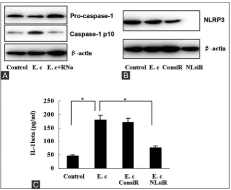
E. coli RNA-induced IL-1β secretion, we detected caspase-1 and NLRP3 expression in HSC-T6 cells. After transfection with E. coli RNA for 12 hours, HSC-T6 cells expressed more caspase-1, whereas less caspase-1 precursor (Pro-caspase-1), indicating E. coli RNA transfection activated caspase-1 (Figure 2A). NLRP3 expression was also found in HSC-T6 cells, but not affected by E. coli RNA (Figure 2B). When NLRP3 expression was silenced by NLRP3 siRNA, IL-1β overproduc-tion by E. coli RNA were almost abolished (Figure 2B and C). These results suggest that NLRP3 inflammasome mediates IL-1β induction by E. coli RNA.
E. coli
RNA induces TGF-β1 secretion by HSCs
To assess the involvement of NLRP3 inflammasome in hepatic fibrosis, we measured TGF-β1 levels in the super-natant of HSCs. It was found that E. coli RNA-transfected HSC-T6 cells secreted more TGF-β1, compared with untrans-fected control cells (Figure 3A). The induction was also abol-ished after E. coli RNA was digested by RNase A (Figure 3A). We further examined the expression of some pro-fibrogenic factors in HSC-T6 cells. As shown by real-time PCR detec-tion, E. coli RNA transfection enhanced the expression ofα-SMA, COL1A1, and TIMP-1 (Figure 3B-D).
IL-1β-nuclear factor κB (NFκB) signaling mediates
TGF-β1 induction by
E. coli
RNA
To determine the role of NLRP3 inflammasome in TGF-β1 induction, we pretreated HSC-T6 cells with recombi-nant IL-1 receptor antagonist (rIL-1Ra, 0.5 µg/ml, Sigma) for 8 hours. As shown by ELISA, TGF-β1 secretion induced by E. coli RNA was inhibited significantly (Figure 4A). We next analyzed the involvement of NFκB in TGF-β1 induc-tion via pretreating HSC-T6 cells with NFκB inhibitor BAY
11-7082 (10 µmol/L, Sigma) for 6 hours. It was found that TGF-β1 induction was suppressed as well (Figure 4A). Consistent with TGF-β1 secretion, TGF-β1 content in HSC-T6 cells was elevated by E. coli RNA transfection, however, the upregula-tion was also blocked by rIL-1Ra or BAY 11-7082, as shown by Immunofluorescence (Figure 4B).
IL-1β and TGF-β1 are involved in the induction of
pro-fibrogenic factors by
E. coli
RNA
To confirm the involvement of NLRP3 inflammasome in the induction of pro-fibrogenic factors by E. coli RNA, we pretreated HSC-T6 cells with rIL-1Ra for 8 hours. Real-time PCR detection showed that E. coli RNA-induced expression
FIGURE 2. Nod-like receptor protein 3 (NLRP3) inflam-masome-dependent interleukin-1 beta (IL-1β) induction, (A) Escherichia coli RNA transfection increased caspase-1 expres-sion, whereas decreased pro-caspase-1 expression in hepatic stellate cells (HSC)-T6 cells, (B) NLRP3 expression was observed in HSC-T6 cells, which was not affected by Escherichia coli RNA. NLRP3 expression was silenced after HSC-T6 cells were trans-fected with NLRP3 siRNA, (C) IL-1β induction by E. coli RNA was suppressed remarkably by NLRP3 siRNA transfection. *p < 0.01. Data are expressed as mean ± standard deviation. E.c: Escherichia coli RNA; RNa: RNase A; ConsiR: Control siRNA; NLsiR: Nod-like receptor protein 3 siRNA.
A B
FIGURE 1. Interleukin-1 beta (IL-1β) induction by Escherichia coli RNA, (A) E. coli RNA stimulated IL-1β secretion by hepatic stellate cells (HSC)-T6 cells, which was abrogated upon RNase A digestion, (B) IL-1β expression was elevated, whereas pro-IL-1β expression was reduced in HSC-T6 cells after E. coli RNA transfection. *p < 0.01. Data are expressed as mean ± standard deviation. E. c: Escherichia coli RNA; RNa: RNase A.
A B
of α-SMA, COL1A1, and TIMP-1 was inhibited remarkably (Figure 5A-C). Moreover, the increased expression of these factors was also suppressed by TGF-β receptor inhibitor A83-01 (1 µmol/L, Sigma), suggesting that the induction was TGF-β-dependent (Figure 5A-C).
DISCUSSION
This study demonstrates E. coli RNA as an activator of NLRP3 inflammasome in HSCs, consistent with several studies on macrophages. Initially, infection of E. coli has been shown to induce caspase-1 activation in macrophage-depen-dent on adenosine triphosphate (ATP) [11]. However, in the
absence of ATP stimulation, direct cytosolic delivery of bac-terial products could also induce caspase-1 activation [12]. Kanneganti et al. observed that E. coli RNA-induced rapid activation of caspase-1 and secretion of IL-1β and IL-18 in macrophages [13]. Furthermore, RNA from E. coli was found to be able to activate the NLRP3 inflammasome not only in macrophages but also in unprimed dendritic cells [14]. It is largely unknown how bacterial RNA-induced NLRP3 inflam-masome activation in these cells.
It has been demonstrated that NLRP3 inflammasome acti-vation is involved in a series of liver diseases and injuries [15]. Recently, hepatitis C virus was reported to activate NLRP3 inflam-masome in chronic hepatitis C patients [16]. Via different animal models, Petrasek et al. identified that alcoholic NLRP3 inflam-masome activation and IL-1β overproduction were crucial in the pathogenesis of alcoholic liver diseases [8]. The mechanisms of
FIGURE 4. Mediation of interleukin-1 beta (IL-1β)-nuclear factor κB (NFκB) signaling in transforming growth factor beta 1 (TGF-β1) induction by Escherichia coli RNA, (A) E. coli RNA-induced TGF-β1 secretion by hepatic stellate cells (HSC)-T6 cells was inhib-ited significantly by recombinant IL-1 receptor antagonist (rIL-1Ra) and NFκB inhibitor BAY 11-7082, respectively, (B) E. coli RNA-induced TGF-β1 production in HSC-T6 cells was suppressed by rIL-1Ra and BAY 11-7082, respectively. *p < 0.01. Data are expressed as mean ± standard deviation. E. c: Escherichia coli RNA.
A
B
FIGURE 3. Induction of transforming growth factor beta 1 (TGF-β1) and other pro-fibrogenic factors by Escherichia coli RNA, (A) E. coli RNA stimulated TGF-β1 secretion by hepatic stel-late cells (HSC)-T6 cells, which was abolished upon RNase A diges-tion, (B-D) E. coli RNA induced expression of for α-smooth muscle actin, collagen Type I α1, tissue inhibitor of metalloproteinases 1 in HSC-T6 cells, respectively. *p < 0.01. Data are expressed as mean ± standard deviation. E. c: Escherichia coli RNA; RNa: RNase A.
B
C
NLRP3 inflammasome-mediated liver damage may be related with hepatocyte pyroptosis, liver inflammation, and fibrosis [17]. Importantly, NLRP3 inflammasome-associated damage can be attenuated by IL-1β antagonist, suggesting a potential role of IL-1Ra in the treatment of these liver diseases [8,17].
Increasing evidence indicates that NLRP3 inflammasome plays an important role in fibrosis [18]. Gasse et al. found that NLRP3 inflammasome was activated and essential in bleomy-cin-induced pulmonary fibrosis [19]. Moreover, asbestos and sil-ica were identified to activate NLRP3 inflammasome [20], and silica-induced pulmonary fibrosis was dependent on NLRP3 inflammasome [21]. The NLRP3 inflammasome mediates fibrosis in systemic sclerosis [22] and myocardial infarction [23]. NLRP3 inflammasome was also involved in liver fibrosis. Study using NLRP3 knock-in mice has revealed that NLRP3 inflam-masome activation induced liver fibrosis [17]. Recently, Wree et al. revealed a crucial role for the NLRP3 inflammasome in the development of fibrosis in non-alcoholic fatty liver disease [24].
The mechanisms underlying liver fibrosis induced by NLRP3 inflammasome activation are not complete understood. Given the crucial role of HSCs in liver fibrosis, it is reasonable to explore the expression and activation of the NLRP3 inflammasome in HSCs. Watanabe et al. identified that NLRP3 was expressed in primary mouse stellate cells and LX-2 HSCs, and activation of NLRP3 inflammasome with monosodium urate crystals upreg-ulated TGF-β and collagen-1 expression [9]. Furthermore, animal studies showed that NLRP3-/- and ASC-/- mice were resistant to
liver fibrosis induced by carbon tetrachloride or thioacetamide, with reduced expression of TGF-β and collagen-1 [9]. On the other side, NLRP3 knock in mice demonstrated HSC activation with collagen deposition in the liver [17].
CONCLUSION
E. coli RNA can induce the expression of TGF-β1 and some pro-fibrogenic factors dependent on NLRP3 inflammasome,
suggesting that NLRP3 inflammasome activation in HSCs plays a role in liver fibrosis under chronic alcohol consumption.
DECLARATION OF INTERESTS
The authors state that there are no conflicts of interest.
REFERENCES
[1] Franchi L, Eigenbrod T, Muñoz-Planillo R, Nuñez G. The inflam-masome: a caspase-1-activation platform that regulates immune responses and disease pathogenesis. Nat Immunol 2009;10(3):241-7. http://dx.doi.org/10.1038/ni.1703.
[2] Cassel SL, Joly S, Sutterwala FS. The NLRP3 inflammasome: a sen-sor of immune danger signals. Semin Immunol 2009;21(4):194-8. http://dx.doi.org/10.1016/j.smim.2009.05.002.
[3] Strowig T, Henao-Mejia J, Elinav E, Flavell R. Inflammasomes in health and disease. Nature 2012;481(7381):278-86. http://dx.doi.org/10.1038/nature10759.
[4] Franchi L, Muñoz-Planillo R, Núñez G. Sensing and react-ing to microbes through the inflammasomes. Nat Immunol 2012;13(4):325-32. http://dx.doi.org/10.1038/ni.2231.
[5] Szabo G, Bala S, Petrasek J, Gattu A. Gut-liver axis and sensing microbes. Dig Dis 2010;28(6):737-44. http://dx.doi.org/10.1159/000324281.
[6] Schnabl B, Brenner DA. Interactions between the intestinal micro-biome and liver diseases. Gastroenterology 2014;146(6):1513-24. http://dx.doi.org/10.1053/j.gastro.2014.01.020.
[7] Rao RK, Seth A, Sheth P. Recent advances in alcoholic liver disease I. Role of intestinal permeability and endotoxemia in alcoholic liver disease. Am J Physiol Gastrointest Liver Physiol 2004;286(6):G881-4. http://dx.doi.org/10.1152/ajpgi.00006.2004. [8] Petrasek J, Bala S, Csak T, Lippai D, Kodys K, Menashy V, et al. IL-1
receptor antagonist ameliorates inflammasome-dependent alco-holic steatohepatitis in mice. J Clin Invest 2012;122(10):3476-89. http://dx.doi.org/10.1172/JCI60777.
[9] Watanabe A, Sohail MA, Gomes DA, Hashmi A, Nagata J, Sutterwala FS, et al. Inflammasome-mediated regulation of hepatic stellate cells. Am J Physiol Gastrointest Liver Physiol 2009;296(6):G1248-57. http://dx.doi.org/10.1371/journal.ppat.1003330.
[10] Son G, Hines IN, Lindquist J, Schrum LW, Rippe RA. Inhibition of phosphatidylinositol 3-kinase signaling in hepatic stellate cells blocks the progression of hepatic fibrosis. Hepatology 2009;50(5):1512-23. http://dx.doi.org/10.1002/hep.23186.
[11] Franchi L, Kanneganti TD, Dubyak GR, Núñez G. Differential
FIGURE 5. Involvement of interleukin-1 beta (IL-1β) and transforming growth factor beta 1 (TGF-β1) in the induction of pro-fibrogenic factors by Escherichia coli RNA, (A) E. coli RNA-induced α-smooth muscle actin expression in HSC-T6 cells was inhibited by recombinant IL-1 receptor antagonist (rIL-1Ra) and TGF-β receptor inhibitor A83-01, respectively, (B) E. coli RNA-induced collagen Type I α1 expression in hepatic stellate cells (HSC)-T6 cells was inhibited by rIL-1Ra and TGF-β receptor inhibitor A83-01, respectively, (C) E. coli RNA-induced tissue inhibitor of metalloproteinases 1 expression in HSC-T6 cells was inhibited by rIL-1Ra and TGF-β receptor inhibitor A83-01, respec-tively. *p < 0.01. Data are expressed as mean ± standard deviation. E. c: Escherichia coli RNA.
Franchi L, et al. Bacterial RNA and small antiviral com-pounds activate caspase-1 through cryopyrin/Nalp3. Nature 2006;440(7081):233-6. http://dx.doi.org/10.1038/nature04517. [14] Eigenbrod T, Franchi L, Muñoz-Planillo R, Kirschning CJ,
Freudenberg MA, Núñez G, et al. Bacterial RNA mediates activa-tion of caspase-1 and IL-1ß release independently of TLRs 3, 7, 9 and TRIF but is dependent on UNC93B. J Immunol 2012;189(1):328-36. http://dx.doi.org/10.4049/jimmunol.1103258.
[15] Szabo G, Csak T. Inflammasomes in liver diseases. J Hepatol 2012;57(3):642-54. http://dx.doi.org/10.1016/j.jhep.2012.03.035. [16] Negash AA, Ramos HJ, Crochet N, Lau DT, Doehle B, Papic N, et al.
IL-1ß production through the NLRP3 inflammasome by hepatic macrophages links hepatitis C virus infection with liver inflamma-tion and disease. PLoS Pathog 2013;9(4):e1003330.
[17] Wree A, Eguchi A, McGeough MD, Pena CA, Johnson CD, Canbay A, et al. NLRP3 inflammasome activation results in hepato-cyte pyroptosis, liver inflammation, and fibrosis in mice. Hepatology 2014;59(3):898-910. http://dx.doi.org/10.1002/hep.26592.
sensing of asbestos and silica. Science 2008;320(5876):674-7. http://dx.doi.org/10.1126/science.1156995.
[21] Cassel SL, Eisenbarth SC, Iyer SS, Sadler JJ, Colegio OR, Tephly LA, et al. The Nalp3 inflammasome is essential for the development of silicosis. Proc Natl Acad Sci U S A 2008;105(26):9035-40. http://dx.doi.org/10.1073/pnas.0803933105.
[22] Artlett CM, Sassi-Gaha S, Rieger JL, Boesteanu AC, Feghali-Bostwick CA, Katsikis PD. The inflammasome activating caspase 1 mediates fibrosis and myofibroblast differentiation in systemic sclerosis. Arthritis Rheum 2011;63(11):3563-74. http://dx.doi.org/10.1002/art.30568.
[23] Shinde AV, Frangogiannis NG. Fibroblasts in myocardial infarction: a role in inflammation and repair. J Mol Cell Cardiol 2014;70:74-82. http://dx.doi.org/10.1016/j.yjmcc.2013.11.015.