R E S E A R C H A R T I C L E
Open Access
Effects of oxytocin and anaesthesia on
vascular tone in pregnant women: a
randomised double-blind
placebo-controlled study using non-invasive pulse
wave analysis
Sofus Rabow
1,3*, Ull Hjorth
2, Sofia Schönbeck
2and Per Olofsson
2Abstract
Background:Oxytocin is an uterotonic drug with profound cardiovascular effects, which in compromised patients could lead to serious events. The objective was to investigate whether oxytocin affects cardiac function and vascular tone in large and small arteries. We hypothesized that oxytocin decreases arterial vascular tone and elevates cardiac output. Methods:51 pregnant women were randomised to treatment with 8.3μg (5 U) oxytocin or placebo injection during first trimester surgical evacuation of the gravid uterus under general anaesthesia. Oxytocin or placebo was administered once either early or late in the procedure, in a double-blind fashion. Digital photoplethysmography pulse wave analysis variables, heart rate, mean arterial blood pressure and electrocardiographic ST index were recorded before and after anaesthesia and after each injection. Non-parametric statistics were used with a two-sidedPvalue < 0.05 considered significant.
Results:Anaesthesia induced a significant fall in blood pressure, heart rate and vascular tone in small and peripheral arteries. Oxytocin had a vasodilatory effect on small and peripheral arteries and increased the left cardiac ventricular ejection time. The ST index decreased.
Conclusions:Pulse wave analysis indicated peripheral vasodilation and increased cardiac output after oxytocin, implying increased myocardial oxygen demand. These effects might have been enhanced by the vasodilating effects of anaesthesia. Previous studies have demonstrated myocardial ischaemia after oxytocin, as reflected by a decrease in ST index in the present study.
Trial registration:Trial registration numberISRCTN17860978, 2018/03/14, Retrospectively registered.
Keywords:Anaesthesia, Arterial stiffness, Oxytocin, Photoplethysmography, Placebo, Pregnancy, Pulse wave analysis, RCT, Vascular tone
* Correspondence:[email protected] 1
Department of Clinical Sciences Lund, Anaesthesiology and Intensive Care, Skåne University Hospital, Lund University, S-22185 Lund, Sweden 3Department. of Intensive and Perioperative Care, Skåne University Hospital,
S-22185 Lund, Sweden
Full list of author information is available at the end of the article
Fig. 1Flow chart showing the steps of the experiment. The first drugs were administered between the T1 and T2 measurement points, and the second drugs between T2 and T3
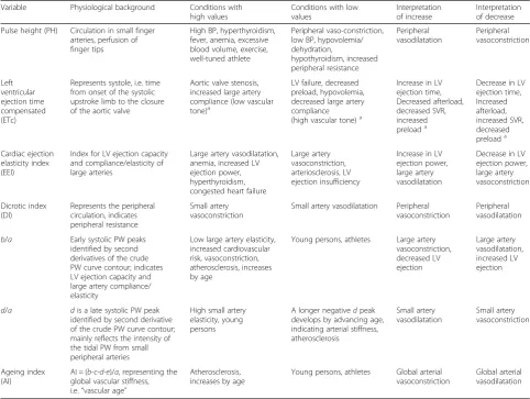
Table 1Description of the digital pulse wave analysis variables used in the study, revised from von Wowern et al. [5]
Variable Physiological background Conditions with high values
Conditions with low values
Interpretation of increase
Interpretation of decrease
Pulse height (PH) Circulation in small finger arteries, perfusion of finger tips
High BP, hyperthyroidism, fever, anemia, excessive blood volume, exercise, well-tuned athlete
Peripheral vaso-constriction, low BP, hypovolemia/ dehydration,
Represents systole, i.e. time from onset of the systolic upstroke limb to the closure of the aortic valve
Aortic valve stenosis, increased large artery compliance (low vascular tone)a
LV failure, decreased preload, hypovolemia, decreased large artery compliance (high vascular tone)a
Increase in LV ejection time, Decreased afterload, decreased SVR, increased preloada
Decrease in LV ejection time,
Index for LV ejection capacity and compliance/elasticity of large arteries
Large artery vasodilatation, anemia, increased LV ejection power, hyperthyroidism, congested heart failure
Large artery vasoconstriction, arteriosclerosis, LV ejection insufficiency
Increase in LV ejection power, large artery vasodilatation
Decrease in LV ejection power, large artery vasoconstriction
Dicrotic index (DI)
Represents the peripheral circulation, indicates peripheral resistance
Small artery vasoconstriction
Small artery vasodilatation Peripheral vasoconstriction
Peripheral vasodilatation
b/a Early systolic PW peaks identified by second derivatives of the crude PW curve contour; indicates LV ejection capacity and large artery compliance/ elasticity
Low large artery elasticity, increased cardiovascular risk, vasoconstriction, atherosclerosis, increases by age
Young persons, athletes Large artery vasoconstriction, identified by second derivative of the crude PW curve contour; mainly reflects the intensity of the tidal PW from small peripheral arteries
High small artery elasticity, young persons
A longer negativedpeak develops by advancing age, indicating arterial stiffness, atherosclerosis global vascular stiffness, i.e.“vascular age”
Atherosclerosis, increases by age
Young persons, athletes Global arterial vasoconstriction
Global arterial vasodilatation
BPblood pressure,SVRsystemic vascular resistance,LVleft heart ventricle,PWpulse wave
Background
Oxytocin is used to contract the uterus for prevention and treatment of post-partum haemorrhage after both
vaginal delivery and Caesarean section (CS) [1]. It is also
used after curettage to prevent post-operative uterine
atony and bleeding [2]. Oxytocin has cardiovascular side
effects such as tachycardia, vasodilation, hypotension and chest pain. This may lead to serious cardiovascular
events in an already compromised patient [3].
We have previously studied the cardiovascular effects of oxytocin using digital photoplethysmographic pulse
wave analysis (DPA) during elective CS [4]. We found
that oxytocin causes arterial vasodilation of both small and large arteries and has a direct negative chronotropic effect with increased left ventricular (LV) ejection power accompanied by electrocardiographic (ECG) ST changes. However, the combination of CS, spinal anaesthesia, intravenous fluids, vasoactive drugs and delivery of the baby and placenta, made it problematic to study the haemodynamic effects of oxytocin exclusively. Therefore, we wanted to conduct a study in a more stable clinical setting. For this purpose, we chose to study the circula-tory effects in first trimester pregnant women undergo-ing uterine evacuation after miscarriage or for surgical termination of pregnancy.
The DPA method can assess cardiac LV ejection time (LVET) and distinguish between tonus changes in large
and small arteries [5]. Using DPA in combination with
ECG ST index and mean arterial blood pressure (MAP) measurements adds information whether there are direct
cardiac effects of oxytocin [5, 6]. The method has been
validated against invasive aortic pressure measurements during vasoconstrictory and vasodilatory manipulations
[7][Takazawa 1998], the physiological background to the
digital photoplethysmographic volume pulse wave
con-tour characteristics has been thoroughly described [8, 9]
and the method correlates well with the gold standard
applanation tonometry method [5, 8]. The DPA has the
advantage of being rapid, non-invasive, pain-free, and user independent.
Methods
The primary objective of the study was to investigate whether oxytocin affects both large and small arteries, and cardiac LV ejection function, using the DPA
method. Based on our own observations [4] and
obser-vations by Weis et al. [10], we hypothesised that
oxyto-cin decreases arterial vascular tone and elevates cardiac output (CO). The regional research ethics committee in Lund approved the study (dnr 2012/649) and all enrolled women gave their informed oral and written consent. The study was performed in accordance with The Code of Ethics of the Declaration of Helsinki. The women were re-cruited consecutively at the Department of Obstetrics and
Gynaecology at Skåne University Hospital, Malmö, during
the spring 2013, as part of the Master’s degree projects by
U. H. and S. S. All women scheduled for termination of pregnancy in the first trimester by vacuum aspiration or curettage, who met the inclusion criteria, were asked to participate. Inclusion criteria were healthy women assessed as American Society of Anesthesiologists physical status classes I-II, with a gestational age below 12 weeks, age above 18 years, and understanding oral and written Swed-ish. In all, 54 women were asked to participate, of whom 3 declined, and the recruitment stopped after 51 included pa-tients. A sample size based on power calculation could not be performed, because in comparison with placebo, the oxytocin effects on DPA variables were unknown.
The women were randomised by P.O. using a
web-based random number generator [11] to first treatment
with either intravenous (IV) oxytocin or placebo. Before the start of anaesthesia, the nurse anaesthetist opened a sealed opaque envelope containing the treatment alloca-tion. The nature of the injection was blinded to the woman, to the researchers, and to the surgeon. Women given oxytocin as the first injection were given placebo as the second injection (oxytocin-placebo sequence, OP group) and vice versa (placebo-oxytocin sequence, PO
group) (Fig. 1). The oxytocin injection comprised 1 mL
of Oxytocin Pilum 8.3 μg/mL (5 U) (Orifarm Generics,
Stockholm, Sweden) and the placebo injection com-prised 1 mL of NaCl 9 mg/mL.
The physiological background to the DPA method has
been described previously [6,8]. The Meridian DPA™
re-ports 17 different variables, but for this study we se-lected those variables with the best repeatability and best correlation to gold standard applanation tonometry: pulse height (PH), ageing index (AI), cardiac LV ejection time (LVET) compensated (ETc), cardiac ejection
elasti-city index (EEI), dicrotic index (DI), and the ratios b/a
andd/a[5]. The variables are described in Table1.
The sequences of monitoring and treatments are shown
in Fig. 2. Three hours prior to scheduled surgery, the
women were pre-medicated with two intravaginal miso-prostol 0.2 mg tablets (Cytotec®, Pfizer AB, Sollentuna, Sweden) and two oral paracetamol 500 mg tablets (Alve-don®, GlaxoSmithKline Consumer Healthcare, Bröndby, Denmark). Upon arrival at the operating room, a peripheral IV catheter was introduced. A 5-lead ECG, a blood pressure
(BP) cuff and a peripheral oxygen saturation (SaO2) probe
were connected to the surveillance monitor (DASH 4000, GE Medical Systems Information Technologies, Danderyd, Sweden). The ECG ST index was automatically derived
from the ECG lead II. Readings of BP, SaO2, and ST index
were manually noted in a case report form.
For the DPA measurements, a customized pulse oxim-etry probe was placed on the right second or third
fin-ger, and then connected to the Meridian DPA™
(Meridian Co. Ltd., Korea, and Salcor AB, Uppsala, Sweden). The Meridian DPA was connected to a laptop (HP 625, Hewlett Packard, Solna, Sweden). Each DPA
measurement takes 70 s to perform, during which sur-gery was halted.
Since the surgery was performed with the woman in the lithotomy position and the DPA measurements are sensitive to body position, the woman was placed in this position on the operating table already before initialising anaesthesia. After 5 min of rest in this position, the baseline measurements (time point T0) were performed
(Fig.2). The woman was then pre-oxygenated with 100%
oxygen through a breathing mask. General anaesthesia
was commenced with an injection of fentanyl 100 μg
(Fentanyl® B. Braun 50μg/mL) followed by a bolus dose
of propofol (Propofol®-Lipuro B. Braun 10 mg/mL), indi-vidually titrated to loss of consciousness and eyelid reflex, after which anaesthesia was maintained with pro-pofol only. The women were kept on mask airway dur-ing anaesthesia.
At each time point, the measurements were started with a DPA recording (lasting 70 s) paralleled by the ECG ST index reading, followed by a BP measurement. The second measurements were performed after the in-duction of anaesthesia, but before the start of surgery
(time point T1, see Fig. 2). The first drug injection was
given when the cervical dilatation was completed and uterine evacuation began. The injection time was one minute. A stopwatch was started and the point T2 DPA recording started 60 s after finishing the injection.
The second drug injection was given at the end of sur-gery but before end of anaesthesia and the point T3
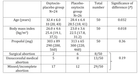
Table 2Demographic characteristics
Figures are mean ± standard deviation and median (range)
measurements were performed in the same way as for
the T2 measurements (Fig.2).
Since the plasma half-time elimination of oxytocin is
3–20 min [12], the comparison of oxytocin vs. placebo
could not be performed in the OP group after the second injection due to a too short wash-out time of oxytocin after the first injection. Thus, the oxytocin effect was in comparison with placebo studied cross-sectionally in the OP group at T1-T2 and
longitudin-ally in the PO group for T1-T2 vs. T2-T3 (Fig.1). For
each studied variable, the difference from before until
after administration was calculated, denoted Δ value.
A positive Δvalue denotes an increase and a negative
value a decrease.
The DPA data were automatically exported to an Excel file in the laptop and later converted to a statistics soft-ware document (StatView version 5.0.1, SAS Institute, Cary, NC, USA). The Meridian manufacturer recom-mends that at least 80% of the pulse waves should be recorded to ensure good quality of the mathematical
analyses of the pulse wave contour, so recordings with < 80% recognition were excluded from statistical analyses.
The Mann-Whitney U test was used to compare continuous variables between groups. For longitudinal comparisons within groups the Wilcoxon signed-ranks matched-pairs test was used. Categorical data were
com-pared with Chi-2 test or Fisher’s exact probability test.
Fisher’s exact test in 2 × 3 tables was performed with
software available on the web [13]. Two-sided P values
< 0.05 were considered statistically significant.
Some of the DPA variables are heart rate (HR) dependent
[5] and the statistical analyses were accordingly performed
with both crude and HR-adjusted DPA values. If simple lin-ear regression analyses yielded a statistically significant
cor-relation (P< 0.05) between HR and a DPA variables at T0,
and the intervention (anaesthesia, oxytocin or placebo administration) also resulted in a significant change in HR, the DPA variables in question was adjusted to a HR of 75 bpm, denoted DPA@75, with the equation DPA@75 =
DPA ±C(75-HR).C denotes the slope constant. The DPA
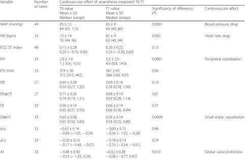
Table 3Haemodynamic effects of general anaesthesia in first trimester pregnant women. For explanation of measurement points T0 and T1, see text and Fig.2
Variable Number
of cases
Cardiovascular effect of anaesthesia measured T0-T1
T0 value
Significance of difference (P)
0.0001 Blood pressure drop
HR (bpm) 33 73 ± 14
70 (44, 96)
65 ± 9 63 (48, 84)
0.002 Heart rate drop
ECG ST index 48 0.15 ± 0.28
0.0001 Peripheral vasodilation
ETc (ms) 33 374 ± 30
372 (313, 442)
361 ± 45 368 (165, 437)
0.56
EEI 27 0.69 ± 0.28
0.59 (0.27, 1.32)
0.60 ± 0.18 0.58 (0.18, 1.00)
0.19
EEI@75 27 0.71 ± 0.26
0.74 (0.19, 1.21)
0.68 ± 0.19 0.69 (0.28, 1.14)
0.61
DI 33 0.66 ± 0.14
0.65 (0.37, 0.93)
0.66 ± 0.14 0.66 (0.36, 0.94)
0.31
DI@75 33 0.65 ± 0.08
0.65 (0.50, 0.83)
0.56 ± 0.14 0.58 (0.25, 0.80)
0.0009 Small-artery vasodilation
b/a 33 −0.63 ± 0.14
0.010 Global vasoconstriction
MAP, mean arterial blood pressure,HRheart rateECG, electrocardiogram,PHpulse height,ETccardiac left ventricular ejection time compensated,EEI, cardiac ejection elasticity index,EEI@75, EEI adjusted to a HR of 75 bpm,DIdicrotic index; DI@75, DI adjusted to a HR of 75 bpm;b/a, ratio between second derivatives of crude pulse wave contour;d/a, ratio between second derivatives of crude pulse wave contour; AI, ageing index
variable ETc and the ECG variable ST index are auto-matically adjusted for HR when reported by the respective apparatus.
Results
One patient in the OP group did not receive the allo-cated intervention due to a surgical complication with perforation of the uterus. Thus, 24 women allocated to the OP group and 26 to the PO group were included in
the study. Their demographic characteristics are shown
in Table2.
At time point T0, in 17/50 (34%) women the PH was below the 80% threshold for receiving adequate signals for pulse wave contour analysis, thus disabling reliable calculations of DPA variables. This excluded the longitudinal comparisons T0-T1 (effects of
anaes-thesia) in 34–46% of cases. For recordings T1-T3, the
recording error rate was 4–8%.
Table 4Haemodynamic effects of 5 U (8.3μg) oxytocin and placebo (NaCl) given intravenously to first trimester pregnant women undergoing surgical evacuation of the uterus. For explanation of time points T1 and T2, see text and Figs.1and2
Variable Effect of drug injection measured T1-T2
Oxytocin (OP group) N = 24
Placebo (PO group) N= 26
Pb Effect T1 value
Mean ± SD
0.0001 BP increase 0.61
HR
0.90 No change 0.020
ECG ST
0.0005 ST increase 0.0008
PH 5.3 ± 2.9
< 0.0001 Peripheral vasodilation
< 0.0001 Peripheral vasodilation
0.021 Decreased afterload, decreased SVR,
0.70 No change 0.069
EEI 0.58 ± 0.19
0.12 No change 0.17
EEI@75 0.67 ± 0.18
0.23 No change 0.43
DI 0.65 ± 0.15
0.84 No change 0.015
DI@75 0.55 ± 0.14
0.97 No change 0.10
b/a −0.59 ± 0.16
0.094 No change 0.19
d/a −0.17 ± 0.14
0.084 No change 0.84
AI −0.31 ± 0.29
0.086 No change 0.27
OPgroup, women given oxytocin at first injection and placebo at second injection; PO group, women given placebo at first injection and oxytocin at second injection
a)
TheΔvalues are not shown, but represent the T2 value minus T1 value.Pdenotes significance of difference inΔvalue between oxytocin and placebo injections, performed with the Mann-WhitneyUtest
b)
At point T0, a significant relationship was found between
HR and DI (P< 0.0001, r20.67) and EEI (P= 0.017, r20.17)
and these variables were accordingly adjusted in the
statis-tical analyses (called DI@75 and EEI@75). AI, b/a
and d/a were not significantly related to HR (P≥
0.50).
Anaesthesia caused a decrease in MAP and HR and a small-artery and peripheral vasodilation, as indicated by PH and DI@75, and a global vasoconstriction was
indi-cated by AI (Table3).
The surgery started after point T1 (Fig.2), and
oxyto-cin and placebo showed from T1 to T2 the following
ef-fects (Table4, Fig.3):
MAP increased by equal magnitudes in the OP and PO groups, i.e. no matter the sort of injection.
An increase in HR was seen only after oxytocin, and the difference to placebo was confirmed by
comparison ofΔHR values.
The ECG ST index changed only after placebo, confirmed by a difference inΔvalues.
PH increased (peripheral vasodilation) by equal magnitudes after oxytocin and placebo, confirmed by the absence of difference inΔPH values.
ETc increased (increase in LVET, increased preload, and/or decreased afterload) only after oxytocin, but the difference inΔETc values did not reach significance (P= 0.069).
EEI and EEI@75 were not affected by oxytocin or placebo.
Changes of DI and DI@75 after oxytocin indicated small-artery vasodilation, and the difference was confirmed by a difference inΔDI values.
b/a,d/aand AI did not change significantly in any group.
Due to a probable insufficient wash-out effect of oxytocin given as the first injection in the OP group,
the effects of placebo given as the second injection
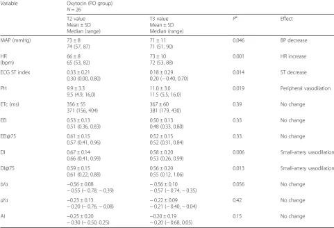
T2-T3 were not analysed (Fig. 1). Table 5 shows the
effects of oxytocin when comparing T2 and T3 values. Effects were found on MAP, HR, ST index, PH, DI and DI@75, whereas no effects were found on
ETc, EEI, EEI@75, d/a and AI. A decrease in b/a did
not reach statistical significance (P= 0.056, which
would indicate large-artery vasodilation and increase in LV ejection if significant).
At point T3 surgery had finished, but the women were
still under anaesthesia (Fig.2). In longitudinal
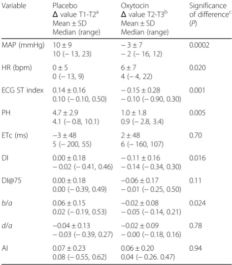
compari-sons in the PO group, the effects of oxytocin measured T2-T3 were compared with the effects of placebo
mea-sured T1-T2 (Table 6, Fig. 1). EEI and EEI@75 statistics
could not be performed due to too few cases in
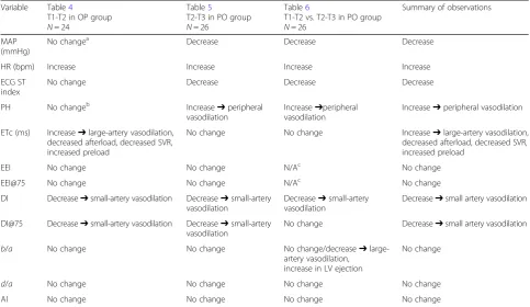
compari-sons (N= 10). The results in Table 6 are interpreted in
Table7, together with an overall interpretation of
oxyto-cin effects displayed in Tables4-6.
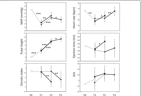
Figure 3 shows the sequential variable changes
throughout the experiment from T0 to T3, and Table7
shows a comprehensive interpretation of oxytocin
effects, where some interpretations need further expla-nations. The MAP increased at T1-T2 in both the oxyto-cin and placebo groups with no significant difference in
magnitude (Table 4), whereas the difference to placebo
in the comparison T1-T2 vs. T2-T3 was significant
(Table 6); as oxytocin at T2-T3 caused a decrease
(Ta-bles 5 and 6) and placebo at T1-T2 an increase (Table
4), the comprehensive interpretation is that MAP
de-creased after oxytocin (Table7).
The ECG ST index was unchanged after oxytocin, whereas
it increased after placebo at T1-T2 comparisons (Table4).
The difference to placebo in the comparison T1-T2 vs. T2-T3 was significant, where oxytocin showed a decrease
and placebo an increase (Tables4-6). The interpretation is
that ECG ST index decreased after oxytocin (Table7).
The ETc increased at T1-T2 after oxytocin whereas
placebo had no effect (Table4). In the longitudinal
com-parison with placebo at T1-T2 vs. T2-T3 (Table6) there
was no difference to placebo, and at T2-T3 oxytocin had
no effect on ETc (Table 5). The findings were then not
unanimous.
Table 5Haemodynamic effects of 5 U (8.3μg) oxytocin given as the second injection at T2-T3 in women given placebo as the first injection
Variable Oxytocin (PO group) N= 26
0.046 BP decrease
HR
0.001 HR increase
ECG ST index 0.33 ± 0.21 0.30 (0.00, 0.80)
0.18 ± 0.29 0.20 (−0.40, 0.70)
0.014 ST decrease
PH 9.9 ± 3.3
9.5 (4.9, 16,0)
11.0 ± 3.0 11.5 (5.5, 16.0)
0.019 Peripheral vasodilation
ETc (ms) 356 ± 55
371 (156, 404)
367 ± 60 381 (179, 430)
0.39 No change
EEI 0.53 ± 0.13
0.51 (0.36, 0.83)
0.50 ± 0.13 0.48 (0.33, 0.80)
0.33 No change
EEI@75 0.61 ± 0.15
0.57 (0.41, 0.96)
0.52 ± 0.15 0.52 (0.31, 0.84)
0.33 No change
DI 0.67 ± 0.14
0.66 (0.41, 0.99)
0.58 ± 0.20 0.53 (0.26, 0.99)
0.006 Small-artery vasodilation
DI@75 0.59 ± 0.15
0.61 (0.22, 0.88)
0.56 ± 0.20 0.55 (0.12, 1.06)
0.013 Small-artery vasodilation
b/a −0.56 ± 0.08
−0.55 (−0.78,−0.39) −
0.56 ± 0.10
−0.57 (−0.74,−0.35)
0.056 No change
d/a −0.23 ± 0.13
−0.20 (−0.76,−0.08)
−0.22 ± 0.09
−0.21 (−0.40,−0.04)
0.42 No change
AI −0.25 ± 0.20
−0.30 (−0.50, 0.25) − 0.20 ± 0.19
−0.20 (−0.68, 0.05)
0.15 No change
a)
No harmful side effects were noted during the study and no woman complained of chest discomfort.
Discussion
This study supports the hypothesis that oxytocin de-creases vascular tone. The changes in DPA variables after an oxytocin bolus suggest vasodilation and de-creased vascular tone of small and peripheral arteries. This was established at both cross-sectional and longitu-dinal comparisons with placebo. The PH, an indicator of the peripheral circulation, increased after anaesthesia and then further after the first drug injection, of equal magnitude after oxytocin and placebo. PH has previ-ously been found to increase with increasing depth of
anaesthesia due to reduced sympathetic activity [14].
Then, a continuously increasing PH with increasing depth of anaesthesia might explain the placebo effect of the first injection.
The second injection was given when surgery was fin-ished and the depth of anaesthesia was at steady state or ceasing. Then, oxytocin caused small-artery and periph-eral vasodilation, fall of MAP and increase in HR, which
further supports the conclusion that oxytocin causes a small-artery and peripheral vasodilation.
The increase in MAP in both groups at the start of surgery might be the result of an increase in sympathetic activity due to painful stimuli. It is, however, possible that a fall in MAP of a very short duration failed to be captured in our study protocol, as MAP was measured after the one-minute DPA recording was completed. The BP measurement takes up to 30 s, meaning that
MAP was measured 2–3 min after the end of the
oxyto-cin injection. It has been demonstrated in previous stud-ies that the MAP reduction reaches its nadir already
within 1–2 min, and returns to normal after another
mi-nute [15,16]. The ECG changes also occur within 1–2 min
[17]. Furthermore, oxytocin has a biphasic effect, with a
rapid initial HR increase and decrease in systolic BP
followed by a fall in HR and increase in BP [3, 18]. We
might then not have captured the rapid initial effect of oxy-tocin in our study. Also the duration of the bolus injection has been attributed to the nadir lag time. Some
re-searchers used statim bolus and others, like us, used a
60 s injection time. A short injection time might maxi-mise the initial effects. Since the BP measurements could interfere with the DPA measurements, we had no choice but to await finishing the DPA measurement be-fore we could record the BP.
The ETc increased after oxytocin at T1-T2 whereas pla-cebo had no effect. Considering the whole experiment, the effect of oxytocin on ETc was not unanimous, but as the T1-T2 comparison was the only one performed during equal conditions in the OP and PO groups, our overall in-terpretation is that oxytocin caused an increase in ETc. According to our previous communications with the South Korean manufacturer of the Meridian DPA, an in-crease in ETc could mean aortic valve stenosis or LV
fail-ure, as previously described [4, 5]. It might be true
concerning aortic valve disease, but is more complex
con-cerning LV failure [19]. Both ETc and the similar measure
Flow Time Corrected, derived from oesophageal Doppler velocimetry of aortic blood flow, correlate strongly with
the LVET [5,20]. Lewis et al. pointed out already in 1977
that LV muscle failure and diminished preload is associ-ated with a shortened LVET, and that a marked increase
in SV would lengthen the LVET [19].
Later on, Singer et al. [21,22] using oesophageal
Dop-pler velocimetry, demonstrated that the LVET was in-creased when preload was inin-creased from a hypovolemic state, but also when preload was decreased from an overload state. Thus, LVET is reaching its maximum at optimal filling pressures. As preload diminishes, com-pensatory peripheral vasoconstriction causing increased systemic vascular resistance (SVR) usually occur. In ef-fect, vasodilating agents alone also produce an increase in LVET, leaving preload unaffected. Taken together, in
Table 6Haemodynamic effects of oxytocin (administered during sequence T2-T3) relative to placebo (sequence T1-T2) in first trimester pregnant women in the placebo-oxytocin group (N= 26). For interpretation and comparisons of oxytocin effects, see Table7 Variable Placebo of differencec (P)
Δvalue calculationsT2value minus T1 value
b)
Δvalue calculations: T3 value minus T2 value
c)
healthy patients without fluid overload, LVET increases not only by increased preload but also by decreased
afterload, i.e. from decreased SVR [23].
From this argumentation, we believe that the rise in ETc after the first oxytocin injection indicates a reduc-tion in SVR, since preload is assumed unaffected. As HR increased without a fall in MAP, an increase in CO is as-sumed. An increase in CO leads to an increase in myo-cardial oxygen demand. Signs of myomyo-cardial ischemia
have previously been noted after oxytocin injection [17],
and that observation could explain the different re-sponses in ECG ST index after oxytocin and placebo.
Thirty-four percent of DPA values were missing at T0 due to cold fingers, probably caused by
pre-opera-tive stress-induced peripheral vasoconstriction [14].
Among confounding factors, sympathetic activity in-duced by noxious stimuli from the surgical procedure might explain changes seen in the placebo group be-tween T1 and T2. Although there were no differences between the groups in the total doses of propofol given, variations in anaesthetic depth is another pos-sible confounding factor.
The cardiovascular effects of anaesthesia included de-creases in BP and HR and dilatation of small and periph-eral arteries. However, the global index AI indicated
vasoconstriction. AI is a composite index of four indices reflecting different phases of the pulse wave from early
systole to early diastole, i.e. b/a, c/a, d/a and e/a [5].
The two indices reported separately in the present study,
i.e. b/a and d/a, did not change significantly, and the
variable reflecting large arteries, EEI, did not change ei-ther. Then, the significant change of AI can be inter-preted as the amalgamated result of non-significant changes of the subset of indices, or it was a result of a type 1 error. Oxytocin has both vasodilatory and
vaso-constrictory properties via the same receptor, V1aR [24],
and various vascular beds and organs display different
distributions and receptor sensitivity [25]. Although not
possible to demonstrate in the present study, vasocon-striction in medium-sized arteries could be a physio-logical response to peripheral vasodilation and falls in BP and HR.
DPA with digital photoplethysmography is a non-in-vasive, operator-friendly and rapid method to study vascular tone and pharmacological hemodynamic ef-fects in a clinical setting where the efef-fects come swift. As illustrated by the DPA measurements before ini-tializing anaesthesia, cold fingers must be avoided as they distort the results. That applies to finger move-ments as well.
Table 7Summary of observations based on haemodynamic effects of oxytocin administered at first injection T1-T2 (Table4) and second injection T2-T3 (Tables5and6). For further explanation of comparisons, see Fig.1, and for explanation of interpretations, see text Variable Table4
T1-T2 in OP group N= 24
Table5
T2-T3 in PO group N= 26
Table6
T1-T2 vs. T2-T3 in PO group N= 26
Summary of observations
MAP (mmHg)
No changea Decrease Decrease Decrease
HR (bpm) Increase Increase Increase Increase
ECG ST index
No change Decrease Decrease Decrease
PH No changeb Increase➔peripheral
vasodilation
Increase➔peripheral vasodilation
Increase➔peripheral vasodilation
ETc (ms) Increase➔large-artery vasodilation, decreased afterload, decreased SVR, increased preload
No change No change Increase➔large-artery vasodilation, decreased afterload, decreased SVR, increased preload
EEI No change No change N/Ac No change
EEI@75 No change No change N/Ac No change
DI Decrease➔small-artery vasodilation Decrease➔small-artery vasodilation
Decrease➔small-artery vasodilation
Decrease➔small artery vasodilation
DI@75 Decrease➔small-artery vasodilation Decrease➔small-artery vasodilation
No change Decrease➔small artery vasodilation
b/a No change No change No change/decrease➔
large-artery vasodilation, increase in LV ejection
No change
d/a No change No change No change No change
AI No change No change No change No change
a)
MAPincreased after both oxytocin and placebo
b)
PHincreased after both oxytocin and placebo, indicating peripheral vasodilation
c)
A strength of the study is the randomised double-blinded placebo-controlled design in a normal clinical setting. In comparison with our previous study on oxytocin during CS, where the procedures with spinal anaesthesia, IV fluids, vasoactive drugs and delivery of
the baby made it problematic to interpret the
hemodynamic effects of oxytocin [4], the present
study was less compromised by confounding factors.
Another strength is the DPA method’s ability to
in-vestigate effects on large and small arteries separately, by using derivative mathematics to analyse the sys-tolic and diassys-tolic segments of the pulse wave con-tour. It is a limitation that the study protocol could not be adapted to capture short biphasic courses of events. As surgery needed to be stopped during DPA and BP measurements, for the safety of the women we could not perform double measurements, as would have been experimentally preferred. Variations in anaesthetic depth and sympathetic activity are pos-sible confounders that could not be controlled for.
Misoprostol 0.4 mg was administered vaginally 3 h prior to scheduled surgery. The drug has a plasma half-time of about 30 min, but due to slow absorption by the vaginal route the bioactivity could remain for a few
hours [26]. It is therefore possible that remainders of
mi-soprostol persisted at the time of the experiments. How-ever, the lasting bioactivity has been shown on the
myometrium [26], whereas no cardiovascular effects of
misoprostol have been demonstrated [27,28]. We
there-fore believe possible remainders of misoprostol did not confound the results. The placebo-controlled design of the study could ensure it was not misoprostol effects that we measured, and the longitudinal measurements
were performed during a short time-span when
steady-state concentrations of misoprostol would not bias the results.
Conclusions
This study confirmed the hypothesis that oxytocin de-creases vascular tone in small and peripheral arteries, resulting in a lower BP and a compensatory increase in HR. These effects might have been enhanced by the vasodilating effects of anaesthesia. We could not confirm any effect on vascular tone in large arteries. The net oxy-tocin effect on the heart is likely to be an increased CO, implying increased myocardial oxygen demand. The demonstrated multiple effects of oxytocin on the cardio-vascular system suggest a cautious use of oxytocin in pregnant women.
Abbreviations
AI:Ageing index; b/a: Second derivative quotient of the acceleration phase the percussion pulse wave; BP: Blood pressure; CO: Cardiac output; CS: Caesarean section; d/a: Second derivative quotient of percussion wave clashing the reflected tidal wave; DI: Dicrotic index; DI@75: DI adjusted to a
heart rate of 75 bpm; DPA: Digital pulse wave analysis; ECG: Electrocardiogram; EEI: Ejection elasticity index; EEI@75: EEI adjusted to a heart rate of 75 bpm; ETc: Left ventricular ejection time compensated (for heart rate); Ftc: Flow time corrected; HR: Heart rate; IV: Intravenous; LV: Left ventricle of the heart; LVET: Left ventricular ejection time; MAP: Mean arterial blood pressure; OP: Oxytocin-placebo sequence; PH: Pulse height; PO: Placebo-oxytocin sequence; ST index: Difference in ECG ST segment from the isoelectric baseline; SVR: Systemic vascular resistance;Δvalue: Difference between measurement at time point Tnminus measurement at point Tn-1
Funding
The study was funded by grants from the Medical Faculty at Lund University (ALF) and from Region Skåne. The funders had no role in study design, data collection and analysis, decision to publish, or preparation of the manuscript.
Availability of data and materials
The datasets used are available from the corresponding author on reasonable request.
Authors’contributions
PO conception and design of the study, PO and SR planning and carrying out the study, analysing data and writing the article, UH and SS carrying out the study, analysing data, reviewing the manuscript. All authors have read and approved the manuscript.
Ethics approval and consent to participate
The regional research ethics committee in Lund approved the study (dnr 2012/649) and all enrolled women gave their informed oral and written consent.
Consent for publication Not applicable.
Competing interests
The authors declare that they have no competing interests.
Publisher’s Note
Springer Nature remains neutral with regard to jurisdictional claims in published maps and institutional affiliations.
Author details
1Department of Clinical Sciences Lund, Anaesthesiology and Intensive Care,
Skåne University Hospital, Lund University, S-22185 Lund, Sweden. 2
Department of Clinical Sciences Malmö, Obstetrics and Gynaecology, Skåne University Hospital, Lund University, Malmö, Sweden.3Department. of Intensive and Perioperative Care, Skåne University Hospital, S-22185 Lund, Sweden.
Received: 25 June 2018 Accepted: 25 September 2018
References
1. Royal College of Obstetrician and Gynaecologists. Prevention and management of postpartum haemorrhage. London: RCOG Greentop Guidel; 2009. p. 1–24. Available from:https://doi.org/10.1111/1471-0528.14178.
2. Nygaard IHH, Valbø A, Heide HC, Kresovic M. Is oxytocin given during surgical termination of first trimester pregnancy useful? A randomized controlled trial. Acta Obstet Gynecol Scand [Internet], vol. 90. Blackwell Publishing Ltd; 2011. p. 174–8. Available from:https://doi.org/10.1111/j. 1600-0412.2010.01025.x
3. Thomas JS, Koh SH, Cooper GM. Haemodynamic effects of oxytocin given as i.v. bolus or infusion on women undergoing caesarean section. Br J Anaesth. 2007;98:116–9.
4. Rabow S, Olofsson P. Pulse wave analysis by digital photoplethysmography to record maternal hemodynamic effects of spinal anesthesia, delivery of the baby, and intravenous oxytocin during cesarean section. J Matern Neonatal Med. 2016;7058:1–26.
5. von Wowern E, Östling G, Nilsson PM, Olofsson P. Digital Photoplethysmography for assessment of arterial stiffness: repeatability and comparison with Applanation tonometry. West J, editor. PLoS One [Internet]. 2015;10:e0135659. Available from:
6. Elgendi M. On the analysis of fingertip photoplethysmogram signals. Curr Cardiol Rev. 2012;8:14–25.
7. Takazawa K, Tanaka N, Fujita M, Matsuoka O, Saiki T, Aikawa M, et al. Assessment of vasoactive agents and vascular aging by the second derivative of Photoplethysmogram waveform. Hypertension [Internet]. 1998; 32:365–70. Available from:https://www.ahajournals.org/lookup/doi/10.1161/
01.HYP.32.2.365. [cited 2013 May 24].
8. Millasseau SC, Ritter JM, Takazawa K, Chowienczyk PJ. Contour analysis of the photoplethysmographic pulse measured at the finger. J Hypertens. 2006;24:1449–56.
9. Millasseau SC, Guigui FG, Kelly RP, Prasad K, Cockcroft JR, Ritter JM, et al. Noninvasive assessment of the digital volume pulse. Comparison with the peripheral pressure pulse. Hypertens. (Dallas, Tex 1979) [Internet]. 2000;36: 952–6.
10. Weis FR, Markello R, Mo B, Bochiechio P. Cardiovascular effects of oxytocin. Obstet Gynecol. 1975;46:211–4.
11. Stat Trek Random Number Generator [Internet]. [cited 2013 Oct 2]. Available from:http://stattrek.com/statistics/random-number-generator.aspx
12. FASS, Farmaceutiska specialiteter i Sverige [Internet] www.fass.se. Available from: http://www.fass.se/LIF/product?userType=0&nplId=20091203000014. [cited 2017 Nov 30].
13. VassarStats: website for statistical computation. Fisher exact probability test: 2x3 [Internet]. [cited 2017 Apr 6]. Available from:http://vassarstats.net/ fisher2x3.html
14. Enekvist B, Johansson A. Pulse perfusion value predicts eye opening after sevoflurane anaesthesia: an explorative study. J Clin Monit Comput. 2015;29:461–5.
15. Johnstone M, Anaesth BJ. The cardiovascular effects of oxytocic drugs. Brit J Anaesth. 1972;44:826–34.
16. Langesaeter E, Rosseland LA, Stubhaug A. Hemodynamic effects of oxytocin during cesarean delivery. Int J Gynaecol Obstet. 2006;95:46–7.
17. Svanström MC, Biber B, Hanes M, Johansson G, Näslund U, Bålfors EM. Signs of myocardial ischaemia after injection of oxytocin: a randomized double-blind comparison of oxytocin and methylergometrine during caesarean section. Br J Anaesth. 2008;100:683–9.
18. Moertl M, Friedrich S, Kraschl J, Wadsack C, Lang U, Schlembach D. Haemodynamic effects of carbetocin and oxytocin given as intravenous bolus on women undergoing caesarean delivery: a randomised trial. BJOG. 2011;118:1349–56.
19. Lewis RP, Rittgers SE, Forester WF, Boudoulas H. Reviews of Contemporary Laboratory Methods Arnold M. Weissler M D, A critical review of the systolic time intervals Circulation. 1977;56:146–158.
20. GSH C, Middleton PM, Celler BG, Wang L, Lovell NH, Asada HH, Shaltis P, RS RA, HRC, et al. Automatic detection of left ventricular ejection time from a finger photoplethysmographic pulse oximetry waveform: comparison with Doppler aortic measurement. Physiol Meas IOP Publishing. 2007;28:439–52. 21. Singer M, Bennett ED. Noninvasive optimization of left ventricular filling
using esophageal Doppler. Crit Care Med. 1991;19:1132–7. 22. Singer M, Allen MJ, Webb AR, Bennett ED. Effects of alterations in left
ventricular filling, contractility, and systemic vascular resistance on the ascending aortic blood velocity waveform of normal subjects. Crit Care Med. 1991;19:1138–45.
23. Deltex Medical CardioQ-ODM Manual [Internet]. [cited 2016 Dec 8]. Available from: http://www.deltexmedical.com/cardioq-odm/flow-parameters/
24. Japundžić-Žigon N. Vasopressin and oxytocin in control of the cardiovascular system. Curr Neuropharmacol. 2013;11:218–30. 25. Thibonnier M, Conarty DM, Preston JA, Plesnicher CL, Dweik RA, Erzurum
SC. Human Vascular Endothelial Cells Express Oxytocin Receptors. Endocrinology. 1999;140:1301–9.
26. Gemzell-Danielsson K, Bygdeman M, Aronsson A. Studies on uterine contractility following mifepristone and various routes of misoprostol. Contraception. 2006;74:31–5.
27. Brecht T. Effect of misoprostol on human circulation. Prostaglandins. 1987; 33:51–60.
28. Ramsey PS, Hogg BB, Savage KG, Winkler DD, Owen J. Cardiovascular effects of intravaginal misoprostol in the mid trimester of pregnancy. Am J Obstet Gynecol. [Internet] Elsevier; 2017;183:1100–2. Available from:https://doi.org/