Svetlana Opric
, Magdalena Stefanovic Radovic
, Ivan Soldatovic
, Biljana Sredic
and Miroslav Granic
Abstract
Background:We present a surgical technique and the preliminary results of breast cancer excision after insertion of a specially constructed marking needle into the tumor, controlled by intraoperative ultrasound.
Resection margins were projected in six directions by ultrasound measurements, determined in relation to the needle, and resection was done in accordance with those measurements. The main objective was to obtain resection margins similar (equal) to those projected by intraoperative ultrasound (10 mm).
Methods:Detailed description of the technique is given. Thirty-two female patients undergoing breast-conserving surgery, up to 30 mm in diameter, for palpable and non-palpable invasive breast cancer, were operated on using this technique. Its feasibility was tested by analyzing the success (rate) of needle placement in the tumor, the measurements executed, and the performance of the excision.
Results:All stages of the technique were successfully performed to completion on all 32 patients. The procedure of needle placement and ultrasound measurement of distances took 11 min on average (between 6 and 20 min). The average distance of the tumor margin from the resection margin was 12.9 mm (2 to 30 mm, 95% confidence interval [11.9, 14.06]). There was one patient with a positive resection margin (3%).
Conclusions:The technique of excising palpable and non-palpable breast cancer by intraoperative ultrasound and an especially constructed marking needle is feasible and comfortable to perform. Preliminary results imply that resection volume can be rationalized, with the same or better oncological safety.
Keywords:Intraoperative ultrasound, Marking needle, Breast-conserving surgery, Breast cancer
Background
Intraoperative ultrasound (IOUS) in breast tumor sur-gery was introduced into clinical practice as a means of localizing non-palpable tumors [1-12] and more recently has been used in the surgery of palpable tumors [13-21], with the aim of optimizing resection procedures and overcoming the shortcomings of classic palpation-guided surgery. In descriptions of the technique of IOUS appli-cation, most authors rely on direct contact between the ultrasound probe and the tumor specimen. Using the
IOUS in this, let it be called, standard way, produces some difficulties that disturb the comfort and accuracy of both procedure and performance: the presence of air and liquid in the wound may create content which sig-nificantly disturb the quality of the ultrasound image; in-sertion of a probe into the wound can mean movement and pressure of the tissue, which may in turn influence the assessment of resection margins; and refraction of ultrasound waves while passing through tissue of irregu-lar shape, such as a tumor, may create a difference be-tween the size of tumor measured by ultrasound and the real size [22].
These difficulties motivated us to create a technique of resection for both palpable and non-palpable breast tu-mors by using a marking needle which is inserted into
* Correspondence:[email protected]
1Department of Surgical Oncology, UMC Bezanijska Kosa, Autoput bb,
Belgrade 11000, Serbia
2Medical Faculty of Belgrade University, Dr Subotica 8, Belgrade 11000, Serbia
Full list of author information is available at the end of the article
the tumor and is guided by ultrasound during the oper-ation, while the patient is anesthetized. Measurements of the distance of resection margins are performed using ultrasound in relation to this needle, in all directions. The resection is thus performed according to these mea-surements, with continual measurement of the resection margins in relation to the needle using a sterile ruler.
Methods
By the end of October 2013, 32 patients with palpable and non-palpable unifocal breast carcinoma (29 palpable and 3 non-palpable) with diameters of up to 30 mm, vis-ible by ultrasound, had been operated on, using the presented technique, in the Department of Surgical On-cology at UMC Bezanijska Kosa. All operations were performed by one surgeon, using a 7.5 MHz US probe, Hitachi EUB 5500 (Tokyo, Japan). The diagnosis of breast carcinoma was made by a frozen section tech-nique in all cases, after tumor excision. The time was
recorded from the beginning of ultrasound measurement to the moment of incision. All patients have signed the informed consent.
The aim was to test the feasibility of the technique. The main parameters of success were:
1. Successful needle insertion into the tumor (that is, the presence of the needle in the tumor at least 2 mm from the closest margin).
2. Successful performance of the resection procedure (the needle at completion of resection should be in the same position as it was at the beginning, checked by ultrasound examination of the specimen at the end of excision).
3. Good correlation between distances from the tumor border to the resection margin projected by ultrasound (always 10 mm in six directions), measured on a histological specimen by a pathologist.
Figure 1Marking needle.
4. An acceptable percentage of positive resection margins (microscopic presence of tumor cells at 2 mm and less from the resection margin).
Technique description
The operation starts by marking the point of tumor pro-jection on the skin by US-imaging. The skin incision is planned according to that point, applying standard di-rections of incision in breast cancer surgery. After the incision line is drawn, a probe is positioned with its lon-ger axis on that line. To visualize the tumor in its largest diameter, the US probe is moved in soft transversal mo-tions, perpendicular to the long axis of the transducer. The tumor diameter (transverse versus vertical), the lesion-to-skin distance, and the lesion-to-fascia distance are measured in millimeters. This process is repeated in the cranial-caudal plane. The probe is again positioned
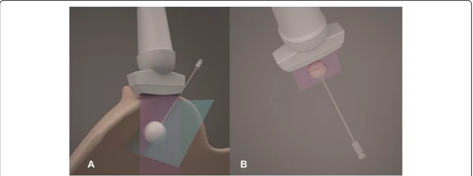
central axis of the tumor (Figure 2A).
The needle is inserted into the central axis of the tumor (or as close as possible to the central axis) as seen from the skin toward the pectoral fascia.
The central position of the needle in the plane orthog-onal to this plane was achieved earlier, by positioning the needle in the plane of the ultrasound wave. The nee-dle passes through the whole length of the tumor. Its tip should be at the distal tumor margin, or a few millime-ters in front of, or beyond the distal margin. When the needle is localized in the desired position, necessary measurements are performed (Table 1, Figure 2B).
By adding of 10 mm to each of these values, all resec-tion margins are precisely determined, that is, their dis-tance from the needle is established (Figure 3).
Resection procedure
Incision of the skin is made along the previously planned line. All measurements (distance from the needle to re-section margins) are performed using a sterile ruler.
The breast tissue is cut along the incision line until the needle fixed in the tissue is reached, proximal from the tumor. The distance of this point from the starting point of needle is determined. Then, by incising the tissue along the length of the needle, we reach the specimen at the
Distance from the needle to tumor borders Needle-surface border (toward skin) Needle-deep border (toward fascia) Needle-cranial border Needle-caudal border
point of the proximal resection margin along the length. The procedure is then continued by incising the breast tis-sue along the whole length of the incision, to the distal re-section margin, and to a depth representing the surface resection margin, at an appropriate distance, from the nee-dle toward the skin.
From this line, resection extends laterally (cranially and caudally) in both directions from the needle until an appropriate distance to resection margins is reached (Figure 4). This continues to the deepest resection margin (toward the pectoral fascia). When resection is completed,
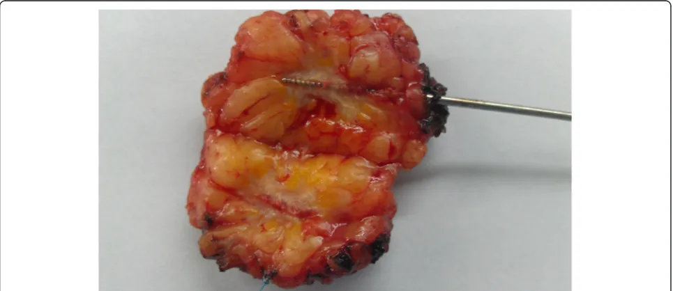
the specimen is released from the surrounding tissue and is positioned on the needle, as on a spit (Figure 5). The specimen is then spatially oriented and sent to histopath-ology for processing and analysis (Table 2).
Results
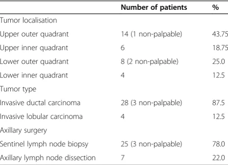
A total of 32 female patients were included in this study (Table 3).
In all cases, the tumor was successfully punctured (100%) and resected (the needle being in the same position
Figure 4Resection procedure.
inside the tumor at the end of procedure as it had been at the beginning (100%)).
The average time from the beginning of measuring to incision was 11 min (6 to 20 min).
The average distance of the tumor margin from the re-section margin was 12.9 mm (95% confidence interval, 11.9 to 14.06). In six cases, when resection margins were 5 mm and less (3 times at 5 mm, twice at 4 mm, once at 3 mm, and once at 2 mm), additional shaving excisions were performed.
There was one patient with a positive resection margin (3%).
Discussion
The technique described here represents an effort to make resection procedures in breast-conserving surgery (both for palpable and non-palpable tumors) more objective and
Studies published thus far have shown extraordinary success with intraoperative ultrasound in localizing non-palpable tumors, as well as advantages in relation to widely accepted WNL [1-12,20,23-26]. More recently, ultrasound measuring of resection margins has proved useful and efficient in planning and performing resections of palpable tumors [14,17,19]. Previously described tech-niques of using ultrasound in planning resection margins in palpable tumors rely on direct contact between the ultrasound probe and the specimen, without a marker visible by ultrasound. In our experience, such an approach is burdened with difficulties, especially inaccuracy in visualization due to non-homogeneous content in the wound and tissue movement during surgical work.
The technique that we propose means introducing a specially designed marking needle into the tumor, visible by ultrasound, in relation to which distances of tumor margins are measured in all directions, as well as the de-sired resection margins. The needle thus placed repre-sents guaranteed unchangeability of dimensions, and visualization is ideal before the incision is made.
A preoperative core biopsy should be avoided because the resulting hematoma could affect the US visualization, and tumor damage could affect stability and firmness when anchoring the marking needle. We therefore did the frozen section procedure in all cases.
Our preliminary results show that the technique is feas-ible. In all cases, tumors were successfully punctured, and all necessary measurements, as well as the resection itself, were performed successfully. The only shortcoming of this procedure may be the additional time needed for measur-ing before incision (about 11 min on average).
Conclusions
Intraoperative US with a marking needle allows real-time localization of breast carcinoma and subsequent planning of surgical margins, thereby resulting in objective, measur-able tumor excision. In a randomized clinical trial, intra-operative US with marking needle guidance for palpable and non-palpable breast cancer will be evaluated for margin clearance, excision volume, cosmetic outcomes, and quality of life. The results will be compared with palpation-guided surgery.
Table 3 Patient and tumor characteristics
Number of patients %
Tumor localisation
Upper outer quadrant 14 (1 non-palpable) 43.75
Upper inner quadrant 6 18.75
Lower outer quadrant 8 (2 non-palpable) 25.0
Lower inner quadrant 4 12.5
Tumor type
Invasive ductal carcinoma 28 (3 non-palpable) 87.5
Invasive lobular carcinoma 4 12.5
Axillary surgery
Sentinel lymph node biopsy 25 (3 non-palpable) 78.0 Axillary lymph node dissection 7 22.0 Mean tumor volume (three diameters multiplied) in cm3
(range): 4.4 (0.28 to 15.12); (volume of non-palpable tumors: 0.28, 0.336, and 0.448 cm3).
Abbreviations
IOUS:intraoperative ultrasound; RM: resection margin; UMC: university medical center; US: ultrasound; WNL: wire needle localization..
Competing interests
The authors declare that they have no competing interests.
Authors’contributions
IN helped in the design of the study and the surgical technique, ZD helped to draft the manuscript, SZ collected, processed, and analyzed data, KJ collected, processed, and analyzed data, CN collected, processed, and analyzed data, SM and OS carried out pathological analysis, SRM supervised echosonography, SI did statistical analysis, SB collected, processed, and analyzed data, and GM acted as general supervisor. All authors read and approved the final manuscript.
Acknowledgements
Esther Helajzen assisted as English language editor.
Author details
1Department of Surgical Oncology, UMC Bezanijska Kosa, Autoput bb,
Belgrade 11000, Serbia.2Medical Faculty of Belgrade University, Dr Subotica 8, Belgrade 11000, Serbia.3Department of Pathology, UMC Bezanijska kosa,
Autoput bb, Belgrade 11000, Serbia.4Department of Radiology, UMC
Bezanijska kosa, Autoput bb, Belgrade 11000, Serbia.5Department of
Statistics, Medical faculty of Belgrade University, Dr Subotica 8, Belgrade 11000, Serbia.
Received: 6 October 2014 Accepted: 4 April 2015
References
1. Schwartz GF, Goldberg BB, Rifkin MD, D’Orazio SE. Ultrasonography: an alternative to x-ray-guided needle localization of nonpalpable breast masses. Surgery. 1988;104:870–3.
2. Rahusen FD, van Amerongen AH T, van Diest PJ, Borgstein PJ, Bleichrodt RP, Meijer S. Ultrasound-guided lumpectomy of nonpalpable breast cancers: a feasibility study looking at the accuracy of obtained margins. J Surg Oncol. 1999;72:72–6.
3. Snider HC, Morrison DG. Intraoperative ultrasound localization of nonpalpable breast lesions. Ann Surg Oncol. 1999;6:308–14. 4. Harlow SP, Krag DN, Ames SE, Weaver DL. Intraoperative ultrasound
localization to guide surgical excision of nonpalpable breast carcinoma. J Am Coll Surg. 1999;189:241–6.
5. Smith LF, Rubio IT, Henry-Tillman R, Korourian S, Klimberg VS. Intraoperative ultrasound-guided breast biopsy. Am J Surg. 2000;180:419–23.
6. Rahusen FD, Bremers AJA, Fabry HFJ, van Amerongen AH, Boom RP, Meijer S. Ultrasound-guided lumpectomy of nonpalpable breast cancer versus wire-guided resection: a randomized clinical trial. Ann Surg Oncol. 2000;9:994–8.
7. Kaufman CS, Jacobson L, Bachman B, Kaufman LB. Intraoperative ultrasonography guidance is accurate and efficient according to results in 100 breast cancer patients. Am J Surg. 2003;186:378–82.
8. Bennett IC, Greenslade J, Chiam H. Intraoperative ultrasound-guided excision of nonpalpable breast lesions. World J Surg. 2005;29:369–74.
9. Haid A, Knauer M, Dunzinger S, Jasarevic Z, Köberle-Wührer R, Schuster A, et al. Intra-operative sonography: a valuable aid during breastconserving surgery for occult breast cancer. Ann Surg Oncol. 2007;14:3090–101. 10. Potter S, Govindarajulu S, Cawthorn SJ, Sahu AK. Accuracy of sonographic
localisation and specimen ultrasound performed by surgeons in impalpable screen-detected breast lesions. Breast. 2007;16:425–8.
11. Ngô C, Pollet AG, Laperrelle J, Ackerman G, Gomme S, Thibault F, et al. Intraoperative ultrasound localization of nonpalpable breast cancers. Ann Surg Oncol. 2007;14:2485–9.
12. Fortunato L, Penteriani R, Farina M, Vitelli CE, Piro FR. Intraoperative ultrasound is an effective and preferable technique to localize non-palpable breast tumors. Eur J Surg Oncol. 2008;34:1289–92.
13. Moore MM, Whitney LA, Cerilli L, Imbrie JZ, Bunch M, Simpson VB, et al. Intraoperative ultrasound is associated with clear lumpectomy margins for palpable infiltrating ductal breast cancer. Ann Surg. 2001;233:761–8.
14. Olsha O, Shemesh D, Carmon M, Sibirsky O, Abu Dalo R, Rivkin L, et al. Resection margins in ultrasound-guided breast-conserving surgery. Ann Surg Oncol. 2011;18:447–52.
15. Davis KM, Hsu CH, Bouton ME, Wilhelmson KL, Komenaka IK. Intraoperative ultrasound can decrease the re-excision lumpectomy rate in patients with palpable breast cancers. Am Surg. 2011;77:720–5.
16. Fisher CS, Mushawah FA, Cyr AE, Gao F, Margenthaler JA. Ultrasound-guided lumpectomy for palpable breast cancers. Ann Surg Oncol. 2011;18:3198– 203.
17. Krekel NM, Lopes Cardozo AM, Muller S, Bergers E, Meijer S, van den Tol MP. Optimising surgical accuracy in palpable breast cancer with intra-operative breast ultrasound - feasibility and surgeons’learning curve. Eur J Surg Oncol. 2011;37:1044–50.
18. Eichler C, Hubbel A, Zarghooni V, Thomas A, Gluz O, Stoff-Khalili M, et al. Intraoperative ultrasound: improved resection rates in breast-conserving surgery. Anticancer Res. 2012;32:1051–6.
19. Krekel NM, Haloua MH, Lopes Cardozo AM, de Wit RH, Bosch AM, de Widt-Levert LM, et al. Intraoperative ultrasound guidance for palpable breast cancer excision (COBALT trial): a multicentre, randomised controlled trial. Lancet Oncol. 2013;14:48–54.
20. Yu CC, Chiang KC, Kuo WL, Shen SC, Lo YF, Chen SC. Low re-excision rate for positive margins in patients treated with ultrasound-guided breast-conserving surgery. Breast. 2013;22:698–702.
21. Gittleman MA. Single-step ultrasound localization of breast lesions and lumpectomy procedure. Am J Surg. 2003;186:386–90.
22. DeJean P, Brackstone M, Fenster A. An intraoperative 3D ultrasound system for tumor margin determination in breast cancer surgery. Med Phys. 2010;37:564–70.
23. James TA, Harlow S, Sheehey-Jones J, Hart M, Gaspari C, Stanley M, et al. Intraoperative ultrasound versus mammographic needle localization for ductal carcinoma in situ. Ann Surg Oncol. 2009;16:1164–9.
24. Bouton ME, Wilhelmson KL, Komenaka IK. Intraoperative ultrasound can facilitate the wire guided breast procedure for mammographic abnormalities. Am Surg. 2011;77:640–6.
25. Barentsz MW, van Dalen T, Gobardhan PD, Bongers V, Perre CI, Pijnappel RM, et al. Intraoperative ultrasound guidance for excision of nonpalpable invasive breast cancer: a hospital-based series and an overview of the literature. Breast Cancer Res Treat. 2012;135:209–19. 11.
26. Ramos M, Díaz JC, Ramos T, Ruano R, Aparicio M, Sancho M, et al. Ultrasound-guided excision combined with intraoperative assessment of gross macroscopic margins decreases the rate of reoperations for non-palpable invasive breast cancer. Breast. 2013;22:520–4.
Submit your next manuscript to BioMed Central and take full advantage of:
• Convenient online submission
• Thorough peer review
• No space constraints or color figure charges
• Immediate publication on acceptance
• Inclusion in PubMed, CAS, Scopus and Google Scholar
• Research which is freely available for redistribution