ISSN(Online): 2319-8753 ISSN (Print): 2347-6710
I
nternational
J
ournal of
I
nnovative
R
esearch in
S
cience,
E
ngineering and
T
echnology
(An ISO 3297: 2007 Certified Organization)
Website: www.ijirset.com
Vol. 6, Issue 5, May 2017
Crack Detection Method on Spinal Canals
Automatically
Mohammed Najeebulla. A 1, Shelmi 2
P.G. Student, Department of Computer Engineering, Cochin College of Engineering & Technology, Valancheri,
Kerala, India1
Assistant Professor, Department of Computer Engineering, Cochin College of Engineering & Technology, Valancheri
Kerala, India2
ABSTRACT: Crack Detection on spinal canal is important in many medical diagnosis and clinical analysis. For detecting the crack on the spinal canal we want to segment our spinal canal from a CT image. For that a segmentation method is also needed here. Here proposes a segmentation method and hence by detecting the crack on the spinal canal. Segmentation of spinal canals have a great role in many medical fields. Spinal canal is one of the best way used to diagnosis and analysis. Accurate segmentation of the spinal canals in computed tomography (CT) images is an important task in many related studies. Many research and clinical studies require the automatic segmentation of the spines, which is capable of facilitating disease diagnosis, treatment, and statistical analysis/evaluation. For example, the segmentation of the spine provides spatial reference to locate and identify other neighbouring anatomical structures in abdomens and chests, thus contributing to the understanding of the full-body scan essentially. In terms of image registration, the segmented spines provide important features that are helpful to the correct alignment of corresponding anatomical structures across individual subjects. Furthermore, it becomes easier to conduct disease-oriented analysis given the segmented topologies/shapes of the spines. In the meantime, based on the segmentation of the spinal canal, the entire spinal cord can easily be delineated, making it possible to count radiotherapy dosages which are crucial to the normal functions of the nerve tracts. Segmented spinal canal is then analysed to detect any crack is present on the surface.
KEYWORDS:Crack detection, Image segmentation, Topology, Random walk, Refinement, Spinal canal, Voxels.
I. INTRODUCTION
Accurate segmentation and crack detection of the spinal canals in computed tomography (CT) images is an important task in many related studies. Many research and clinical studies require the automatic segmentation of the spines, which is capable of facilitating disease diagnosis, treatment, and statistical analysis/evaluation. In terms of image registration, the segmented spines provide important features that are helpful to the correct alignment of corresponding anatomical structures across individual subjects. Furthermore, it becomes easier to conduct disease-oriented analysis given the segmented topologies/shapes of the spines. Although relatively more work in the literature focus on the segmentation of the spines in magnetic resonance (MR) images, we will investigate the automatic segmentation of the spinal canals from highly varying computed tomography (CT) images in this paper. Accurately segmenting the spinal canal facilitates the computer-aided detection process of anomalies, such as epidural masses on CT scans.
ISSN(Online): 2319-8753 ISSN (Print): 2347-6710
I
nternational
J
ournal of
I
nnovative
R
esearch in
S
cience,
E
ngineering and
T
echnology
(An ISO 3297: 2007 Certified Organization)
Website: www.ijirset.com
Vol. 6, Issue 5, May 2017
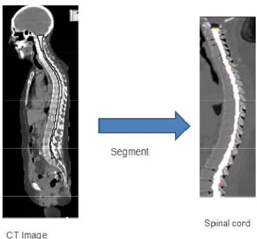
Fig -1: Segmenting the spinal canal
Figure 1 shows CT image and Spinal cord. From the CT images we can segment Spinal cord by some methods. If any problem or crack is exists, it results “crack detected” message.
Here propose an automatic segmentation and crack detection method and apply it to our highly challenging image cohort that is acquired from multiple clinical sites and from the CT channel of the PET-CT scans. Although relatively more work in the literature focus on the segmentation of the spines in magnetic resonance (MR) images, we will investigate the automatic segmentation of the spinal canals from highly varying computed tomography (CT) images. As mentioned earlier, many previous work on the segmentation of the spinal canal/cord in the literature are devoted to the modality of MR images, partly due to the better capability of MR imaging in rendering and differentiating soft tissues. One method utilized the gradient vector flow field to segment the spinal cord for a computer- aided diagnosis system. A semi automatic method was proposed ,such that the cord surface is acquired based on the manually specified cord center line and through the active surface model with intrinsic smoothness constraints.
Another method combined deformed atlas and topology preserving classification to address the segmentation of the spinal cord. Recently next model applied the gradient competition anisotropy technique to segment the spinal cord as well as to extract its center line. Instead of the MR modality, tremendous efforts are also devoted to segmenting the spines in CT images, which is the focus of this paper as well. Several related literature reports fall into the category of semi-automatic segmentation , which may not be easy to be applied upon large-scale image cohorts. Alternatively, presented an automatic segmentation pipeline by parsing anatomical objects in a recursive and top-down manner. Similarly, a top-down parcellation strategy was also adopted, which utilized watershed and graph search to segment the spinal canal. In general, the top-down solution relies on the identification of the spine column for rough but necessary spatial reference, while the detection of all these anatomical structures can be non-trivial nevertheless.
II. RELATEDWORK
ISSN(Online): 2319-8753 ISSN (Print): 2347-6710
I
nternational
J
ournal of
I
nnovative
R
esearch in
S
cience,
E
ngineering and
T
echnology
(An ISO 3297: 2007 Certified Organization)
Website: www.ijirset.com
Vol. 6, Issue 5, May 2017
seeding voxels of different labels, which correspond to \spinal-canal" and \not-spinal-canal" in our case. Human experts can further edit the placement of seeds until the desired quality of the segmentation is achieved. Although it is convenient to apply the random-walk method for interactive segmentation of the spinal canals, the interaction may cost high in terms of both expert training and lengthy processing of each image dataset. So there will arise some research questions that whether segmentation can be made automatic? if yes then how? Which segmentation will be select? etc. Our research aim is the automatic segmentation of spinal canal from CT images and with that make a full body scan.
2.1SEGMENTATION OF SPINE
A method for automatic segmentation is based on the watershed[2] technique and morphological operators. Watershed has been used in combination with other techniques in cardiology on ultrasound images, in neurology on MR images and recently we have used this technique on spine MR images. The principle of watershed transform is based on the detection of ridges and valleys. The image is viewed as a topological image where intensity represents the altitude of the pixels. The image is flooded from its minimum and it allows the delimitation between the catchment basins and the ridges (watershed lines). Hence the catchment basins represent region of homogeneous intensity representing the region of interest. The results based on the watershed technique showed that the technique is able to cope with variation of topologies and shape and that it was possible to use the algorithm in the sagittal and coronal plane. However, no merging of information (sagittal and coronal plane) was done to encompass the problem of bad boundaries detection in images located at the extreme lateral sides. Also, no 3D quantitative evaluation of this automatic segmentation technique was yet performed.
Also we model lung segmentation as an optimization problem that takes properties of lung boundaries, regions, and shapes into account. In general, segmentation in medical images has to cope with poor contrast, acquisition noise due to hardware constraints, and anatomical shape variations. Lung segmentation is no exception in this regard. We therefore incorporate a lung model that represents the average lung shape of selected training masks. We select these masks according to their shape similarity as follows. We first linearly align all training masks to a given input CXR. Then, we compute the vertical and horizontal intensity projections of the histogram equalized images. To measure the similarity between projections of the input CXR and the training CXRs, we use the Bhattacharyya coefficient. We then use the average mask computed on a subset of the most similar training masks as an approximate lung model for the input CXR. In particular, we use a subset containing the five most similar training masks to compute the lung model. This empirical number produced the best results in our experiments. Increasing the subset size to more than five masks will decrease the lung model[3] accuracy because the shapes of the additional masks will typically differ from the shape of the input X-ray. The pixel intensities of the lung model are the probabilities of the pixels being part of the lung field. Note that the ground-truth masks do not include the posterior inferior lung region behind the diaphragm. Our approach, and most segmentation approaches in the literature, exclude this region because manifestations of TB are less likely here. In a second step, we employ a graph cut approach and model the lung boundary detection with an objective function. The clustering approaches can be categorized into two general groups: partitional and hierarchical clustering algorithms. Partitional clustering algorithms such as K-means[4] and EM clustering are widely used in many applications such as data mining , compression, image segmentation and machine learning. Therefore, the advantage of clustering algorithms is that the classification is simple and easy to implement. In some cases we treat segmenting the spinal canal as a typical binary segmentation[5] problem. Let p(x) denote the probability of the voxel being foreground (i.e., inside the spinal canal) and p'(x) the probability for being background (i.e., outside the spinal canal), respectively. In general, we have p(x)+p'(x)=1 after proper normalization. The binary segmentation of the spinal canal can then be acquired by applying a confidence threshold to the map of p(x) within the entire image space.
III.PROPOSED METHOD
ISSN(Online): 2319-8753 ISSN (Print): 2347-6710
I
nternational
J
ournal of
I
nnovative
R
esearch in
S
cience,
E
ngineering and
T
echnology
(An ISO 3297: 2007 Certified Organization)
Website: www.ijirset.com
Vol. 6, Issue 5, May 2017
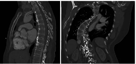
Fig -2: Spines of a normal and abnormal person
Figure 2 shows that spines of a normal and abnormal person. A normal person does not contain any crack or ills. But in abnormal person there exists some cracks, problems etc.
We treat segmenting the spinal canal as a typical binary segmentation problem. Let p(x) denote the probability of the voxel x being foreground (i.e., inside the spinal canal) and the p'(x)probability for being background (i.e., outside the spinal canal), respectively. We have p(x)+p'(x)=1. The binary segmentation of the spinal canal can then be acquired by applying a confidence threshold to the map of p(x) within the entire image space. The probability map can be generated in different ways. For example, by providing foreground and background seeding voxels automatically, we can acquire the probability map through the random-walk solver as in our method. The seeding voxels are determined according to the iteratively refined topology of the spinal canal. Specifically, to initiate the random-walk-based segmentation, we start from the supervised voxelwise classification to identify a small set of voxels, which are assigned with very high classification confidences and thus most likely to be foreground. The identified voxels act as positive seeds and are fed into the random-walk solver to generate a conservative binary segmentation with relatively low sensitivity. Concerning the fact that the spinal canals are generally tubular structures even though their shapes vary significantly across the patient population, both geometric and appearance constraints that are anatomically meaningful are then enforced to extract and to refine the topology of the segmented spinal canal. The refined topology, which is continuous and smooth, thus leads to the updated placement of the seeding voxels, which in turn increases the sensitivity in segmenting the spinal canal via random walk.
We categorize our method as being a bottom-up solution, which is significantly different from the conventional top-down methods. Specifically, we identify a set of seeding voxels where the tentative segmentation is mostly reliable, instead of detecting the anatomical objects of much larger sizes.
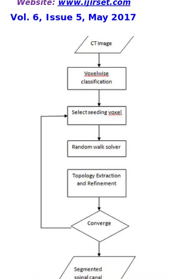
The seeding voxels are initially detected through appearance-based discriminative learning and then updated according to the (tentative) spine topology. The segmentation result, as well as the spine topology, can be improved in subsequent iterations. In this way, the segmentation of the spinal canal can accumulate or propagate from a few voxels, until reaching satisfactory result throughout the entire image space. So the method adopted can be explained as follows. Identify the most reliable seeding voxel to initiate the segmentation by voxelwise appearance features and discriminative learning. Compute the binary segmentation of the spinal canal based on tentative seeding voxels. Extract spine topology, which is represented by it's medial line, based on the result from random walk solver. Refine the medial line of the spinal canal based on the geometric and appearance constraints. Segmentation procedure reaches its convergence once the topology of the spinal canal stops growing.
ISSN(Online): 2319-8753 ISSN (Print): 2347-6710
I
nternational
J
ournal of
I
nnovative
R
esearch in
S
cience,
E
ngineering and
T
echnology
(An ISO 3297: 2007 Certified Organization)
Website: www.ijirset.com
Vol. 6, Issue 5, May 2017
Fig -3: Flow chart of spinal canal segmentation
3.1 VOXELWISE CLASSIFICATION
In order to identify highly reliable foreground voxels as positive seeds for the random-walk solver, we turn to voxelwise classification based on discriminative learning. The classification consists of the training and the testing stages. In the training stage, we utilize the PBT-based classifiers to establish the dependence between the visual (appearance) features of voxels and their segmentation labels. Each new subject is then passed into the testing stage for determining the segmentation of its spinal canal. In particular, we have manually annotated the medial lines of the spinal canals on the training set of 20 images. The training images are randomly selected from our dataset, while each of the eight clinical sites contributes at least one training image. The medial line is defined to connect the medial points of the spinal canal on all traverse slices. Voxels exactly along the medial lines are used for the samples of the foreground, while background samples are obtained from a constant distance away to the medial lines.
3.2 SPINE REFINEMENT AND RANDOM WALK SOLVER
ISSN(Online): 2319-8753 ISSN (Print): 2347-6710
I
nternational
J
ournal of
I
nnovative
R
esearch in
S
cience,
E
ngineering and
T
echnology
(An ISO 3297: 2007 Certified Organization)
Website: www.ijirset.com
Vol. 6, Issue 5, May 2017
Fig -4: Topology refinement
Topology refinement is as in figure 4. In this process, voxels that push off from normal line is neglected. Then draw a line that meet all corrected voxels.
To acquire the complete segmentation of the spinal canal, we introduce the topology constraints. Specifically, we use the medial line of the spinal canal to represent its topology. After calculating all segments of the medial line given the tentative segmentation, we can interleave them into a single connected curve. After the topology of the spinal canal has been extracted and refined, we are able to provide better seeds for the random-walk solver to use. All voxels along the refined medial line, including the newly admitted virtual segments, act as foreground seeds. By repeating the refinement of the spine topology and the placement of the seeding voxels, we can cascade several random-walk solvers for the sake of the spinal segmentation result. Each random-walk solver in the automatic pipeline is able to improve the tentative segmentation, based on the spine topology refined from the previous segmentation. The iterative procedure terminates when the topology of the spinal canal, or the length of the medial line, becomes stable. In particular, we allow the medial line to grow at its both ends. The growth of the segmentation can stop automatically at the inferior end, since the medial line cannot penetrate the bones due to the appearance constraint. Similar to the PBT-based voxel-wise classification, the random-walk solver also produces voxelwise probability of being foreground/background. After users have specified foreground/background seeds, the random-walk solver departs from a certain non-seeding voxel and calculates its probabilities to reach foreground and background seeds. we are able to identify the initial seeding voxels through the voxelwise classification step.
IV.EXPERIMENTAL RESULT
ISSN(Online): 2319-8753 ISSN (Print): 2347-6710
I
nternational
J
ournal of
I
nnovative
R
esearch in
S
cience,
E
ngineering and
T
echnology
(An ISO 3297: 2007 Certified Organization)
Website: www.ijirset.com
Vol. 6, Issue 5, May 2017
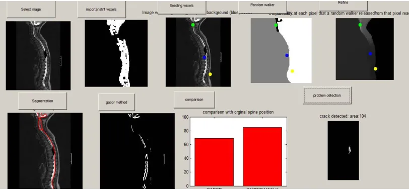
In this figure, experimental results are shown. This is the output and shows many steps. Select image, Important voxels, seeding voxels, random walker, refine, segmentation, gabor methor, comparison, problem detection are steps. It should be checked by using spinal canal having cracks and also a normal person.
V. CONCLUSION
Here an automatic method to segment the spinal canals from highly varying CT images is proposed. With initial seeding voxels that are provided by PBT-based voxelwise classification, we introduce the topology constraints into the segmentation via random walk. Our iterative optimization has successfully enhanced the capability of a single random-walk solver in dealing with tubular spinal canals, in that the boundary conditions (i.e., the placement of the seeding voxels) can be iteratively improved to provide better segmentation results. Our large-scale evaluation shows that the proposed method is highly accurate and robust even if the datasets are very diverse and challenging. Due to limited accesses to state-of-the-art methods, rigorously fair comparisons can hardly be conducted. Though we compared our method with four other methods reported in the literature, it is worth noting that the comparisons were based on different datasets that were used by individual papers. State-of-the-art methods typically reported failures when segmenting certain outlier images. However, it is worth noting that our method has been successfully validated on all testing images. That is, no failure case has been generated through the proposed method. Concerning the challenges caused by our large-scale cohort of diverse images, we argue that our method is very robust and accurate for the segmentation of the spinal canal. Although our method has demonstrated its robust and accurate segmentation capability upon the large-scale dataset, we are aware that the proposed method could be challenged in certain situations. In general, we conclude that the proposed method can well handle our large-scale dataset, which is collected from clinical routines of multiple sites. There are two directions in our future work. First , we will improve the speed performance of our method.In our current implementation particularly, the computation of each random walk solver in the cascaded pipeline is independent. In fact, only boundary conditions of two consecutive random-walk solvers change during the segmentation process. Therefore, we would be able to reduce the redundancy in computation. Second, we will apply our method to more related segmentation problems. In particular, we will probe the possibility of segmenting spinal canals/cords from magneticresonance (MR) images via the proposed method. Also relative position of other body parts can be determined by segmenting the spinal canal, since body parts like heart, lungs are situated in a specified measuring distance from spinal canal.
REFERENCES
[1] F. Zhao and X. Xie, “An overview of interactive medical image segmentation”, BMVA vol. no. 7, pp. 1–22, 2013.
[2] A. W. Akshaya Mishra, “Intervertebral disc segmentation” , IEEE conference on Computational linguistics-Volume 1, pp. 453-459, 2000. [3] Y. B. O. V. R. Zabih, “Fast approximate energy minimization via graph cuts", IEEE International Conference in image processing, , pp.
647-648, 2001
[4] A. S. B. Samma and R. A. Salam, “Adaptation of k-means algorithm for image segmentation", IEEE International Conference in Data Mining, pp. 198-248, 2009.