ISSN(Online): 2319-8753 ISSN (Print) : 2347-6710
I
nternational
J
ournal of
I
nnovative
R
esearch in
S
cience,
E
ngineering and
T
echnology
(An ISO 3297: 2007 Certified Organization)
Vol. 4, Issue 9, September 2015
An Efficient Segmentation Algorithm Using
LDWT and MHWT Technique
Nosthush Kenjige 1, Srinivas Halvi 2, Narayan D 3
P.G. Student, Dept. of Medical Electronics, Dayananda Sagar College, Bangalore, India1
Associate Professor, Dept. of Medical Electronics, Dayananda Sagar College, Bangalore, India2
Associate Professor, Dept. of E&C, BMSCE, Bangalore, India3
ABSTRACT: Digital images play an important role in daily life applications such as satellite monitoring, magnetic resonance imaging (MRI), and ultrasound as well as in areas of research and technology such as geographical information systems (surveys, extraction of information about boundaries). Object segmentation is a very basic and essential task in computers and object recognitions. In this project, we present an image segmentation technique that extract edge information from wavelet coefficients and uses mathematical morphology to segment the image. Initially, the threshold is taken and using the threshold, we get its binary version. Once the binary image is obtained, the modified Haar technique and the lifting DWT is performed on the image to get a high pass image with its edges. This is followed with morphological closing operations which dynamically adjusts the structural element as per the orientation of its local edge. The holes that are obtained are then subsequently filled by fill operations. For comparison; we compare our results with that of the DWT method. The quality of the images obtained using the different methods suggest that the modified Haar technique and lifting DWT provides superior images.
KEYWORDS: Image processing, Modified Haar DWT, segmentation, Lifting DWT
I. INTRODUCTION
Image segmentation is a route in which the digital image is broken down or divided or detached into its regions. It can also be divided into a set of pixels based on the texture or color. These divided portions represent the different objects present in the image. Segmentation is a necessity to obtain the features present in the image and also to classify the resultant features. There are various processes using which the segmentation can be realized. This includes practices like thresholding, labeling of components, boundary based segmentation, template matching and texture segmentation. Segmentation also consists of procedures that enhances the image, restores and also helps in simple representation of data.
Edge detection is a very well-studied field within image processing. The edges present in the image and the regions are closely related as there is a sharp change in Intensity levels at the boundaries of the regions. Due to this close relation, edge Detection techniques have been used as a completely different unit under segmentation. When an image is analyzed, it can be found that the edges of the images are present in the higher frequency components of the image and in order to unearth the edge details we need to study the high frequencies of that particular image. Wavelet domain delivers frequency data that is mappable in localization in the corresponding spatial domain, commanding us to trust that high frequency details pertain to high pass data. In this paper, we extract the edge details of an image by engaging the Modified Haar technique and the Lifting discrete wavelet transform (LDWT). This image is then subjected to the application of adaptive morphological closing that scans the orientation of the local edges and adjusts the structural element as per the orientation.
ISSN(Online): 2319-8753 ISSN (Print) : 2347-6710
I
nternational
J
ournal of
I
nnovative
R
esearch in
S
cience,
E
ngineering and
T
echnology
(An ISO 3297: 2007 Certified Organization)
Vol. 4, Issue 9, September 2015
In the perceptual properties the homogeneity in the resulting segment and the heterogeneity across the neighborhood segment can be studied.
In the computer vision, the process of segmentation and clustering have always has been a great challenge.
Presently, powerful segmentation techniques are present that includes spatial domain filters such as Sobel, Prewitt, canny, Roberts, etc.
In segmentation, to distinguish the object from the background, two techniques can be used, namely the discontinuity and the similarity systems.In the discontinuity detection process, the changes in the intensity level (grey level) is detected and based on these abrupt changes, the segments are formed.In the similarity detection technique, based on the thresholding and the region growing, image segmentation is performed.
Motivation: Image segmentation is a basic necessity in real time applications in industries such as healthcare, security services and military services. All of these services make use of image sensors to record the image and subsequently the image is transferred to the control base. These images contain unwanted information and noise, and more often than not, they have redundant information.
Since it is real time processing, it would be convenient to send the image faster and in a much lesser bandwidth. The process of segmentation allows us to send the images at a much rapid pace and also over a less bandwidth.
Contribution: There are numerous techniques which have been employed for the segmentation of images but none
of them are applicable to all the types of segmentation needs. They may be suited best for one particular application but not so much for another.
In the case of low textured images in the background, traditional methods also under segment the images on various occasions. Ultimately, the motto is to increase the image quality performance of the segmented image using our technique.
II. RELATEDWORK
Mohammad Talebi, Ahamd Ayatollahi, Ali Kermani [3] proposed that the process of image segmentation is very challenging in the case of ultrasound imaging due to the speckle noises present in the image. And also, since the ultrasound images are of lower contrast, it adds another level of toughness in order to segment the image using the conventional technique.
In their paper, they have presented a method which removes the disadvantages of active contour method. They have used the genetic active contour method and the results obtained by them are far superior as compare to that of an active contour process.
Ali Kermani, Ahmad Ayatollahi, Ahmad Mirzaei, Mohammad Barekatain[4] proposed that the ultrasound images cannot be segmented reliably and quickly enough using the conventional methods due to the speckle noises, and also the undesirable artifacts present in the images. They proposed a different tool that mainly depends on the evaluation of the tissues using a histogram. In this method, the histogram is modified using the discrete wavelet transform. This method is called the modified local histogram range image method [MLHRI].
Suhuai Luo, Xuechen Li, Jiaming Li [5] presented a segmentation technique by blending High Order Statistical Texture Features for the segmentation of anatomical structures such as the kidney. It processes the texture in order to extract the high order structural features and next, it distributes the image by segmenting the pixels using vectors. It is then operated morphologically to get an accurate image.
The unique selling point of this paper is that it considers both the local and global textures in the neighborhood and thus increases the grayscale distributions.
ISSN(Online): 2319-8753 ISSN (Print) : 2347-6710
I
nternational
J
ournal of
I
nnovative
R
esearch in
S
cience,
E
ngineering and
T
echnology
(An ISO 3297: 2007 Certified Organization)
Vol. 4, Issue 9, September 2015
The region that is under investigation should appear with the highest quality and the other parts could appear in lower quality under this method.
III. PROPOSED METHOD
Figure 1.Block Diagram of Proposed Model
The wavelet domain analysis provides us information about frequency that can be mapped to the corresponding spatial domain. The high frequency components of the image are concentrated at the edges and if we can extract the high frequencies from the image, then we can acquire the edge information. As shown the above block diagram, we extract the edge information using MHWT and LDWT methods.
The following steps are taken to segment the image:
1. The original image is first subjected to preprocessing in which the noises present are removed and mainly, the image is resized as per convenience.
2. The resized image has to be converted to a binary image and this is done by normalizing the image using the gray value limits. The threshold is chosen suitably and as per the threshold value, the image gets converted to black and white image (to get good subset of contrast). In case of color images, using the level setting, the normalization can be done using 10-90% black and white, in order to extract the edge information.
3. The binary image is now decomposed to wavelet domain using the modified Haar technique and the Lifting DWT to extract the edge information.
4. Once the wavelet is applied, the approximate coefficients are set and the image is subjected to inverse DWT. In this particular process of edge extraction, the lowest energy band is set to 0. The image obtained at this step contains a good deal of edge information.
5. The image obtained thus far consists of many gaps and these gaps have to be connected. The process of connecting the image is done by 8- connected neighbors. In The 8-connected neighbor method, a structural element scans the pixels and thus connects the image.
6. The morphological close operator is applied to the connected image in order to remove the boundary distortion.
The 3*3 elements are used to scan the pixels in horizontal, vertical and diagonal manner.
7. Once the boundaries are less distorted and the image obtained is a fully connected image, the images are filled. I.e. the morphological fill operator is applied.
8. The boundaries of the objects are extracted after the holes are filled. This image consists of a good amount of edge information and it is beneficial for many specific cases.
ISSN(Online): 2319-8753 ISSN (Print) : 2347-6710
I
nternational
J
ournal of
I
nnovative
R
esearch in
S
cience,
E
ngineering and
T
echnology
(An ISO 3297: 2007 Certified Organization)
Vol. 4, Issue 9, September 2015
In the splitting stage, the wavelet filters h (z) and g (z) is taken in a poly phase matrix. The poly phase matrix is then renewed into an elementary matrix by means of Euclidean algorithm. The matrix that is achieved is then approved to prediction and updation. These two steps are explicit to different functions.
The various examples of predictions are CDF(which is named after Cohen, Daubechies and Feauveav), 5/3 integer wavelet transform etc
The 5/3 integer wavelet transform is given by the expression:
The 2D DWT is used as an extensive tool in the discipline of image and video compression due to its suitability, amid the most admired practical blueprints, the row-column, line and block based are the most used.[7][8]
Modified Haar wavelet transform:This is the mother wavelet and to carry out the transform operator, the Haar transform utilizes translations and dilutions of the functions.
In Haar transforms, the data or the nodes gets employed at n level but by captivating average and difference via the previous level nodes, the process gets much quicker than the average Haar method and thus this system is called the fast Haar transform.[8][9]
The Modified fast Haar transform can be processed by (4/ x+y+w+z) instead of (2/x+y) for averaging and (4/x-y-w-z) as a substitute of (2/x-y) for differencing process. In this routine 4 nodes are considered instead of two and hence it is less time consuming and processes much faster.
The assessment between the typical Haar transform and the modified Haar transform results show a significant improvement for the MFHT as illustrated in the fig 4.
By using the MFHT, the number of approximate coefficients and the number of division operations can be reduced. Nonetheless, the negative aspect is that the number of addition and subtractions cannot be favored by decreasing the division operator.[8]
This morphological operator can be used to determine the perimeter of the objects. For the perimeter to be perceived, it has to follow two rules:
1. The pixels should be in on state
2. One or more pixels in the neighborhood should be in the off state.
The perimeter detection is very helpful in cases where we have to determine the boundaries of the objects.
Reconstruction: The LL sub band obtained after filtering and LH, HL, HH sub-bands obtained after LDWT/MHWT is reconstructed by using inverse MHWDT and inverse DWT to the original image.
2 1 ) ( 2 1 )
(Si Si Si1 P 2 1 ) ( 4 1 )
ISSN(Online): 2319-8753 ISSN (Print) : 2347-6710
I
nternational
J
ournal of
I
nnovative
R
esearch in
S
cience,
E
ngineering and
T
echnology
(An ISO 3297: 2007 Certified Organization)
Vol. 4, Issue 9, September 2015
IV.EXPERIMENTAL RESULTS
Problem definition: The use of wavelet transform to segment the image ensures that the extra components present in the image are removed and it also ensures that the characteristics of the signals are readable and stored.
The lifting discrete wavelet transform and modified Haar transform techniques preserve the unique characteristics and remove the redundant information and thus can be used in the field of medical image investigations.
Objective:
To increase PSNR
To decrease RMSE
Table 1: Proposed Algorithm
We select subjective evaluation to quantify the quality of an image. Various parameters are used to measure the image quality in objective evaluation of the image such as Mean square error (MSE), Peak signal to noise ratio (PSNR), Root mean square error (RMSE).
Peak signal to noise ratio (PSNR)
PSNR is extensively drawn or exploited in the field of engineering to determine the excellence of the restructured image. It is the percentage of highest power of a signal and the power of the crooked noise that upsets the representation of the image. It is usually symbolized in logarithmic decibels. It is calculated with respect to the mean square error (MSE) which is the error between the average square of the estimated image and the original image. The expression is given below:
= . . [( ( , )− ^( , )) ]
Where,
( , )− Original image
^( , )− Estimated image
MN - size of the image
And the PSNR is specified by:
= . .
∑ .∑ ( ( , )− ^( , ))
MN - size of the image
Input: Original image
Output: Segmented image
Step 1: Read the original image.
Step 2: Apply the different preprocessing techniques
Step 3: Apply Modified Haar DWT/LDWT and get LL, LH, HL, HH sub-bands.
Step 4: suppress the LL band and then obtain the inverse of the transforms.
Step 5: apply 8-connectivity for the reconstructed image.
Step 6: apply morphological operators to close the edges.
ISSN(Online): 2319-8753 ISSN (Print) : 2347-6710
I
nternational
J
ournal of
I
nnovative
R
esearch in
S
cience,
E
ngineering and
T
echnology
(An ISO 3297: 2007 Certified Organization)
Vol. 4, Issue 9, September 2015
Root mean square error (RMSE) is the other quality examining metric of the image. It is obtained by taking square root over mean square error (MSE).
Root mean square error is defined as,
= ∑ .∑ . [( ( , )−
^( , )) ]
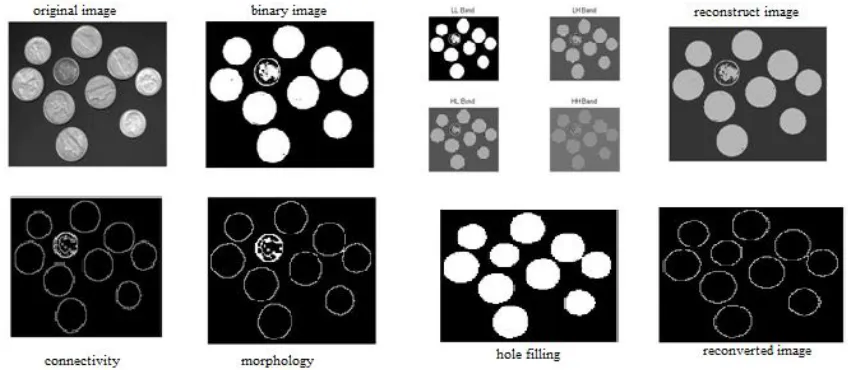
The images of the results obtained at every stage during MHWT are shown below:
Fig 2 Example of phase wise Application of the Proposed Algorithm (MHWT)
The stepwise application of the Lifting DWT applied to an image is shown below in Fig 3.
ISSN(Online): 2319-8753 ISSN (Print) : 2347-6710
I
nternational
J
ournal of
I
nnovative
R
esearch in
S
cience,
E
ngineering and
T
echnology
(An ISO 3297: 2007 Certified Organization)
Vol. 4, Issue 9, September 2015
The comparison between the existing DWT and the proposed method of LDWT and MHWT is shown below.
Fig 4: Comparison of reconstructed images
The following tables below compares the PSNR values and RMSE (root mean square error) values we obtained with MHWT, lifting DWT with the canny edge detection and the existing DWT method.
Peak signal to noise ratios (PSNR) of different techniques with different images:
Table 2: PSNR values of different techniques
Although the PSNR values of canny are higher than the MHWT values, the MHWT is preferred because the canny edge detection method does not eliminate the unwanted edges, unlike the MHWT.
The root mean square error values obtained are shown below: Image Canny
method
DWT Proposed Method MHWT LDWT Coins.png 3.7786 5.2688 5.2549 2.0185 Air.jpg 13.1700 13.8863 13.8839 5.2342
Table 3: RMSE values of different techniques
V. CONCLUSION
The results obtained by the method of Modified Haar and Lifting DWT have proved that the edges obtained are of higher quality with less distortion and also lesser noises. The closure of image and also the connectivity employed in our method has worked better in terms of both PSNR and the RMSE. The method we have employed can be used for different image modalities to manipulate and visualize the data as per their applications like differentiating the objects from the background in case of face detection, iris detection and fingerprint detection. In geology, it can be used in locating objects such as crops, forests or any other images from the satellites.
In the field of medical technology, it can be used to calculate the tissue volumes, localize tumors present, help in planning of surgeries, navigation during surgeries and also helps in studying anatomy.
Image Canny method
ISSN(Online): 2319-8753 ISSN (Print) : 2347-6710
I
nternational
J
ournal of
I
nnovative
R
esearch in
S
cience,
E
ngineering and
T
echnology
(An ISO 3297: 2007 Certified Organization)
Vol. 4, Issue 9, September 2015
REFERENCES
[1]www.cs.auckland.ac.nz/courses/compsci773s1c/lectures/ImageProcessing-html/topic3.htm [2]-http://in.mathworks.com/help/images/morphology-fundamentals-dilation-and-erosion.html
[3]Mohammad Talebi, Ahamd Ayatollahi, Ali ermaniMedical ultrasound image segmentation using genetic active contour Journal of Biomedical Science and Engineering Vol.4 No.2, Pub. February 25, 2011
[4]Ali Kermani, Ahmad Ayatollahi, Ahmad Mirzaei, Mohammad Barekatain Medical ultrasound image segmentation by modified local histogram range image methodJournal of Biomedical Science and EngineeringVol.3 No.11, Pub. November 18, 2010
[5]SuhuaiLuo, Xuechen Li, Jiaming LiImprovement of Liver Segmentation by Combining High Order Statistical Texture Features with Anatomical Structural Features
EngineeringVol.5 No.5B, Pub. July 26, 2013 [6]EnginMendi, Mariofanna Milanova
Contour-Based Image Segmentation Using Selective Visual AttentionJournal of Software Engineering and ApplicationsVol.3 No.8, Pub.: August 26, 2010
[7] AN EFFICIENT NEIGHBOURHOOD PIXEL FILTERING ALGORITHM FOR WAVELET- BASED IMAGE DENOISING", International Journal of Computers and Applications, 2012