Motor Coordinate Transformations:
A Multi-Level Analysis of Neuronal
Activity in Cortical Area MSTd
Lukas A. Brostek
Motor Coordinate Transformations:
A Multi-Level Analysis of Neuronal
Activity in Cortical Area MSTd
Lukas A. Brostek
Dissertation
an der Graduate School of Systemic Neurosciences
der Ludwig–Maximilians–Universit¨
at
M¨
unchen
vorgelegt von
Lukas A. Brostek
aus Danzig
’Gain-field-like’ tuning behavior is characterized by a modulation of the neuronal response
depending on a certain variable, without changing the actual receptive field characteristics
in relation to another variable. Eye position gain fields were first observed in area 7a of
the posterior parietal cortex (PPC), where visually responsive neurons are modulated by
ocular position. Analysis of artificial neural networks has shown that this type of tuning function might comprise the neuronal substrate for coordinate transformations.
In this work, neuronal activity in the dorsal medial superior temporal area (MSTd) has
been analyzed with an focus on it’s involvement in oculomotor control. MSTd is part of
the extrastriate visual cortex and located in the PPC. Lesion studies suggested a
partici-pation of this cortical area in the control of eye movements. Inactivation of MSTd severely
impairs the optokinetic response (OKR), which is an reflex-like kind of eye movement that
compensates for motion of the whole visual scene.
Using a novel, information-theory based approach for neuronal data analysis, we were able
to identify those visual and eye movement related signals which were most correlated to
the mean rate of spiking activity in MSTd neurons during optokinetic stimulation. In a majority of neurons firing rate was non-linearly related to a combination of retinal image
velocity and eye velocity. The observed neuronal latency relative to these signals is in
line with a system-level model of OKR, where an efference copy of the motor command
signal is used to generate an internal estimate of the head-centered stimulus velocity signal.
Tuning functions were obtained by using a probabilistic approach. In most MSTd neurons
these functions exhibited gain-field-like shapes, with eye velocity modulating the visual
response in a multiplicative manner. Population analysis revealed a large diversity of
tuning forms including asymmetric and non-separable functions. The distribution of gain
fields was almost identical to the predictions from a neural network model trained to
per-form the summation of image and eye velocity. These findings therefore strongly support the hypothesis of MSTd’s participation in the OKR control system by implementing the
transformation from retinal image velocity to an estimate of stimulus velocity. In this
sense, eye velocity gain fields constitute an intermediate step in transforming the
Another aspect that was addressed in this work was the comparison of the irregularity of
MSTd spiking activity during optokinetic response with the behavior during pure visual
stimulation. The goal of this study was an evaluation of potential neuronal mechanisms
underlying the observed gain field behavior. We found that both inter- and intra-trial variability were decreased with increasing retinal image velocity, but increased with eye
velocity. This observation argues against a symmetrical integration of driving and
modu-lating inputs. Instead, we propose an architecture where multiplicative gain modulation
is achieved by simultaneous increase of excitatory and inhibitory background synaptic
input. A conductance-based single-compartment model neuron was able to reproduce
realistic gain modulation and the observed stimulus-dependence of neural variability, at
the same time.
In summary, this work leads to improved knowledge about MSTd’s role in visuomotor transformation by analyzing various functional and mechanistic aspects of eye velocity
Summary v
1 Introduction 1
1.1 The Visual System . . . 2
1.2 Processing of Visual Motion . . . 3
1.2.1 The Middle Temporal Cortex . . . 3
1.2.2 The Medial Superior Temporal Cortex . . . 4
1.3 Tracking Eye Movements . . . 5
1.3.1 Smooth Pursuit Eye Movements . . . 5
1.3.2 Optokinetic Response . . . 7
1.4 Neuronal Data Analysis . . . 9
1.4.1 Regression Analysis . . . 9
1.4.2 Information-theoretic Approaches . . . 10
1.4.3 Neuronal Variability . . . 11
1.5 Aim of this Thesis . . . 12
2 Cumulative Thesis 15 2.1 The Response of MSTd Neurons to Perturbations in Target Motion During Ongoing Smooth-Pursuit Eye Movements . . . 17
2.2 An Information-theoretic Approach for Evaluating Probabilistic Tuning Functions of Single Neurons . . . 19
2.3 A Method for Evaluating Tuning Functions of Single Neurons based on Mutual Information Maximization . . . 21
2.4 Neuronal Variability of MSTd Neurons Changes Differentially With Eye Movement and Visually Related Variables . . . 23
2.5 Eye Velocity Gain Fields in MSTd for Visuomotor Coordinate Transfor-mations . . . 25
2.6 Gain Modulation from Balanced Excitatory-Inhibitory Synaptic Input . . . 27
3 Discussion 33 3.1 Comparison with previous MSTd studies . . . 35
3.2 Gain Fields for Sensorimotor Coordinate Transformations . . . 36
3.3 Underlying Neuronal Structure . . . 37
3.4 Temporal Coding in MSTd? . . . 38
3.5 Is MSTd Involved in Smooth Pursuit Control? . . . 39
3.6 Further Investigations . . . 41
Bibliography 47
Acknowledgements 55
Vision is the sense humans most rely on. Seeing enables us to sense and perceive the world
around us. By identifying the color, shape and movement of countless objects, our visual
sense allows us to distinguish important from irrelevant things, as well as their position
and velocity relative to us.
In humans, like in all primates and a number of other mammals and birds, eyes have
developed in such a way that a relatively small part of the retina is populated by a
com-paratively high density of photoreceptor cells. This area is called fovea. Due to that, only
a small part of our visual field is perceived in full sharpness and color. For compensation
we perform frequent eye movements, capturing all interesting details of the visual scene.
The composition of the full picture of the perceived environment from all these ’snapp-shots’ happens subconsciously.
While enabling us to see the world in high detail, foveal vision also poses a number of
challenges for the visual system. To prevent loss of small moving objects from our sight
they need to be tracked by our eyes. On the other hand, we often see objects in motion not
because they move in front of us, but because we move our eyes. Therefore, compensation
of eye-movement induced visual motion is crucial for proper perception. Furthermore, to
avoid blurred vision during self-motion through space, the visual image needs to be
stabi-lized on the fovea. This task is accomplished by compensating optokinetic eye movements
into the opposite direction of self-movement.
These examples demonstrate that both physiological systems for vision and the generation
of eye movements need to be coupled. Today, there is strong evidence for the existence
of specialized brain regions where this coupling might occur. This doctoral thesis focuses on the Medial Superior Temporal Cortex, a region assumed to perform such function.
In the following sections an introduction to the current state of scientific knowledge about
the visual system, processing of visual motion, smooth pursuit, and optokinetic eye
1.1 The Visual System
The visual system begins with the eyes, where photoreceptor cells in the retina
trans-form light into electric signals. The density of photoreceptor cells is not unitrans-form, but concentrated around the fovea, wich is an area of about one square millimeter diameter.
Being much more than just a simple organ for sensing light, the eye already extracts an
enormous amount of information about different facets of the visual image by it’s retinal
neural networks. Important parts of signal processing for visual motion detection, for
in-stance, are realized by the network of retinal ganglion cells already (Gollisch and Meister,
2010).
The neuronal pathway that leaves the eye is called retinofugal projection (Fig. 1.1). Most
of the optic nerve neurons innervate the lateral geniculate nucleus (LGN) of the dorsal
thalamus. Neurons in the LGN give rise to axons that project to the primary visual cortex
(V1) in the occipital lobe, which is also called striate cortex. V1 is organized
retinotopi-cally, meaning that neighboring cells in the retina feed information to neighboring places
in the primary visual cortex (Hubel and Wiesel, 1962). Many neurons in V1 respond best to an elongated bar of light moving across their receptive fields. The greatest response is
given to a bar with a particular orientation.
Signals from the striate cortex are projected to more than two dozens of different
extra-striate cortical areas in the temporal and parietal lobes (Felleman and Van Essen, 1991).
The extrastriate areas are functionally and anatomically subdivided into two major
path-ways. The ventral pathway is assumed to be involved in the perception of the visual
world and the recognition of objects (Mishkin et al., 1983). It runs from the primary
visual cortex into the infratemporal cortex. Neurons in area V4 have larger receptive
fields than cells in the striate cortex and are selective for orientation and color. The
infe-rior temporal lobe (IT) lies behind V4 and has complex spatial receptive fields. Neurons
in this area respond to a variaty of colors and abstract shapes. A percentage of neu-rons responds even strongly to stimuli as complex as pictures of faces (Desimone, 1991).
Therefore, this area is presumed to be important for visual perception and visual memory.
The dorsal pathway, on the other hand, projects from the primary visual cortex into the
posterior parietal cortex. This pathway is assumed to carry information regarding the
MT MST
V1 V4
IT Retina
LGN
Figure 1.1: The visual system. The retinofugal projection innervates the lateral geniculate nucleus (LGN) and then projects to primary visual cortex (V1). From there the ventral pathway goes to V4 and the inferior temporal lobe (IT). The dorsal pathway projects to the middle temporal (MT) and medial superior temporal (MST) areas. (According to Mishkin et al. (1983))
1.2 Processing of Visual Motion
Two areas in the dorsal pathway are assumed to be crucial for visual motion processing:
the middle temporal (MT) and medial superior temporal (MST) areas in the parietal
cortex.
1.2.1 The Middle Temporal Cortex
MT is located in the posterior bank of the superior temporal sulcus and is one of the most
studied areas in macaque cortex. MT is also known as visual area 5 (V5) and receives
retinotopically organized input from a number of other cortical areas such as V2 and V3,
and is also directly innervated by cells in the striate cortex. Cells in this area have larger
receptive fields than V1.
Visual responses of MT neurons are determined principally by several properties of the
stimulus: retinal position, direction and speed of motion (Maunsell and Van Essen, 1983a),
stimulus size (Born and Tootell, 1992), and binocular disparity (Maunsell and Van Essen, 1983b). Whether or not MT receives other than retinal input, is still disputed (Newsome
et al., 1988). A recent work suggests that MT neurons use eye movement signals to code
Lesions in macaque cortical area MT produce motion-perceptual (Newsome and Pare,
1988) and oculomotor (D¨ursteler and Wurtz, 1988) deficits. Electrical microstimulation
of MT neurons influences perceptuel judgements of motion direction (Salzman et al.,
1990). Further support for the idea of MT’s involvement in the processing of visual
mo-tion comes from the finding that in a momo-tion direcmo-tion discriminamo-tion task the trial-to-trial variability in MT neuronal signals is correlated with the choices the monkey makes
(Brit-ten et al., 1996).
MT’s key output target stuctures are implicated in the analysis of optic flow and the
generation of eye movements (Mishkin et al., 1983). Further along the dorsal pathway
lies a region with more specialized types of movement selectivity: area MST, which is
reviewed in the following paragraph.
1.2.2 The Medial Superior Temporal Cortex
The medial superior temporal cortex (MST) is part of the posterior parietal lobe and
receives direct projections from adjacent area MT. MST is usually divided into two
sub-regions with different functional properties: the dorsal (MSTd), and the ventrolateral region (MSTl).
Many MST neurons respond to visual stimuli and have large, often bilateral, receptive
fields exceeding 15 degree of the visual field (Komatsu and Wurtz, 1988). The neuronal
latency to visual stimulation is about 50 ms (Kawano et al., 1994). MSTd neurons
re-spond to rotating, expanding and planar large field motion. The neuronal response is
invariant of the position, form and size of these optic patterns (Duffy and Wurtz, 1991).
MST neurons modulate their visual response when the field of expansion is shifted (Duffy and Wurtz, 1995) or when the rate of expansion changes (Duffy and Wurtz, 1997). As in
area MT, neurons in MST are selective for binocular disparity (Roy et al., 1992).
There is strong evidence that, aside from retinal input, MST receives extraretinal input
as well. Many MSTd and some MSTl cells show strong modulation during smooth
pur-suit eye movements and continue firing during blinking of the target. In MSTl neuronal
response starts before the onset of eye movements (Ilg et al., 2004). On the contrary, in most of the MSTd neurons the pursuit response begins after the onset of eye
move-ments (Newsome et al., 1988). Many neurons also respond to an imaginary target (Ilg
and Thier, 2003). During fixation as well as smooth pursuit eye movements, the response
1997). Another extraretinal input is provided by the vestibular system. Two thirds of
MSTd neurons that are sensitive to optic flow also show spatial tuning for inertial motion
without optic flow, pointing out to vestibular input (Gu et al., 2007).
Due to these characteristics, three functional roles have been proposed for this area. First,
MST is probably involved in the perception of motion. Lesions in MST produce similar
motion-perceptual deficits as in area MT (Rudolph and Pasternak, 1999). Second, MST
might be involved in the generation of smooth pursuit and optokinetic eye movements.
This assumption is also supported by lesion studies (D¨ursteler and Wurtz, 1988). Third,
MST is probably involved in the integration of visual and vestibular motion cues for the
perception of heading direction during self-motion. This was shown by studies, where the behavioral estimates of direction of self-motion were affected after electrical stimulation
of MST neurons (Britten and van Wezel, 1998). In regard of this view, it is supposed
that MSTd might compensate for self generated eye movements in heading perception
(Bradley et al., 1996).
1.3 Tracking Eye Movements
A distinction is made between the voluntary tracking or pursuit of small moving objects
and the involuntary tracking of a moving large-field visual scene.
1.3.1 Smooth Pursuit Eye Movements
Developed in mammals with frontal eyes only, the smooth pursuit eye movement (SPEM)
system is an evolutionary young feature. SPEMs are used for tracking small moving
objects within the high-acuity region near the fovea. Pursuit usually only occurs in
re-sponse to a moving visual stimulus. Pursuit eye movements are most effective when the target speed is relatively slow. Like saccades, SPEMs are voluntary eye movements. The
movement initiation latency is usually about 100–150 ms, which is generally shorter than
for saccades. Both humans and monkeys can reach maximum pursuit velocities of about
80−100◦/s (Lisberger et al., 1981).
SPEMs are already present in 4-week-old infants and are fully developed at about 3 months of age (Phillips et al., 1997). Even at higher ages, the SPEM system stays
adap-tive. When patients with ocular muscle weakness are forced to view monocularly with
their weak eye for several days, the pursuit system shows changes in the movements of the
it takes to bring the target’s image onto the fovea of the weak eye and to keep it there
(Optican et al., 1985). Another form of adaptation in the pursuit system is the so called
smooth pursuit gain modulation. When a stimulus is presented with brief perturbations
superimposed on the target movement, pursuit of these perturbations gets better with
increasing eye velocity (Churchland and Lisberger, 2001, 2002). Furthermore, the SPEM system is highly predictive. When subjects follow a target moving on a periodically
re-peating trajectory, they are able to anticipate changes in target motion after one period
only and follow them without further delay (Barnes and Asselman, 1991).
In the view of control system theory, the tracking eye movement system can be seen as
a negative feedback servo control (Fig. 1.2) whose function is to pursue the image of a
small moving object on the fovea. The difference between stimulus and eye velocity is
called retinal slip or image velocity. It is sensed by the retina and then further processed
by the dorsal pathway of the visual system. In a simple negative feedback system, this
image velocity signal could serve already as control command to the eye plant. However,
to replicate the actual temporal characteristics observed in humans and monkeys, the
implemantation of an additional internal feedback has been proposed (Robinson et al., 1986). The addition of an efference copy of the oculomotor command allows the
estima-tion of the stimulus velocity signal, which then serves as the central control command.
The efferent pathways project this signal to motoneurons, and the eyeball, including the
retina, is moved in an effort to match eye and stimulus motion.
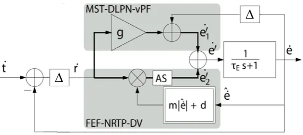
The neuronal basis of the pursuit control circuit is shown in Fig. 1.3. It is assumed that
two parallel pathways are involved in the generation of SPEMs (B¨uttner and B¨
uttner-Ennever, 2006; Nuding et al., 2008). One path includes the dorsal visual stream, pons and
cerebellum. As described in the previous sections, areas MT and MST are the sources of
visual motion information. Lesion studies (D¨ursteler and Wurtz, 1988) and
microstimu-lation (Komatsu and Wurtz, 1989) give strong evidence for the involvement of areas MT
and MSTl in the generation of SPEMs. It is assumed that the dorsal subpart MSTd plays
a minor role in pursuit (Newsome et al., 1988). Area MST projects to the dorsolateral pontine nuclei (DLPN) (Distler et al., 2002). Lesions in this area produce mainly
ipsi-lateral SPEM deficits (May et al., 1988). DLPN projects to the flocculus (FL) in the
cerebellum, where lesions also impair the pursuit system (Zee et al., 1981). From the FL
originate projections to the vestibular nuclei (VN) in the brainstem, from where SPEM
signals can reach the oculomotor nuclei (OMN).
1 0.2 s + 1
Δ
20Δ
600.9 0.2 s + 1
+ +
est stim vel
stimulus
vel
eye vel
est eye vel
MSTd Eye plant
Retina →MT
Internal model
image vel
A
B
0 0.5
0 5 10 15 20
velocity
time [s] stimulus eye (data) eye (model)
-Figure 1.2: (A) Computational model of the tracking eye movement control system. Image velocity is the difference between stimulus and eye velocity. The internal eye plant model provides an efference copy, which is used to estimate stimulus velo-city. This signal serves as control signal for the eye plant. This simple model does not account for any latencies, nor adaptive and predictive mechanisms. Latencies were chosen to fit OKR charakteristics. (B) The step function re-sponse of the model (blue) for a sudden increase in stimulus velocity (brown). The light blue trace shows example eye velocity data. (see section 2.5)
cerebellum. Areas MT and MST have reciprocal connections with the frontal eye fields
(FEF). Lesions in the FEF in monkeys (Shi et al., 1998) and humans (Morrow and Sharpe,
1995) cause severe ipsidirectional deficits particularly in predictive aspects of SPEM. FEF
projects mainly to the nucleus reticularis tegmenti pontis (NRTP) (Ono et al., 2005).
NRTP has neuronal connections to the ocular vermis (OV) and the paraflocculus (Glick-stein et al., 1994) in the cerebellum. Lesions in OV lead to SPEM deficits (Takagi et al.,
2000). OV projects to the caudal part of the fastigial nucleus in the vermis, where lesions
also impair SPEM (Robinson et al., 1997). In addition to the two pathways mentioned,
it is known that the basal ganglia (Basso et al., 2005) and thalamus (Tanaka, 2005) are
involved in the SPEM system.
1.3.2 Optokinetic Response
The optokinetic response (OKR) describes reflex driven eye movements that compensate
for motion of the entire visual scene as occurs with self-motion through space. OKR eye
movements move in the direction of visual motion, thus improving image stabilization on
the retina. The combination of OKR and fast resetting saccades during unidirectional stimulation is called optokinetic nystagmus (OKN), which plays an important role in
maintaining maximal periods of clear vision during continuous uni-directional movement
MT MST FEF
NRTP DLPN
OV FL
VN OMN
Figure 1.3: The two cortical pathways of the tracking eye movement system. The first pathway is shared by both SPEM and OKR. It originates from areas MT/MST and projects to the dorsolateral pontine nuclei (DLPN). From there go con-nections to the flocculus (FL), which projects to the vestibular nuclei (VN) and oculomotor nuclei (OMN). The second pathway is assumed to be part of the SPEM system only. It originates in the frontal eye fields (FEF) and goes to the nucleus reticularis tegmenti pontis (NRTP). From there go connec-tions to the ocular vermis (OV), which projects to the fastigial nucleus in the vermis. Areas MT/MST and the FEF are connected reciprocally. (According to B¨uttner and B¨uttner-Ennever (2006))
In the generation of OKR two components can be distinguished. The direct component
occurs shortly after the onset of the optokinetic stimulus and is also known as ocular
fol-lowing response (OFR). The term OFR generally refers to the immediate OKR response
after the motion onset of a large visual stimulus (Miles, 1998). The indirect component leads to a more gradual increase in slow-phase eye velocity during continuous stimulation.
OFR has a short latency of about 50 ms in monkeys (Miles et al., 1986) and 70 ms in
hu-mans (Gellman et al., 1990), which is shorter than the 100-150 ms for SP eye movements.
This in addition to the size of the visual stimulus and the involuntary character of the eye
movements are major features to differentiate between OKR and SPEM. For extended
stimulation, OKN can reach velocities of about 180 deg/s in monkeys, and about 120
deg/s in humans (Cohen et al., 1977). These velocities are the sum of the direct and
indirect components of OKN.
The major neuronal pathways for generation of the indirect component of OKR seem to
lie in the brainstem. Fibers from the retina terminate in the nuclei of the accessory optic
OKN also the vestibular nuclei are activated (Waespe and Henn, 1987). The cerebellum
is not thought to be involved in the indirect component of OKR (B¨uttner and Waespe,
1984). Although the indirect component is basically transmitted via brainstem pathways,
these pathways are under cerebral cortical control, particularly in humans and monkeys.
Accordingly, bilateral occipital lesions impair the indirect OKR component (Zee et al., 1981).
For the direct component of OKR, there is strong evidence for involvement of similar
neuronal areas as used for the generation of SPEM (Fig. 1.3). Both, pursuit and optoki-netic eye movements are severely impaired following lesions of areas MT/MST (D¨ursteler
and Wurtz, 1988; Takemura et al., 2007), DLPN (May et al., 1988), flocculus (Zee et al.,
1981) or Vermis (Takagi et al., 2000). The involvement of MST in the direct component
of OKR has also been shown in a number of other studies. The response latencies of MST
neurons change in parallel with the response latencies of the simultaneously observed eye
movements when different visual stimuli are used to elicit OKR (Kawano et al., 1994).
Moreover, many MSTd neurons reflect the post-saccadic enhancement of the OKR/OFR
in their neuronal response (Takemura and Kawano, 2006). The fronto-cortical SPEM
pathway, however, seems to be less involved in the generation of OKR. Bilateral lesions
of the FEF slowed SPEM but did not affect the OKR (Keating et al., 1996).
1.4 Neuronal Data Analysis
This section provides a brief introduction to selected techniques in neuronal data analysis.
The objective of all these methods is a characterization and identification of the underlying
neural information processing system.
1.4.1 Regression Analysis
Regression analysis is the estimation of the dependency of a dependent variable on some
otherindependent variables. In the most commonly used form of linear regression analysis,
the maximum likelihood estimate of parametersβfrom observationsxis determined using
a linear modelC according to
x=C·β+r, (1.1)
withr denoting normally distributed residual error. The solution is given by the ’pseudo
inverse’ of C
In neural data analysis, the observations x are typically given by the neuronal firing
rate (F R), whereas the linear model is composed of n stimulus- and/or behavior-related
variables (var):
F R =β0+var1·β1+. . .+varn·βn. (1.3)
The dependency of the firing rate on the different independent variables is then expressed by parameters β.
1.4.2 Information-theoretic Approaches
Information-theory based methods offer an alternative to model-based approaches of
sys-tem identification. This technique allows the estimation of dependency of neuronal acti-vity on certain independent variables without further assumptions on the exact form of
this dependency.
Entropy H(X) is a measure for the uncertainty of a single random variable X. The
reduction in uncertainty due to another random variable is called ’mutual information’
(Shannon and Weaver, 1949; Cover and Thomas, 1991). For two random variablesX and
Y with probability distributions p(x) and p(y) the mutual information is
I(X;Y) =H(Y)−H(Y|X). (1.4)
The entropy of Y and the conditional entropy of Y given X are defined as
H(Y) =− Y
p(y) logp(y) (1.5)
H(Y|X) =− X
p(x) Y
p(y|x) logp(y|x), (1.6)
with p(y|x) being the conditional probability distribution of Y given X. The mutual
information is a measure for the dependence between the two random variables. It is
symmetric in X and Y, always non-negative, and equal to zero only if X and Y are
mu-tually independent.
In analysis of neuronal data, mutual information is usually used to determine how much
information spiking activity carries about some stimulus- or behavior-related variable. One means of estimating the information contained in the neuronal response is comparing
the occurence of specific spiking patterns over a large number of trials where the same
stimulus was presented and the same behavior was consistently observed (Rieke et al.,
be determined, ignoring certain patterns of spiking activity. The latter approach will be
further elaborated in sections 2.2 and 2.3.
1.4.3 Neuronal Variability
The term ’neuronal variability’ usually refers to the regularity of spiking activity.
Investi-gating the temporal structure of the neuronal response allows a characterization of the
spiking process and puts certain constraints on the amount of transmitted information
and the underlying neuronal structure. Analysis of neuronal variability is therefore one
of the elementary system identification techniques in neuroscience.
Neuronal variability can be measured in various ways. The two most commonly used
measures are the Fano factor (F F) and the coefficient of variation (CV). The F F (Fano, 1947) measures the variability of the spike countacross trials which were recorded during
identical conditions according to
F F = V ar[SC]
E[SC] , (1.7)
withE and V ar symbolizing mean and variance, respectively, and SC denoting the spike
counts of the trials. The F F is usually determined for time intervals of 50 or 100 ms
length and analyzed over time.
The CV (Cox and Lewis, 1966), on the other hand, determines the variability of the
inter-spike intervalswithin a single trial:
CV =
V ar[ISI]
E[ISI] , (1.8)
with ISI = [isi1, isi2, . . . , isin] denoting the inter-spike intervals of each analyzed spike
train.
For a stationary renewal process, in which inter-spike intervals are assumed to be
inde-pendent and identically distributed, it holds that
F F =CV2 (1.9)
The special case where the number of spikes in non-overlapping intervals is independent
for all intervals, and the probability to obtain an event in the interval [t, t+ Δt] equals
λ·Δt, is called ’Poisson process’. The inter-spike interval distribution of this homogeneous
point process has the exponential form
P(ISI) =λ·e−λ·ISI. (1.10)
For the Poisson process, it holds that
F F =CV = 1 (1.11)
for the limit of long observations (Cox and Lewis, 1966).
1.5 Aim of this Thesis
During the recent decades a lot of knowledge about eye movements has been gained. The
psychophysical properties of smooth pursuit and optokinetic eye movements have been
thoroughly investigated. Based on these findings and control system theory,
computa-tional models were developed to simulate the different oculomotor systems. From these models we can learn which signals need to be provided to the oculomotor system to
pro-duce the observed behaviour. Furthermore, computational models propose architectures
for the processing of signals and give an idea which mathematical operations could be
performed by the neuronal structures.
From the anatomical and physiological point of view, a variety of brain regions is
as-sumed to be involved in visual processing and control of eye movements. Focal lesion and
electrical microstimulation studies not only determine whether some area is involved in a
specific task or not. Often the observed deficits allow conclusions about the function of
the analyzed area. Recording the neuronal activity in a specific brain area allows further
analysis. Electrophysiological studies enable us to determine constraints on the kind and
amount of information coded by a neuronal population. The onset latency of neuronal activity allows conclusions about the signal flow.
Today much is known about the early parts of the visual system and the first steps in processing of visual inputs. There is strong evidence that areas MT and MST are involved
in the perception of visual motion. Yet, their exact function in participation and control
The aim of this doctoral thesis is analyzing monkey neuronal recordings from area MSTd
during optokinetic eye movements. Using system identification techniques and
informa-tion theory, the goal is to better understand the informainforma-tion coded by MSTd neurons in
the context of eye movements. MSTd’s functional role will be evaluated and a link between
MSTd and the optokinetic system established. A substantial part of this work focuses on the question of how information might be encoded in general by MSTd neurons, as
the analysis of electrophysiological data requires understanding of neuronal coding
mech-anisms. Trying to understand neuronal processes in a deeper level, we will not only be
focusing on the analysis of neuronal firing rates, but further will investigate aspects like
the intra- and inter-trial variability of the spiking activity. The following section presents
This cumulative thesis consists of four studies that were peer-reviewed and accepted
for publication in scientific journals. Furthermore, one submitted manuscript, and an
additional results section were included. In the following, the abstracts of the papers
are presented. The contribution of the author of this doctoral thesis to the respective
publications is indicated. The full published papers and the submitted manuscript are
2.1 The Response of MSTd Neurons to Perturbations in Target
Motion During Ongoing Smooth-Pursuit Eye Movements
Ono S, Brostek L, Nuding U, Glasauer S, B¨uttner U, Mustari MJ (2010). The Response
of MSTd Neurons to Perturbations in Target Motion During Ongoing Smooth-Pursuit
Eye Movements. J Neurophysiol 103: 519-530.
Several regions of the brain are involved in smooth-pursuit eye movement (SPEM)
con-trol, including the cortical areas MST (medial superior temporal) and FEF (frontal eye
field). It has been shown that the eye-movement responses to a brief perturbation of the
visual target during ongoing pursuit increases with higher pursuit velocities. To further
investigate the underlying neuronal mechanism of this nonlinear dynamic gain control
and the contributions of different cortical areas to it, we recorded from MSTd (dorsal
division of the MST area) neurons in behaving monkeys (Macaca mulatta) during
step-ramp SPEM (5−20◦/s) with and without superimposed target perturbation (one cycle,
5 Hz, 10◦/s). Smoothpursuit related MSTd neurons started to increase their activity
on average 127 ms after eye-movement onset. Target perturbation consistently led to larger eye-movement responses and decreasing latencies with increasing ramp velocities,
as predicted by dynamic gain control. For 36% of the smooth-pursuitrelated MSTd
neu-rons the eye-movement perturbation was accompanied by detectable changes in neuronal
activity with a latency of 102 ms, with respect to the eye-movement response. The
re-maining smooth-pursuitrelated MSTd neurons (64%) did not reflect the eye-movement
perturbation. For the large majority of cases this finding could be predicted by the
dy-namic properties of the step-ramp responses. Almost all these MSTd neurons had large
visual receptive fields responding to motion in preferred directions opposite to the optimal
SPEM stimulus. Based on these findings it is unlikely that MSTd plays a major role for
dynamic gain control and initiation of the perturbation response. However, neurons in MSTd could still participate in SPEM maintenance. Due to their visual field properties
they could also play a role in other functions such as self-motion perception.
The author of this doctoral thesis contributed to this work by performing the data
2.2 An Information-theoretic Approach for Evaluating Probabilistic
Tuning Functions of Single Neurons
Brostek L, Eggert T, Ono S, Mustari MJ, B¨uttner U, Glasauer S (2011). An
Information-Theoretic Approach for Evaluating Probabilistic Tuning Functions of Single Neurons.
Front Comput Neurosci 5: 15.
Neuronal tuning functions can be expressed by the conditional probability of observing a
spike given any combination of explanatory variables. However, accurately determining
such probabilistic tuning functions from experimental data poses several challenges such
as finding the right combination of explanatory variables and determining their proper
neuronal latencies. Here we present a novel approach of estimating and evaluating such
probabilistic tuning functions, which offers a solution for these problems. By maximizing
the mutual information between the probability distributions of spike occurrence and the
variables, their neuronal latency can be estimated and the dependence of neuronal
acti-vity on different combinations of variables can be measured. This method was used to
analyze neuronal activity in cortical area MSTd in terms of dependence on signals related to eye and retinal image movement. Comparison with conventional feature detection and
regression analysis techniques shows that our method offers distinct advantages, if the
dependence does not match the regression model.
The author of this doctoral thesis contributed to this work by developing the novel data
analysis approach, performing the data analysis, writing the manuscript and designing
2.3 A Method for Evaluating Tuning Functions of Single Neurons
based on Mutual Information Maximization
Brostek L, Eggert T, Ono S, Mustari MJ, B¨uttner U, Glasauer S (2011). A Method for
Evaluating Tuning Functions of Single Neurons based on Mutual Information
Maximiza-tion. AIP Conf Proc 1305: 423–429.
We introduce a novel approach for evaluation of neuronal tuning functions, which can be
expressed by the conditional probability of observing a spike given any combination of
independent variables. This probability can be estimated out of experimentally available
data. By maximizing the mutual information between the probability distribution of the
spike occurrence and that of the variables, the dependence of the spike on the input
vari-ables is maximized as well. We used this method to analyze the dependence of neuronal
activity in cortical area MSTd on signals related to movement of the eye and retinal image
movement.
2.4 Neuronal Variability of MSTd Neurons Changes Differentially
With Eye Movement and Visually Related Variables
Brostek L, B¨uttner U, Mustari MJ, Glasauer S (2012). Neuronal Variability of MSTd
Neu-rons Changes Differentially With Eye Movement and Visually Related Variables. Cereb
Cortex in press.
Neurons in macaque cortical area MSTd are driven by visual motion and eye movement
related signals. This multimodal characteristic makes MSTd an ideal system for studying
the dependence of neuronal activity on different variables. Here we analyzed the
tempo-ral structure of spiking patterns during visual motion stimulation using two distinct
be-havioural paradigms: fixation and optokinetic response. For the fixation condition
inter-and intra-trial variability of spiking activity decreased with increasing stimulus strength,
complying with a recent neurophysiological study reporting stimulus-related decline of
neuronal variability. In contrast, for the optokinetic condition variability increased
to-gether with increasing eye velocity while retinal image velocity remained low. Analysis
of stimulus signal variability revealed a correlation between the normalized variance of image velocity and neuronal variability, but no correlation with normalized eye velocity
variance. We further show that the observed difference in neuronal variability allows
clas-sifying spike trains according to the paradigm used, even when mean firing rates were
similar. The stimulus-dependence of neuronal variability may result from the local
net-work structure and/or the variability characteristics of the input signals, but may also
reflect additional timing-based mechanisms independent of the neuron’s mean firing rate
and related to the modality driving the neuron.
The author of this doctoral thesis contributed to this work by designing the experiment,
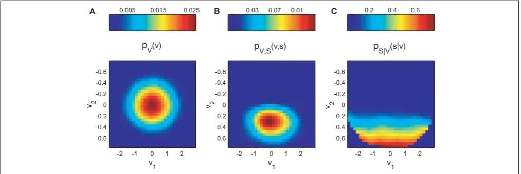
2.5 Eye Velocity Gain Fields in MSTd for Visuomotor Coordinate
Transformations
Brostek L, B¨uttner U, Mustari MJ, Glasauer S. Eye Velocity Gain Fields in MSTd for
Visuomotor Coordinate Transformations. Submitted.
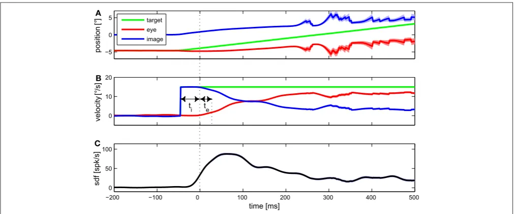
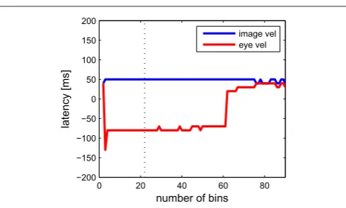
Lesion studies argue for an involvement of cortical area MSTd in the control of optoki-netic response (OKR) eye movements. Neurons in this area respond to visual motion and
eye movement related signals. However, MSTd’s function in visuomotor transformation is
still unclear. Using a novel approach for characterizing neural tuning with high resolution,
we show that during optokinetic stimulation the majority of MSTd neurons exhibits gain
field-like tuning functions. Rather than coding one variable, neural responses showed a
large diversity of tuning to combinations of retinal and extra-retinal input. Eye velocity
related activity was observed prior to the actual eye movements, reflecting an efference
copy. The observed tuning functions resembled those emerging in a network model trained
to perform summation of two population-coded signals. Together, our findings support
the hypothesis that MSTd implements the transformation from retinal to head-centered stimulus velocity signals for the control of OKR.
The author of this doctoral thesis contributed to this work by designing the
experi-ment, performing the data analysis, developing the computational models, writing the
2.6 Gain Modulation from Balanced Excitatory-Inhibitory Synaptic
Input
In this section additional results are presented which have not been published or
submit-ted for publication yet.
Cortical neuronsin vivo continuously receive input from thousands of excitatory and in-hibitory synapses (Kandel et al., 2000). Given a ’spontaneous’ average firing rate of 5-20
Hz in neocortical neurons, the resulting synaptic currents present a significant influence
on the integrative properties of the target neuron. Here, we investigated whether this
synaptic background activity may explain the gain-field-like tuning behavior we observed
in MSTd neurons. First, we present a point-conductance neural model proposed by
Des-texhe et al. (2001) to analyze the influence of balanced excitatory and inhibitory input
on neuronal gain. After this, we compare the predictions from this model with our MSTd
data shown in Brostek et al. (2012).
To represent the currents generated by thousands of stochastically releasing synapses a
point-conductance model was used. Synaptic activity was represented by two independent
fast glutamatergic and GABA-ergic conductances described by stochastic random-walk
processes (Destexhe et al., 2001).
The total synaptic current Isyn was decomposed into a sum of two independent
conduc-tances:
Isyn =ge(t)(V −Ee) +gi(t)(V −Ei) (2.1)
where ge(t) and gi(t) are time-dependent excitatory and inhibitory conductances, and
Ee and Ei their reversal potentials, respectively. The conductances ge(t) and gi(t) were described by a one-variable stochastic process similar to the Ornstein-Uhlenbeck process
(Uhlenbeck and Ornstein, 1930):
dge(t)
dt =−
1
τe[ge(t)−ge0] +
√ 2σe
√
τe χ1(t) (2.2)
dgi(t)
dt =−
1
τi[gi(t)−gi0] +
√ 2σi
√
τi χ2(t) (2.3)
where ge0 and gi0 are average conductances, τe and τi are time constants, σe and σi are ’diffusion’ standard deviations, andχ1(t) andχ2(t) are Gaussian white noise of zero mean
and unit standard deviation. The parameters for this random-walk process were adapted
The stochastic point-conductance model of background synaptic input activity was
in-serted into a single compartment Hodgkin-Huxley-type model (Hodgkin and Huxley,
1952), shown in Fig. 2.1A:
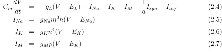
CmdV
dt = −gL(V −EL)−INa−IK−IM − 1
aIsyn−Iinj (2.4)
INa = gNam3h(V −ENa) (2.5)
IK = gKn4(V −EK) (2.6)
IM = gMp(V −EK) (2.7)
where Cm is the specific membrane capacitance, gL is the leak conductance density, and
EList the leak reversal potential. INa is the voltage-dependentN a+ current andIK is the
’delayed-rectifier’ K+ current responsible for action potentials. IM is a non-inactivating
K+ current responsible for slow afterhyperpolarization and spike frequency adaptation. The parameters of the gating variables m, h, n, and p were the same as in Destexhe et
al. (2001). All other parameters were also adapted for the originally modeled layer VI
pyramidal cell from cat parietal cortex and are shown in table 2.2. a = 34636μm2 is the
total membrane area of the modeled neuron, and Iinj is an additionally injected input
current.
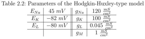
To analyze in what way the level of background synaptic input is modulating the gain of a
neuronal response to an excitatory ’driving’ signal, we determined firing rate and
trial-to-trial variability of the model neuron for different values ofIinj andIsyn (Fig. 2.1B). In the
latter case, excitatory and inhibitory currents were scaled with the same factor, denoted at the ordinate of the figure. The scaling of Isyn affects both mean and standard
devia-tion of synaptic input currents, which corresponds to increases of overall synaptic activity
and higher correlation of synaptic inputs, respectively (Fellous et al., 2003). For each
condition 100 trials of 1000 ms length were simulated. The Fano factor was determined
for a window length of 100 ms and averaged over the whole trial. As the figure shows,
the firing rate increased with an increase of Iinj, which was enhanced for higher values
of Isyn. This tuning strongly resembles the gain-field-like behavior we have observed in
MSTd neurons. In this sense, Iinj would correspond to the ’driving’ image velocity input,
Table 2.1: Parameters of the two random-walk processes
Ee 0 mV Ei −75 mV
Table 2.2: Parameters of the Hodgkin-Huxley-type model ENa 45 mV gNa 120 cmmS2
EK −82 mV gK 100 cmmS2 EL −80 mV gL 0.045 cmmS2
gM 1 cmmS2
whereas different levels of Isyn could be interpreted as changes of the ’modulatory’ eye
velocity signal.
In contrast to the firing rate, trial-to-trial variability of spiking activity showed
differen-tial behavior for increasing values of Iinj and Isyn (Fig. 2.1B2). An increase of synaptic
background activity caused a strong increase of the Fano factor, whereas neural
vari-ability exhibited little dependency or even a small decline for increasing values of Iinj.
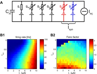
This finding is in good accordance with our data. In Brostek et al. (2012) we analyzed
neuronal variability of MSTd neurons during two different conditions: fixation with
vi-sual stimulation (FIX) and optokinetic response (OKR). For FIX, the monkey’s task was
to fixate a a small target spot located at the center of the screen. After some random time the LF stimulus started to move with constant velocity in the neurons preferred
direction for a period between 1000 and 1800 ms. During OKR, the spot was turned
off and the monkey’s eye movements followed the visual motion. As shown in Fig. 2.2,
the Hodgking-Huxley-type neural model exhibits very similar behavior in firing rate and
trial-to-trial variability of spiking activity like MSTd neurons when either the ’driving’
signal, or ’modulatory’ signal is increased, while the other signal is held close to zero.
In the following section we will discuss the implications of this finding on a potentially
C
mgL gNa+ gK+ g
e gi
I
inj gMI
syn0 2 4 6 8 10
0
0.5
1
1.5 0
20 40 60 80 100
Iinj [μA]
Isyn
firing rate [Hz]
0 2 4 6 8 10
0
0.5
1
1.5 0
0.5 1 1.5 2 2.5 3
Iinj [μA]
Isyn
Fano factor
B1
B2
A
0 50 100
Model Data
0 50 100
1 1.5 2 2.5
5 10 15 20 25
0.5 1 1.5 2 2.5
5 10 15 20 25
image velocity [°/s] eye velocity [°/s]
Fano factor
firing rage [Hz]
Fixation with visual
stimulation (FIX) Optokinetic response (OKR)
A1
A2
B1
B2
Within the framework of this doctoral thesis several aspects of neuronal activity in cortical
area MSTd have been investigated. Focusing on the neuronal responses during optokinetic
response (OKR) and smooth pursuit (SP), we were able to improve scientific knowledge
regarding the involvement of MSTd in these kinds of tracking eye movements. We
ana-lyzed the function of eye velocity gain fields in the context of tracking eye movements on
both systems and network level. Furthermore, we explored potential underlying mecha-nisms of gain modulation on the neuronal level.
To allow a model-free analysis of neuronal tuning behavior, we developed a novel
mutual-information-based approach for the evaluation of multi-dimensional probabilistic tuning
functions (Brostek et al., 2011a,b). Traditional model-based approaches like regression
analysis critically depend on the validity of their assumptions. Simple linear models often
are not sufficient for analyzing neuronal data. Our information-theoretic approach
over-comes these difficulties by maximizing the mutual information between stimulus variables
and neuronal response. It allows us to estimate the neuronal latency and to compare the
correlations between spiking activity and certain explanatory variables. This technique
can be applied in unbalanced designs and allows quantification of any possible dependence of neuronal activity on selected explanatory variables. However, the length of the
neu-ronal recording sets limiting constraints on the dimension of the analyzed tuning function.
We applied this novel approach to analyze MSTd neuronal activity during visual
stim-ulation using a so called ’white noise motion’ paradigm. In this experimental setting a
large-field random-dot pattern moves continuously and randomly in the axis of each
neu-ron’s preferred direction. The monkey’s task is to follow the stimulus as well as possible,
performing OKR eye movements. However, using stimulus velocities above maximal eye
velocity allowed us to cover wide ranges of both eye velocity and retinal image velocity
values at the same time. We found that neuronal responses showed a large diversity of
tuning to combinations of retinal and extra-retinal input instead of coding one of these variablesexplicitely. The majority of MSTd neurons exhibited rather gain-field-like tuning
functions. Analysis of neuronal latency revealed a leading of eye velocity related activity
relative to the actual eye movements. This signal can therefore not be of sensory origin
The distribution of eye velocity gain fields we found closely resembled the predictions
from a neural network model trained to perform the summation of image and eye
velo-city. The diversity of MSTd gain field shapes including asymmetric and non-separable
tuning functions was almost identical to the model results after completion of the
learn-ing process. Some neurons exhibited sharp, vertical tunlearn-ing functions, whereas other units
showed rather horizontal, image velocity related tuning. Together with the measured
neuronal latencies, these results provide strong evidence for MSTd participating in the OKR control system by implementing the transformation from retinal image velocity to
a head-centered stimulus velocity signal.
Beside the mean rate, there are more features of neuronal responses that may depend
systematically on certain stimuli. One of these is the regularity or variabitity of
spik-ing activity. In Brostek et al. (2012) we analyzed the variability of neuronal activity
in MSTd neurons during fixation with large-field visual stimulation and optokinetic eye
movements. Our analysis revealed two major features: first, in MSTd neurons the
trial-to-trial variability of neuronal activity, expressed by the Fano factor, is quenched by the
onset of visual stimulation. This change in variability is not necessarily directly related
to stimulation, as proposed earlier (Churchland et al., 2010). During visual stimulation
and fixating eye movements we found a sustained low level of variability. For optokinetic
response, however, only a transient decline of the Fano factor was observable. Second, and more remarkable, the relationship between spiking irregularity and the two stimulation
variables, image and eye velocity, was opposite. Both variables, which were uncoupled by
using two orthogonal paradigms, affected the intra- and inter-trial variability of neuronal
activity, meaning that the change in variability did not dependent on the task. All three
measures of neuronal variability we analyzed, Fano factor, squared coefficient of variation,
and ’Varience of the Conditional Expectation’ (Churchland et al., 2011), were negatively
correlated with retinal image velocity and positively correlated with eye velocity.
Our finding of decreasing spiking variability with an increasing ’driving’ signal image
velocity and increasing variability with an increase of the ’modulatory’ signal eye
velo-city puts certain constraints on the underlying neuronal structure. A conductance-based
single-compartment model neuron where multiplicative gain modulation is achieved by a simultaneous increase of excitatory and inhibitory background synaptic input yields
realistic increases of firing rate and can reproduce the observed stimulus-dependence of
Figure 3.2 provides a summary of our main results, illustrating the three different levels
that were analyzed within the scope of this doctoral thesis. In the following sections
certain aspects of our results will be discussed in more detail and compared with previous
findings. This chapter ends with an outlook on potential further investigations.
3.1 Comparison with previous MSTd studies
Most MSTd neurons show different behavior during smooth pursuit and OKR (Kawano
et al., 1994), as well as for radial and planar visual stimulation (Duffy and Wurtz, 1991),
respectively. Prior studies in area MSTd, which were focusing on its role in perception of
self-motion and heading direction, generally usedradial visual stimulation in combination with small target pursuit eye movements. Our OKR results, which were recorded
dur-ingplanar visual stimulation, may therefore not be directly comparable to these previous
studies. Nevertheless, those studies that were using smooth pursuit and radial stimulation
also found that visual responses of MSTd neurons are modulated during eye movements
(Bradley et al., 1996; Ben Hamed et al., 2003; Bremmer et al., 2010). In this sense, our
results are in compliance with these earlier studies.
A number of other prior studies investigated neuronal tuning in MSTd during planar
visual stimulation with combined small target pursuit and yielded diverging conclusions.
Kawano and colleagues, for instance, suggested that MSTd neurons might directly encode the velocity of a large-field visual stimulus in head or world-centered coordinates (Inaba et
al., 2007). A similar study by Chukoskie and Movshon 2009 could only partially confirm
this hypothesis. They found some neurons in MSTd that encoded stimulus velocity. Most
of the neurons, however, exhibited a variety of different other tuning behaviors ranging
from pure retinal to head-centered stimulus velocity coding. This finding has remarkable
similarity to our results, considering the difference of paradigms. We found only few
neu-rons with a transformation index close to zero, which could also be interpreted as coding
stimulus velocity in a restricted range of stimulus space. Nevertheless, instead of smooth
pursuit, we were using an OKR paradigm and could therefore assume an involvement of
the analyzed neurons in oculomotor control (D¨ursteler and Wurtz, 1988). This allowed us to shift the focus from the question ’which signals are coded?’ to ’what functions are
implemented?’. Our coordinate transformation hypothesis offers a straightforward
3.2 Gain Fields for Sensorimotor Coordinate Transformations
Numerous other studies in the posterior parietal cortex (PPC) have found gain-fields-like
tuning behavior before. For instance, visual responses of neurons in the lateral
intra-parietal area (LIP) and cortical area 7A are gain-modulated by eye and head position
signals (Snyder et al., 1998). The activity of neurons in the parietal reach region (PRR)
is modulated by eye and limb position (Chang et al., 2009). Zipser and Andersen 1988
were the first to show that eye position gain fields might be used to transform the reference
frame of eye-centered visual responses into head-centered responses. Beyond sensorimo-tor transformations, the computational function of gain fields might be a general method
for neural computation when transformations between different brain representations are
required (Salinas and Thier, 2000).
Yet, all previous studies were limited in a certain sense: the characterization of neuronal
responses was incomplete as only very few and specific combinations of visual input and
motor output could be tested. Our novel approach overcomes these limitations and
al-lows us to characterize neural tuning with high resolution and over a large input-output
range by dissociating visual motion and eye movements without additional task
require-ments. This enabled us to analyze novel aspects as for instance the distribution of gain
field types, which could not have been investigated using any of the traditional approaches.
It is common practice to correlate neuronal activity with certain variables, assuming a direct encoding of sensory or motor signals by different neuronal populations. This
ap-proach may be appropriate for the early input or output stages of neuronal processing.
It poses, however, serious difficulties when intermediate processing steps of
sensorimo-tor transformation are analyzed. Theoretical studies have shown that a neural coding
scheme where each object in each reference frame is represented by a different set of
neurons quickly will reach limitations due to the combinatorial explosion in the number
of required cells (Poggio, 1990). It was therefore suggested that a much more efficient
scheme for neuronal representation might be used: instead of representing each variable
by a certain pool of neurons, one set of basis functions can represent a number of
differ-ent variables simultaneously. Arbitrary variables are then represdiffer-ented by a simple linear combination of these basis functions (Girosi et al., 1995; Pouget and Sejnowski, 1997).
Gain fields, as demonstrated by Pouget and Sejnowksi 1997, exhibit all characteristics
necessary to form a set of basis functions. The diversity of tuning functions we observed
in our data is consistent with this theory. Hence, eye velocity gain fields in MSTd could be
es-timate of heading direction (Ben Hamed et al., 2003) or perceived self-motion velocity. In
our case of planar visual stimulation, perceived self-motion velocity is simply the stimulus
velocity signal directed towards the opposite side. Such inversion can be easily obtained
by changing the weights of the connections to the output layer in our neural network
model. The self-motion signal might be generalized for head- and body-motion by the inclusion of vestibular information (Gu et al., 2007). Our results are therefore compatible
with the idea of area MSTd serving various functions in self-motion perception, as well
as in oculomotor control.
3.3 Underlying Neuronal Structure
It is generally assumed that neuronal activity arises from an interaction between ongoing spiking generated spontaneously by neuronal circuits and responses driven by external
stimuli (Dayan and Abbott, 2001). In this view the observed variability in neuronal
activity is generally interpreted as noise, interfering with the actual signal coded by the
neurons (Shadlen and Newsome, 1998). Recurrent networks are a kind of topological
structure that is presumed to be found in many cortical areas. A general feature of this
type of networks is the stimulus-driven suppression of chaotic, spontaneous activity. The
decline in variability depends on stimulus frequency and amplitude (Rajan et al., 2010).
The decline of spiking irregularity with visual stimulation we observed in MSTd (Brostek et al., 2012) may be explained by the presence of recurrent circuitry. However, the increase
in neuronal variability with higher eye velocity remains unexplained by the recurrent
net-work hypothesis. The change in neuronal variability in our data is not just related to the
presence of stimulation. A network topology that could explain the observed behavior is
therefore probably asymmetrical and processes both input signals, image velocity and eye
velocity, differently.
Anatomical observations led Sherman and Guillery (1998) propose that neurons might
have two classes of inputs, one responsible for driving neural responses and the other for
modulating those responses. Based on this idea, Chance et al. (2002) suggested a gain
field mechansism where the gain of a neuronal response to excitatory drive can be mod-ulated by varying the level of balanced excitatory and inhibitory synaptic input. Using
both, an in vitro neuron model, and an analytic firing rate model, they could show that
simultaneously increasing the background firing rates in a balanced manner results in a
analyzing divisivegain modulation, where the gain decreases with an increase of the
mod-ulatory signal. Interestingly, the authors did not observe changes of neuronal variability
with gain modulation in their in vitro model.
Using a far more detailed Hodgking-Huxley-type model with inserted stochastic conduc-tances mimicking balanced excitatory and inhibitory synaptic input, we could show that
an increase of backgound activity can actually result in multiplicative gain modulation,
as well. Furthermore, our model is able to reproduce the differential behavior in
neu-ral variability we have observed in MSTd neurons. A comparatively simple feedforward
mechanism allowed us to explain both, a decrease of spiking irregularity with increase of
image velocity, and an increase with eye velocity, without the inclusion of recurrent or
feedback circuitry.
3.4 Temporal Coding in MSTd?
A great amount of knowledge about neuronal information processing has been gained by
relating the mean neuronal firing rate to any variables supposed to be coded in the
an-alyzed area. Beside the mean rate, however, there are more features of neural responses
that may depend systematically on certain stimuli. Theoretical and experimental
stud-ies in numerous cortical and sub-cortical regions indicated that the temporal pattern of
spiking activity carries important information as well (Buracas and Albright, 1999; Rieke
et al., 1997). For instance in auditory neurons the mean firing rate represents some
com-bination of amplitude and frequency of a tone. At the same time there is the tendency
for inter-spike intervals (ISIs) to cluster around integer multiples of the stimulus period,
allowing the separation of frequency and amplitude information. Also in cortical areas spiking irregularity has been used as an evidence to support the temporal coding
hypothe-ses (Softky and Koch, 1993).
As we have shown, in MSTd neurons the mean firing rate, which is the reciprocal of
the mean inter-spike interval, codes some non-linear combination of visual and eye
move-ment related signals (Brostek et al., 2011a). At the same time the variance of the ISI
decreases with visual and increases with oculomotor stimulation (Brostek et al., 2012).
This independent temporal code may allow the separation of the two signals, similar to phase-locking in auditory neurons.
1962). The approximate one-to-one relation between F F and CV2 observed in our data
argues for the renewal assumption. Both across-trial and within-trial variability are
de-termined by the distribution of ISIs of the corresponding renewal process. The gamma
distribution is an appropriate approximation for the distribution of ISIs in most neuronal
systems (Stein, 1965). A change in spiking irregularity is associated with a modification of the ISI distribution. This again may result from changing membrane properties in
single neurons, circuit properties of networks of neurons, or a combination of both. Miura
et al. 2007 for instance proposed a network architecture, where the firing rate could be
decoupled from the ISI distribution by proper balance of excitatory and inhibitory inputs.
However, the questions whether the change of the ISI distribution in dependence of visual
and oculomotor input has a functional meaning, and whether the additional information,
embodied in changing spiking irregularity, is actually used by MSTd and subsequent
ar-eas, or reflects just an epiphenomenon, remain to be solved by future investigations.
3.5 Is MSTd Involved in Smooth Pursuit Control?
There is strong evidence from lesion studies that the dorsal visual pathway, which is
re-sponsible for motion processing, is involved in the generation of SPEMs (D¨ursteler and
Wurtz, 1988). Microlesions in areas MT and MSTl lead to two kinds of pursuit deficits: the retinotopic deficit, which describes problems of matching eye speed to target speed
when the target is moving in any direction in the visual field contralateral to the side of
the brain with the lesion, and the directional deficit, which is the inability to match eye
speed to target speed when the target is moving towards the side of the lesion. Lesions
in MSTd, however, only lead to retinotopic deficits, indicating a minor role of this area
in pursuit control. This view is supported by electrical microstimulation studies, as in
MSTd only few neurons were found, where stimulation produced an acceleration of
pur-suit (Komatsu and Wurtz, 1989).
Nevertheless, many neurons in MSTd show significantly increased activity during SPEMs
in the abscence of other visual stimulation (Komatsu and Wurtz, 1988). In Ono et al.
(2010) we found that about one third of MSTd neurons belong to the subpopulation of
the so called ’smooth pursuit neurons’. These neurons continue to respond during pursuit despite a blink (Newsome et al., 1988) or even complete disappearance (Ilg and Thier,
2003) of the pursuit target. Furthermore, the pursuit related activity in MSTd has been
reported to be higher than the visually induced activity in many neurons (Churchland