Article
1
Surface Plasmon Resonance (SPR) Computational
2
Study of Hemoglobin (Hb) in Human Blood
3
Detection
4
Wida Yanti 1, Kamsul Abraha 2,*and Agung Bambang S.U. 2
5
1 Departement of Physics, Faculty of Science and Technology, Sunan Kalijaga Islamic State University, Jl
6
Marsda AdiSucipto No.1, Yogyakarta, Indonesia; [email protected]
7
2 Departement of Physics, Faculty of Mathematic and Natural Science, Gadjah Mada University, Jl Sekip
8
Utara BLS.21, Yogyakarta, Indonesia; [email protected]; [email protected]
9
* Correspondence: [email protected]; Tel.: +62-812-271-0165
10
Abstract: A theoretical analysis of haemoglobin (Hb) concentration detection is presented in this
11
work with the objective of achieving more sensitive detection and monitoring low concentrations.
12
Surface-enhanced SPR spectroscopy on silver nanoparticles was employed for recording Hb
13
concentrations less than 10 g/L. In this paper, Fe3O4@Au core-shell, nanocomposite spherical
14
nanoparticle consisting of a spherical Fe3O4 core covered by Au shell, was used as an active
15
material for biomolecules detection in the Surface Plasmon Resonance (SPR)-based biosensor in the
16
wavelength 632.8 nm. We present the simulation of detection amplification technique through
17
Attenuated Total Reflection (ATR) spectrum in the Kretschmann configuration. The system consists
18
of a four-layer material i.e prism/Ag/Fe3O4@Au+Hb/air. Dielectric function determination of the
19
core-shell nanoparticle (Fe3O4@Au) and the composite (Fe3O4@Au+Hb) was done by applying the
20
Effective Medium Theory approximation and the calculation of the reflectivity is carried out by
21
varying the size of core-shell (r0). In this simulation, the refractive index of the BK7 prism is 1.51;
22
the refractive index of Ag thin film is 0.13455+3.98651i with the thickness of 40 nm, and the
23
refractive index of the composite is varied depending on the size of nanoparticle core-shell. Our
24
results show that by varying the radius of the core and the shell thickness, the dipof the reflectivity
25
(ATR) spectrum is shifted to the larger angle of incident light and the addition of core-shell in the
26
conventional SPR-based biosensor leads to enhancement of the SPR biosensor sensitivity, for the
27
core-shell radius 10 nm, the sensitivity increased by 1.35% for F = 0.1, and by 4.89% for F =0.8
28
compared to the sensitivity of the conventional SPR-based biosensor without core-shell addition.
29
Keywords: Haemoglobin detection; SPR spectroscopy; Biosensors; Computer simulation;
30
Core-shell Fe3O4@Au.
31
32
1. Introduction
33
Currently, there is increasing interest in the development of magnetic and plasmonic
34
nanoparticles as the active materials for biomolecule detection. The new nanoparticle that combines
35
multiple functions or properties not obtainable in the individual material has attracted
36
considerable attention because of its revolutionary technology for sensitivity enhancement of surface
37
plasmon resonance (SPR)-based biosensor [1]. Optical sensor based on SPR is one of the sensitive
38
methods that detects biomolecules and works on the changes of the material refractive index,
39
having fast response, real-time, biospecific interaction analysis and being the label-free technique [2].
40
SPR is a physical process that occurs when the wave vector of the evanescent wave (EW) matches the
41
wave vector of the surface plasmon (SP) under the total internal reflection condition. This resonance
42
condition is expressed as
43
1/2 2
0 0
2
sin
m dp SPR
m d
n
n
c
c
n
ω
θ
ω
ε
ε
=
+
(1)44
45
The variable on left hand side is the propagation constant of a light beam incident at a
46
resonance angle
θ
SPRthrough the light coupling device (prism) of refractive indexn
p. While the47
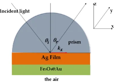
right-hand is the propagation constant with
ε
m as real part of the metal permittivity andn
d as the48
refractive index of dielectric material or sensing medium.
ω
0 andc
are the light frequency and49
the speed of the light in the vacuum respectively. The evanescent wave occurs at the metal-dielectric
50
interface when a p-polarized wave passes a prism through a metallic layer into a dielectric media.
51
The wave vector of the evanescent wave is a function of refractive indices of the dielectric,
52
metal and analyte i.e the sensing medium. Therefore, if there is a local change in the refractive index
53
of the sensing medium near the metal surface, it will in turn lead to a change in the propagation
54
constant of SP and in the angle of incidence light in order to satisfy the resonance. For applying SPR
55
biosensor, the Kretschmann geometry [3]of Attenuated Total Reflection (ATR) has been found to be
56
very suitable for the sensing and has become the most widely used geometry in SPR biosensor.
57
Mostly, the metallic layer that is used in SPR biosensor measurement consists of either gold or silver.
58
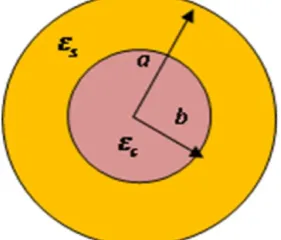
The first demonstration about SPR-based sensor for bio-sensing was reported in 1983 by Liedberg et
59
al [4]. Several ways to enhance sensitivity of SPR biosensor for detecting biomolecules have been
60
explored for the detection of DNA hybridization [5], acetylcholinesterase [6], membrane protein [7]
61
and human blood-group [8]. SPR can also be a potential candidate for bio-sensing other biological
62
properties such as haemoglobin concentration.
63
From some of researches it is acquired that the conventional SPR-based biosensor was not
64
capable of sensing the small amount biomolecules such as DNA, virus or bacteria [9] due to the poor
65
attachment of biomolecule on the metal surface and the low concentration of it is difficult to detect
66
directly [10]. It happens sincethe changes in the refractive index of the medium [11] under a thin
67
metal layer are very small. Therefore, the enhancement of sensitivity for detecting small
68
biomolecules can be developed by several approaches such as by involving nanoparticle core-shell
69
as the active material in the conventional SPR-based biosensor. Comparing with nanoparticle which
70
has a spherical shape, the involvement of core-shell aims to avoid polar resonance [12]and to obtain
71
some plasmonic wavelength by varying the radius of the core and the thickness of the shell. A
72
core-shell was said to be a unique material since it is a combination of magnetic and plasmonic
73
materials which has different optical properties between the core and the shell. Some studies have
74
observed, either experimentally or theoretically, about the optical properties of the core-shell with
75
its involvement in the SPR-based biosensor. It is observed that the optical response or resonance
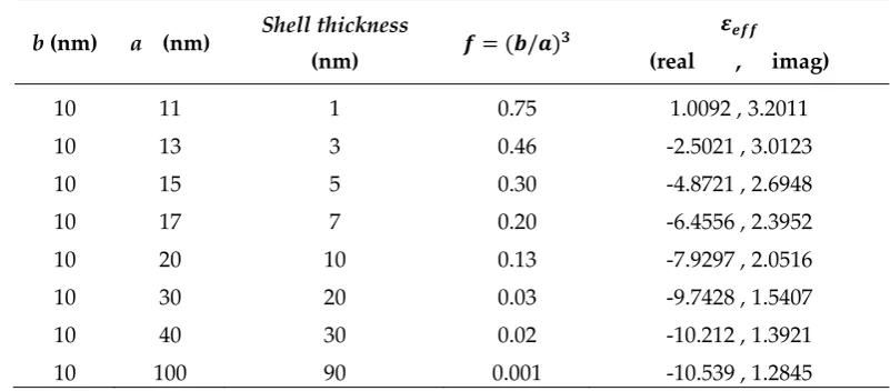
76
core-shell can be used for tuning the plasmonic wavelength [13], e.g AgSiO2 [12], TiO2@Au and
78
TiO2@Ag [14]. The study of the optical response of Fe3O4@Au core-shell was performed by varying
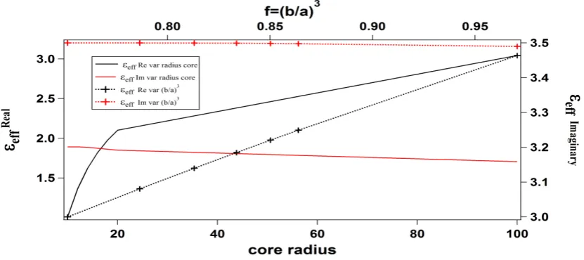
79
the radius of Fe3O4 and the thickness of Au. There was shift resonance spectrum due to the changes
80
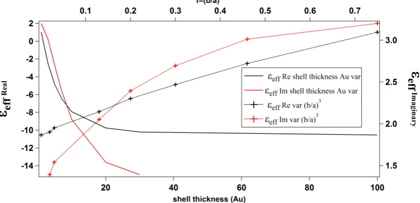
of the size of the core and the shell [15]. The core Fe3O4 could make the biomolecule attachment
81
easier by the help of its magnetic property, while the shell Au exhibits nontoxicity and compatible
82
property. Furthermore, the performance of the SPR-based biosensor can be enhanched by using the
83
nanoparticle core-shell Fe3O4@Au rather using only Fe3O4 or only Au. The presence of Fe3O4@Au is
84
also capable of enhancing the immobilization of biomolecules e.g Haemoglobin [16], detecting
85
antibody IgG [17] and detecting the DNA of chum salmon [18].. Whereas, involvement of the
86
Fe3O4@Au in the SPR-based biosensor was performed for enhancement detection of thrombin [19],
87
and protein concentration of interleukin IL17 [20]. Detection of the haemoglobin concentration has
88
been explored by SPR-based biosensor for three wavelengths (401.5 nm, 589.3 nm and 706.5 nm)
89
with haemoglobin concentration varying between 0 and 140 g/l [21].
90
Due to the coating of Au shell for the magnetic core that protected it from oxidation and
91
aggregation, the stabilization of the core-shell (Fe3O4@Au) was enhanced obviously. And the current
92
SPR technology has so many advantages as well, i.e processing high sensitivity and selectivity,
93
non-destructive, large tunability from the visible into the infrared (IR) spectrum region, label-free
94
analysis and being capable of real-time monitoring. SPR is a kind of electromagnetic resonance that
95
exists when there is an interface between metal and dielectric. This system has been used for sensing
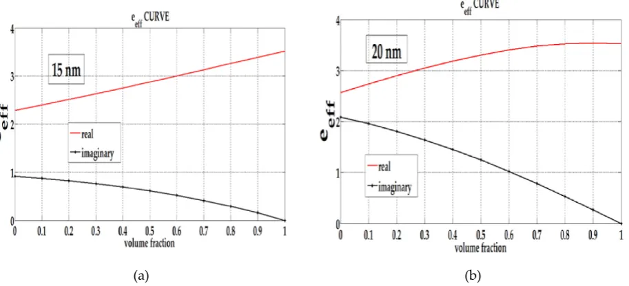
96
various biomolecules [22],[23]. This method is very sensitive to size, shape and the refractive index
97
of surrounding medium or the medium that kept contact with the thin metal layer. When the
98
biomolecule comes in contact with the metal thin film, it is adsorbed on its surface and hence
99
increasing the refractive index at the interface and resulting in a change of the resonance angle.
100
In this paper, we have been investigating the ATR spectrum of three and four multilayer
101
biosensor based on SPR system with Fe3O4@Au core-shell addition and the biology element is
102
hemoglobin in the human blood with the refractive index is 1.338 [24]. The different size of
103
Fe3O4@Au+Hb composites leads to a change in the SPR resonance angle.
104
This study was focused on the simulation of the effects of the size of the core radius and shell
105
thickness to the effective permittivity of Fe3O4@Au. Also, the effects of the volume fraction and the
106
size of the core-shell on the composite effective permittivity and on the SPR-based biosensor
107
reflectivity were investigated. Then, the enhancement of the sensitivity of SPR configuration was
108
estimated.
109
2. Materials and Methods
110
2.1. Kretschmann configuration with four layers
111
Here, we applythe analytical and computational approximation to calculate reflectivity in the
112
Attenuated Total Reflection (ATR) method and determine the effective permittivity of the composite
113
(the mixture of Fe3O4@Au and Hb embedded in the water). In this study, we used the Kretschmann
114
configuration [3]with four layers. i.e prism/Ag/composite/air shown in Figure 1. The angle
θ
i and115
r
θ
are the incident and the reflection angle respectively,k
z is the wave vector component along116
Figure 1. Kretschmann configuration for SPR-based biosensor with the inclusion of
118
Fe3O4@Au core-shell.
119
Figure 2 shows the model of the composite layer containing the inclusion material
120
(Fe3O4@Au+biomolecule) and the host material (water). The inclusion material consists of the
121
scattered grain material (Fe3O4@Au) and the interfacial shell material (Hb).
122
Figure 2. The composite model contains the complex particle (inclusion)
123
and the host material.
124
In this SPR configuration, the refractive index of the BK7 glass prism is 1.510, the wavelength of
125
the electromagnetic wave is 632.8 nm, the complex refractive index of silver 0.13455+3.98651i [25],
126
and dielectric constant of Hb ( ) 1.338for the concentration is less than 10 g/L.The refractive index
127
of water and air is 1.33 and 1.0 respectively [9]. The thickness of Ag film was d=40 nm, and the
128
composite was d=20 nm. The ATR reflectivity R is given by the Fresnel equation [26].
129
2 2 2
2
1
j z j
j z j
ik d i j j k
i j k ik d
i j j k
r
r e
R
r
r r e
+
=
=
+
(2)130
i j j i i j
i j j i
k
k
r
k
k
ε
ε
ε
ε
−
=
+
(3)132
here, is the surface reflectivity coefficient between medium i and medium j.
k
i j is the wave133
vector component perpendicular to the surface, is the wave vector component parallel to the
134
surface, whereas and are respectively the j-th layer thickness and the i-th medium dielectric
135
constant.
136
2.2. The effective permittivity of the spherical Fe3O4@Au core-shell
137
Our simulation of the Fe3O4@Au core-shell is performed on the model as shown in Figure 3.
138
The magnetic nanoparticle core-shell consists of a Fe3O4 core of radius b coated by a metallic Au of
139
thickness ( − ). The dielectric constants of the magnetic nanoparticle and the metallic Au are ,
140
and , respectively.
141
Figure 3. The model of nanoparticle Fe3O4@Au core-shell.
142
whereas, the value of complex is adopted from Schlegel [27] through reflectivity measurement
143
and Kramers-Kronig relation and can be quoted from Johnson and Christy work [25]. The
144
effective permittivity
(
ε
eff)
of Fe3O4@Au core-shell is derived from the internal homogenization for145
plasmonic and dielectric constituent material [28], namely
146
(
)
(
)
(
)
(
)
3 3
eff 3 3
2
2
2
c s c s
s
c s c s
a
b
a
b
ε
ε
ε ε
ε
ε
ε
ε
ε ε
+
+
−
=
+
−
−
(4)147
2.3. Calculating the effective permittivity of the composite
148
The effective permittivity of the composite
(
ε
eff)
is calculated by neglecting the correlation149
between the inclusion material (complex material or Fe3O4@Au+Hb) and host material (water), using
150
the Maxwell Garnett formula [29]
151
eff eff
eff eff
(1
)
2
m n
m n
F
ε
ε
F
ε
ε
ε
ε
ε
ε
−
−
−
+
+
+
(5)152
(
)
(
)
(
1 2) (
2 1)
11 2 2 1
2
2
2
n
ε ε
α ε ε
ε
ε
ε ε
α ε ε
+
+
−
=
+
−
−
(6)154
where
3 0
r
D
α
=
,r
0the radius of Fe3O4,D
the radius of the complex particle (Fe3O4@Au+Hb),F
155
the volume fraction of the inclusion material to the host material,
ε
n the dielectric constant of the156
complex particle and
ε
m the dielectric constant of the host material.ε
1 is the Hb dielectric157
constant as the interfacial shell, while
ε
2 is the dielectric constant of scattered grain (Fe3O4@Au158
core-shell).
159
2.4. Biosensor sensitivity from ATR spectrum
160
The calculation of the sensitivity of SPR-based biosensor is written as [30]
161
162
SPR
S
n
θ
Δ
=
Δ
(7)163
where
Δ
θ
SPRis the difference of the SPR angle and ∆ is the change in refractive index.164
3. Results and Discussion
165
The changes of the radius of the Fe3O4 coreand the thickness of the Au shell lead to the change
166
in the effective permittivity of Fe3O4@Au core-shell While the change of the inclusion material to
167
host material leads to the change in the effective permittivity of the composite. Therefore, if the
168
complex particle is applied to SPR-based biosensor system, this change leads to the enhancement of
169
the sensitivity of this biosensor. We can show from the reflectivity spectrum that the resonant angle
170
shift to the right.
171
Table 1. The effective permittivity of Fe3O4@Au core-shell for the shell thickness variation.
172
b (nm) a (nm) Shell thickness
(nm) = ( / ) (real , imag)
10 11 1 0.75 1.0092 , 3.2011
10 13 3 0.46 -2.5021 , 3.0123
10 15 5 0.30 -4.8721 , 2.6948
10 17 7 0.20 -6.4556 , 2.3952
10 20 10 0.13 -7.9297 , 2.0516
10 30 20 0.03 -9.7428 , 1.5407
10 40 30 0.02 -10.212 , 1.3921
10 100 90 0.001 -10.539 , 1.2845
Table 2. The effective permittivity of Fe3O4@Au core-shell for the core radius variation.
174
b (nm) a (nm) = ( )
(real , imag)
10 11 0.75 1.0092 , 3.2011
12 13 0.78 1.3637 , 3,2017
14 15 0.81 1.6230 , 3.2000
16 17 0.83 1.8209 , 3.1975
18 19 0.85 1.9768 , 3.1947
20 21 0.86 2.1028 , 3.1921
100 101 0.97 3.0427 , 3.1587
Table 3. The effective permittivity of Fe3O4@Au core-shell for the = ( ⁄ ) variation.
175
b (nm) a (nm) = ( )
(real , imag)
19 20 0.85 2.04008 , 3.19339
18 20 0.73 0.77837 , 3.19889
17 20 0.61 -0.49070 , 3.16118
16 20 0.51 -1.74550 , 3.08115
15 20 0.42 -2.96636 , 2.96227
14 20 0.34 -4.13333 , 2.81043
13 20 0.27 -5.22780 , 2.63368
Table 1shows the effective permittivity of core-shell (Eq. 4) for variation in the shell thickness.
176
Table 2 shows the effective permittivity of core-shell for variation inthe core radius and Table 3
177
shows the effective permittivity of core-shell for ( ) variation. The data in Tabel 1, , Table 2 and
178
Table 3are presented in Figure 4, Figure 5 and Figure 6 respectively.
179
Figure 4. The effective permittivity of core-shell for shell thickness variation and
180
= ( ⁄ ) variation.
Figure 5. The effective permittivity of core-shell for the radius of the core variation
182
and = ( ⁄ ) variation.
183
Figure 6. The effective permittivity of core-shell for = ( ⁄ ) variation.
184
Figure 4 shows that the increasing shell thickness for fixed core radius (10 nm), leads to the
185
decreasing of the real and imaginary part of the core-shell effective permittivity. But, the real part
186
tends to be constant at the shell thickness above 30 nm. While Figure 5 shows that increasing in the
187
core radius for fixed the shell thickness (1 nm) leads to increasing in the real part of the core-shell
188
effective permittivity while the imaginary part tends to be constant. If the core-shell effective
189
permittivity is viewed only from the = ( ⁄ ) variation, it shows that the increasing = ( ⁄ )
190
leads to increasing in the real and imaginary parts of the effective permittivity (Figure 6). Different
191
radius of core-shell a (15 nm, 10 nm, 5nm) with the same = ( ⁄ ) variation shows the same
192
effective permittivity value. And then the effective permittivity of the composite can be obtained
193
from Eq. 5.
194
The thicknesses of Ag metal in this SPR system is the other parameter that must be carefully
195
controlled in order to obtain an optimum performance for surface plasmon excitation. Therefore the
196
choise of the metal thickness is of greatest importance. By varying the Ag thickness, the ATR
197
spectrum shows where the Ag metal film thickness yields the most desirable resonance peak. As the
198
Ag thickness increases (50 -60 nm), the depth of the resonance peak decrease. This is indicate the
199
acting as a reflectance plane when its thickness increases to a point where light cannot couple into
201
the surface charge oscillations that make up the plasmon mode. Whereas, if the Ag thin film is very
202
thin (20-30 nm), it result in more coupling into the SP mode but due to light scattering, the sensitivity
203
was redused. Obviously, from these effects, a compromise must be reached to obtain a satisfactory
204
SPR system. Figure 7 was shows the optimal thickness to support SPR system determined to lie at 40
205
nm.
206
Figure 7. The ATR spectra for the Ag metal thickness varied from 20 nm to the 60 nm.
207
Based on the above results, we can choose the values of the core radius, the shell thickness and
208
the ratio of the core to the core-shell radius = ( ⁄ ) to obtain the desirable effective permittivity
209
of the Fe3O4@Au core-shell. Figure 8 and Figure 9 shows the effective permittivity of Fe3O4@Au+Hb
210
composite (inclusion material) with volume fraction (F) variation given by the size variation of the
211
core-shell a from 5 nm to 20 nm for fixed = ( ) = 0.73. Here, F is the ratio between the amount of
212
the inclusion material to the host material.
213
(a) (b)
Figure 8. The composite effective permittivity with variation in the volume
(a) (b)
Figure 9. The composite effective permittivity with variation in the volume fraction (F) of
214
composite for fixed size of the core-shell (a) 15 nm(b) 20 nm.
215
The reflectivity from the SPR-based biosensor consisting of nanoparticle core-shell is shown in
216
Figure 10 and Figure 11. If the layer only consists of a prism, a thin film of metal (40 nm Ag) and 20
217
nm biomolecule (conventional SPR), the dip of the ATR curve occurs at the incident angle 45.17o
218
(solid line). After the composite had been deposited onto the surface of the Ag thin film, the dip of the
219
reflectivity curve was shifted to the larger angle. Referring to Figure 10 for the volume fraction (F)=
220
0.1 and the Fe3O4@Au radius was varied from 5 nm to 20 nm, the SPR angle was shifted to the larger
221
angle. By increasing the radius of Fe3O4@Au, the angle of resonance increases as well. It can be seen
222
in Figure 10 that the minimum reflectivities are seen at 45.75o for thickness 5 nm and at 45.78o for 10
223
nm thickness, while from Figure 11, for the volume fraction(F)= 0.8 and the Fe3O4@Au radius was
224
varied from 5 nm to 20 nm, the SPR angle is shifted to the larger angle as well. The figure shows that
225
the minimum reflectivities occured at 47.38o for thickness 10 nm and at 47.55o for thickness 15 nm.
226
Figure 10. The ATR spectra for the volume fraction of the composite F=0.1. The radius of
227
inclusion material at R=20 nm and the radius of core-shell varied from 5 nm to the 20 nm
228
with fixed ( / )3=0.73.
Figure 11. The ATR spectra for the volume fraction of the composite F=0.8.
230
The radius of inclusion material at R=20 nm and the radius of core-shell
231
varied from 5 nm to the 20 nmwith fixed ( / )3=0.73.
232
Therefore, it can be obtained from Eq. 7 that for the core-shell radius 10 nm, the sensitivity
233
increased by 1.35 % for F=0.1, and by 4.89 % for F =0.8 compared to the sensitivity of the conventional
234
SPR-based biosensor without core-shell addition
235
5. Conclusions
236
In summary, we have presented a simulation for the size effect of the inclusion material in the
237
SPR-based biosensor through the ATR spectra. Our calculations confirm that the property
238
combination of the magnetic and plasmonic materials leads to the enhancement of the SPR-based
239
biosensor sensitivity that applies to detect the existence of Hb as analyte. By varying the radii of the
240
core (Fe3O4) and the shell (Au), the refractive index of the core-shell changes and leads to the change
241
in the composite (core-shell+Hb+water) permittivity. The change in this effective permittivity results
242
in the change of the dip position in the reflectivity spectrum to the larger angle. The SPR dips were
243
shifted when the core-shell was added to the composite as the active material. This large shift in the
244
dip angle suggests the potential for its application in the highly sensitive biosensor, in this case,
245
sensing Hb as the analyte.
246
Author Contributions: Widayanti conceived the idea for the study, conceptualization, data curation, funding
247
acquisition, methodology, software, visualization and writing original draft of the manuscript. Kamsul Abraha
248
contributed to investigated, validated, writing-review & editing draft of the manuscript and Agung Bambang
249
S.U contribute to helped for supervision and validation. Kamsul Abraha as the corresponding author for this
250
manuscript. All author contributed significant effort to the manuscript preparation.
251
Conflicts of Interest: The authors declare no conflict of interest.
252
References
253
1. Wang, J.; Sun, Y.; Wang, L.; Zhu, X.; Zhang, H.; and Song, D. Surface plasmon resonance biosensor based
254
on Fe3O4/Au nanocomposites. Colloids and Surfaces B: Biointerfaces.2010, 81, 600-606.
255
2. Agbor, N.E.; Cresswell, J.P.; Petty, M.C.; and Monkman, A.P. An optical gas sensor based on polyaniline
257
Langmuir-Blodgett films. Sensors and Actuators B: Chemical. 1997, 41, 137-141.
258
https://doi.org/10.1016/S0925-4005(97)80286-9.
259
3. Kretschmann, E.; Raether, H. Notizen: Radiative Decay of Non Radiative Surface Plasmons Excited by
260
Light.. Zeitschrift Naturforsh. 1968, 23 A, 2135-2136, https://doi.org/10.1515/zna-1968-1247.
261
4. Liedberg, B.; Nylander, C.; Lunström, I. Surface plasmon resonance for gas detection and biosensing.
262
Sensors and Actuators. 1983, 4, 299-304. https://doi.org/10.1016/0250-6874(83)85036-7.
263
5. He, L.; Musick, M.D.; Nicewarner, S.R; Salinas, F.G.; Benkovic, S.J.; Natan, M.J.; Keating, C.D. Colloidal
264
Au-Enhanced Surface Plasmon Resonance for Ultrasensitive Detection of DNA Hybridization. Journal of
265
the American Chemical Society. 2000, 122, 9071-9077. https://pubs.acs.org/doi/abs/10.1021/ja001215b.
266
6. Milkani, E.; Lambert, C.R.; Mc Gimpsey, W.G. Direct detection of acetylcholinesterase inhibitor binding
267
with an enzyme-based surface plasmon resonance sensor. Anal Biochem. 2011, 408, 212-219.
268
https://doi.org/10.1016/j.ab.2010.09.009.
269
7. Salamon, Z.; Macleod, H.A.; Tollin, G. Surface plasmon resonance spectroscopy as a tool for investigating
270
the biochemical and biophysical properties of membrane protein systems. II: Applications to biological
271
systems. Biochimica et biophysica acta. 1997, 1331, 131-152. https://www.ncbi.nlm.nih.gov/pubmed/9325439..
272
8. Sharma, A.K. Plasmonic biosensor for detection of hemoglobin concentration in human blood: Design
273
considerations. Journal of Applied Physics. 2013, 114, 044701. https://doi.org/10.1063/1.4816272.
274
9. Wu, L.; Chu, H.S.; Koh, W.S.; Li, E.P. Highly sensitive graphene biosensors based on surface plasmon
275
resonance. Opt. Express. 2010, 18, 14395-14400. https://doi.org/10.1364/OE.18.014395.
276
10. Liang, R. P.; Yao, G. H.; Fan, L. X.; Qiu, J. D. Magnetic Fe3O4@Au composite-enhanced surface plasmon
277
resonance for ultrasensitive detection of magnetic nanoparticle-enriched α-fetoprotein. Anal Chim Acta.
278
2012, 737, 22-28https://doi.org/10.1016/j.aca.2012.05.043.
279
11. Frasconi, M.; Tortolini, C.; Botrè, F.; Mazzei, F. Multifunctional Au Nanoparticle Dendrimer-Based Surface
280
Plasmon Resonance Biosensor and Its Application for Improved Insulin Detection. Analytical Chemistry.
281
2010, 82, 7335-7342. https://doi.org/10.1021/ac101319k.
282
12. Baida, H.; Billaud, P.; Marhaba, S.; Christofilos, D.; Cottancin, E.; Crut, A.; Lermé, J.; Maioli, P.; Pellarin,
283
M..; Broyer, M.; Del Fatti, N.; Vallée, F.; Sánchez-Iglesias, A.; Pastoriza-Santos, I.; Liz-Marzán, L.M.
284
Quantitative Determination of the Size Dependence of Surface Plasmon Resonance Damping in Single
285
Ag@SiO2 Nanoparticles. Nano Letters. 2009, 9, 3463-3469. https://doi.org/10.1021/nl901672b.
286
13. Zhu, J. Surface Plasmon Resonance from Bimetallic Interface in Au–Ag Core–Shell Structure Nanowires.
287
Nanoscale Research Letters. 2009, 4, 977-981. https://dx.doi.org/10.1007%2Fs11671-009-9344-4.
288
14. Pathak, N.; Ji, A.; Sharma, R.P. Tunable Properties of Surface Plasmon Resonances: The Influence of
289
Core–Shell Thickness and Dielectric Environment. Plasmonics. 2014, 9,651-657.
290
https://doi.org/10.1007/s11468-014-9677-4.
291
15. Ahmadi, N.; Poursalehi, R.; and Farshi, M.K.M. The Interparticle Coupling Effect on Plasmon Resonance
292
Properties of Magnetite@Au Magnetoplasmonic Nanoparticles. Procedia Materials Science. 2015, 11,
293
254-258. https://doi.org/10.1016/j.mspro.2015.11.131.
294
16. Liu, Y.; Han, T.; Chen, C.; Bao, N.; Yu, C. M.; Gu, H. Y. A novel platform of hemoglobin on core–shell
295
structurally Fe 3O 4@Au nanoparticles and its direct electrochemistry. Electrochimica Acta. 2011, 56,
296
17. Wang, J.; Song, D.; Zhang, H.; Zhang, J;, Jin, Y.; Zhang, H.; Zhou, H.; and Sun, Y. Studies of Fe3O4/Ag/Au
298
composites for immunoassay based on surface plasmon resonance biosensor. Colloids and surfaces. B,
299
Biointerfaces. 2013, 102, 165-170. https://doi.org/10.1016/j.colsurfb.2012.08.040.
300
18. Zhou, H.; Lee, J.; Park, T.J.; Lee, S.; and Park, J. Ultrasensitive DNA monitoring by Au-Fe3O4
301
nanocomplex. Sensors and Actuator B: Chemical.2012, 163, 224-232,
302
http://dx.doi.org/10.1016%2Fj.snb.2012.01.040.
303
19. Chen, H.; Qi, F.; Zhou, H.; Jia, S.; Gao, Y.; Koh, K.; and Yin, Y. Fe3O4@Au nanoparticles as a means of
304
signal enhancement in surface plasmon resonance spectroscopy for thrombin detection. Sensor and
305
Actuator B: chemical. 2015, 212, 505-511, https://doi.org/10.1016/j.snb.2015.02.062.
306
20. Guo, X. Fe3O4@Au nanoparticles enhanced surface plasmon resonance for ultrasensitive immunoassay.
307
Sensor and Actuator.2014, 205, 276-280, http://dx.doi.org/10.1016%2Fj.snb.2014.08.055.
308
21. Sharma, A.K.; Jha, R.; Pattanaik, H. S. Design considerations for surface plasmon resonance based
309
detection of human blood group in near infrared. Journal of Applied Physics, 2010, 107, 034701.
310
https://doi.org/10.1063/1.3298503.
311
22. Stuart, D.A.; Haes, A.J.; Yonzon, C.R.; Hicks, E.M.; Van Duyne, R.P. Biological applications of localized
312
surface plasmonic phenomenae. IEE proceedings. Nanobiotechnology. 2005, 152, 13-32. doi:
313
10.1049/ip-nbt:20045012.
314
23. Jain, P.K.; Huang, X.; El Sayed, I.H.; El-Sayed, M.A. Review of some interesting surface plasmon
315
resonance-enhanced properties of noble metal nanoparticles and their applications to biosystems’.
316
Plasmonics. 2007, 2, 107-118. https://doi.org/10.1007/s11468-007-9031-1.
317
24. Zhernovaya, O.; Sydoruk, O.; Tuchin, V.; Douplik, A. The refractive index of human hemoglobin in the
318
visible range. Physics in Medicine and Biology. 2011, 56, 4013-4021.
319
https://doi.org/10.1088/0031-9155/56/13/017.
320
25. Johnson, P.B.; and Christy, R.W. Optical Constants of the Noble Metals. Physical Review B. 1972, 6,.
321
4370-4379. https://doi.org/10.1103/PhysRevB.6.4370.
322
26. Rather, H. Surface Plasmons on Smooth and Rough Surfaces and Gratings,; Springer-Verlag, Berlin. 1986, ISBN
323
978-3-540-47441-8. https://books.google.co.id/books?id=3rsdAQAAMAAJ.
324
27. Schlegel, A.; Alvarado, S.F.; and Wachter, P. Optical properties of magnetite (Fe3 O 4 ). Journal of Physics C:
325
Solid State Physics.1979, 12, 1157-1164. https://doi.org/10.1088/0022-3719/12/6/027.
326
28. Chettiar, U.K.; Engheta, N. Internal homogenization: Effective permittivity of a coated sphere. Opt.
327
Express. 2012, 20, 22976-22986. https://doi.org/10.1364/OE.20.022976.
328
29. XUE, Q. Effective-medium theory for two-phase random composites with an interfacial shell. J. Mater.
329
Sci. Technol. 2000, 16, 367-369. http://www.jmst.org/CN/Y2000/V16/I04/367.
330
30. Verma, R.; Gupta, B.D.; Jha, R. Sensitivity enhancement of a surface plasmon resonance based
331
biomolecules sensor using graphene and silicon layers. Sensors & Actuators: B. Chemical. 2011, 160,