Scholarship@Western
Scholarship@Western
Electronic Thesis and Dissertation Repository
8-14-2019 9:30 AM
A role for Shh and Bmp4 in regulating the dorsal-ventral patterning
A role for Shh and Bmp4 in regulating the dorsal-ventral patterning
of the developing pharyngeal region
of the developing pharyngeal region
Alex Szpak
The University of Western Ontario
Supervisor
Drysdale, Thomas A.
The University of Western Ontario Graduate Program in Biology
A thesis submitted in partial fulfillment of the requirements for the degree in Master of Science © Alex Szpak 2019
Follow this and additional works at: https://ir.lib.uwo.ca/etd
Part of the Developmental Biology Commons
Recommended Citation Recommended Citation
Szpak, Alex, "A role for Shh and Bmp4 in regulating the dorsal-ventral patterning of the developing pharyngeal region" (2019). Electronic Thesis and Dissertation Repository. 6673.
https://ir.lib.uwo.ca/etd/6673
This Dissertation/Thesis is brought to you for free and open access by Scholarship@Western. It has been accepted for inclusion in Electronic Thesis and Dissertation Repository by an authorized administrator of
ii
connecting the nasal and oral cavity to the larynx and digestion by connecting the oral
cavity to the esophagus. The developing pharyngeal region displays dorsoventral
patterning, and currently there is little information identifying the underlying mechanisms
that regulate this patterning. This is in part due to the complexity of the developing
pharyngeal region that requires contributions from all three germ layers along with neural
crest cells. The expression profiles of Sonic Hedgehog (Shh) and Bone Morphogenetic
Protein 4 (Bmp4) adjacent to the developing pharyngeal region are reminiscent of their
expression around the developing neural tube where they regulate dorsoventral patterning.
By pharmacologically altering these signalling pathways I was able to support the
hypothesis that the correct dorsoventral gene expression pattern observed in the developing
pharyngeal region is regulated by opposing gradients of Shh and Bmp4.
Keywords: Sonic Hedgehog, Bone Morphogenetic Protein 4, hand1, pharyngeal region,
iii
The pharynx is the part of the throat that connects the mouth and nasal cavity to the
larynx and esophagus. The purpose of the pharynx is to facilitate respiration by
connecting the mouth and nasal cavity to the larynx and allow digestion by connecting
the mouth to the esophagus. The developing pharyngeal region can be observed on the
lateral side of vertebrate embryos just below the developing head and can be identified by
a series of tissue outgrowths called pharyngeal arches. During early development, the
pharyngeal region displays patterning of genes along the anteroposterior axis (front to
back) and dorsoventral axis (top to bottom). The signaling molecule retinoic acid
regulates the patterning of genes along the anteroposterior axis, however, the signaling
molecules that regulate the gene pattern along the dorsoventral axis remains unknown.
The main aim of this thesis is to uncover those signaling molecules that regulate the
dorsoventral patterning of the developing pharyngeal region. The signaling molecules
Sonic Hedgehog (Shh) and Bone Morphogenetic Protein 4 (Bmp4) have been shown to
regulate the dorsoventral patterning of the neural tube and are expressed later in
development on the dorsal and ventral sides of the developing pharyngeal region,
respectively. Therefore, I hypothesized that Shh and Bmp4 work in opposing gradients to
pattern genes along the dorsoventral axis of the developing pharyngeal region. The
hypothesis was tested by chemically inhibiting or activating Shh and Bmp4 signaling,
staining the mRNA of genes located within the developing pharyngeal region and
assessing the localization of the genes’ expression domains along the dorsoventral axis
following treatment of the Xenopus laevis embryos. The results were able to support the hypothesis that the correct gene expression along the dorsoventral axis of the developing
iv
The initial observations that Shh signalling was regulating the dorsoventral patterning
of the developing pharyngeal region in X. laevis was investigated by Kevin Fan and Dr. Thomas A. Drysdale. I have personally performed all of the writing and experiments
defined within this thesis. Dr. Drysdale is attributed with co-authorship of this thesis due
to his supervision, assistance in designing experiments, examining data, and editing the
v
I would like to first thank my supervisor, Dr. Thomas Drysdale, for taking me on as a
fourth-year research project student and introducing me to the developmental biology field,
and the many benefits of using Xenopus laevis as model organisms. Throughout my fourth-year and master’s project Tom has been very supportive, encouraging, inspiring and
fostered my growing interest in the developmental biology field, allowing me to learn many
essential laboratory techniques, and to become a better student. I am grateful for all the
time, effort, patience, and wisdom that he has devoted to me while in his laboratory. These
are just a few of the reasons why I decided to stay to continue my studies at Western
University and complete a master’s project. I have gained so many life and science skills
from my time as one of Dr. Drysdale’s students.
I would next like to thank the professors that provided valuable feedback, assistance,
and criticism throughout my time as a master’s student. First, I would like to thank my
advisory committee, Dr. Sashko Damjanovski, and Dr. Anthony Percival-Smith for their
incredible feedback, and criticism during committee meetings. I would like to thank fellow
lab members Victoria Deveau, and Lucimar Teodoro for assistance with lab experiments,
and advice for troubleshooting and learning new protocols. I would also like to additionally
thank Dr. Sashko Damjanovski again for reviewing this thesis before its submission. And
lastly, I would like to thank my parents for all the endless support they have given me
throughout my time as a graduate student.
The funding for this thesis project, and opportunity to present at multiple conferences
was provided by Western University, Department of Biology, NSERC, and Children’s
Health Research Institute. I would also like to thank the Collaborative Program in
Developmental Biology for providing funding allowing me to present at the 2018
vi
Page
Abstract ... ii
Summary for Lay Audience ... iii
Co-Authorship... iv
Acknowledgements ... v
Table of Contents ... vi-viii List of Tables ... ix
List of Figures ... x-xii
List of Abbreviation ... xiii-xiv
Chapter 1: Introduction
1.1 Xenopus laevis as a model for development ... 1-3
1.2 Early embryonic development of the pharyngeal region ... 3-9
1.3 Axial Patterning in the embryo ... 10-13
1.4 Sonic hedgehog (Shh) signalling ... 14-17
1.5 Bone morphogenetic signalling 4 (Bmp4) signalling ... 18-19
1.6 Regional dorsoventral pattern within the developing pharyngeal
complex ... 20-24
1.6.1 Ventral pharyngeal region marker ... 20-21
1.6.2 Intermediate pharyngeal region markers ... 21-22
1.6.3 Dorsal pharyngeal region markers ... 22-24
vii Chapter 2: Materials and Methods
2.1 Generation of X. laevis embryos ... 34
2.2 Shh activator and inhibitor ... 34
2.3 Bmp4 inhibitors ... 34-35
2.4 Embryo fixation ... 35
2.5 Plasmid transformations to prepare probes ... 35
2.6 Restriction digest to prepare probes ... 35-37
2.7 Probe synthesis for in situ hybridization ... 38
2.8 Whole-mount in situ hybridization ... 38-39
2.9 Single guide RNA (sgRNA) of the hand1 gene synthesis ... 39-43
2.10 Microinjection of the hand1 guide RNA and Cas9 protein ... 44
2.11 T7E1 assay to determine efficiency of CRISPR/Cas9 ... 44-47
2.12 Imaging and statistical analysis ... 48-51
Chapter 3: Results
3.1 Altering Shh signalling caused a disruption in the dorsoventral
patterning of the developing pharyngeal region ... 52-61
3.2 Shh signalling regulated the expression of the pax1 in the 5th pharyngeal
arch... 62-63
3.3 Inhibiting Bmp4 signalling resulted in an abnormal dorsoventral
patterning of the developing pharyngeal region ... 64-71
3.4 hand1 played an active role in dorsoventral patterning of the developing
viii
4.1 Shh signalling regulates the dorsoventral patterning of the developing
pharyngeal region ... 79-82
4.2 Bmp4 signalling regulates the dorsoventral patterning of the developing
pharyngeal region ... 82-83
4.3 Shh signalling is required for pax1 expression in the 5th pharyngeal arch ... 83-84
4.4 hand1 gene regulates the dorsoventral patterning of the developing
Pharyngeal region downstream of Shh and Bmp4 signalling ... 85-86
4.5 Future investigations of signalling pathways which regulate craniofacial
morphogenesis and patterning ... 87-89
4.6 Conclusions ... 89
Appendix – Early X. laevis embryogenesis and supplementary figures ... 90-100
References………...101-111
ix
Page
Table 1. List of the antisense RNA probes used for marking the ventral,
intermediate and dorsal regions of the pharyngeal complex. ... 37
Table 2. The sgRNA template synthesis PCR cycling conditions. ... 41
Table 3. Primer sequences for the synthesis of sgRNA. ... 42
Table 4. T7 endonuclease assay primer sequences targeting the area
around the hand1gene on the short and long chromosome... 46
Table 5. PCR cycling conditions for amplifying the area of the
x
Page
Figure 1. Diagrams displaying where the pharyngeal arches are located in
X. laevis and the organization of the germ layers and neural crest cells within the pharyngeal region. ... 6
Figure 2. Diagrams displaying the localization of expression of Shh and
Bmp4 in the head region of X. laevis. ... 9
Figure 3. Diagrams demonstrating the expression localization of Shh and
Bmp4 in the neural tube. ... 13
Figure 4. Shh signalling pathway... 17
Figure 5. Bmp4 signalling pathway. ... 19
Figure 6. Diagram displaying the expression domains of the markers of
ventral, intermediate and dorsal regions of the developing
pharyngeal complex ... 24
Figure 7. Diagram depicting how CRISPR/Cas9 genome editing technology
introduces insertion or deletion into gene of interest ... 27
Figure 8. Diagram depicting the prediction of the change in the mRNA
localization when inducing Shh signalling by exposing
the X. laevis embryos to purmorphamine. ... 30
Figure 9. Diagram depicting the predictions of the change in the mRNA
localization following cyclopamine inhibition of Shh. ... 31
Figure 10. Diagram depicting the predictions of the change in the mRNA
localization when inhibiting Bmp4 signalling by exposing
xi
Figure 11. Diagram depicting the predictions of the change in the mRNA
localization after hand1 mutation by CRISP/Cas9 technology. ... 33
Figure 12. Diagram showing the location of the forward and reverse
primers as well as the gRNA PAM sites ... 43
Figure 13. Images of X. laevis embryos displaying the markers to determine
the effect of the reagents and mutation of the hand1 gene. ... 50-51
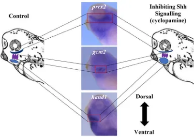
Figure14. Inhibiting Shh signalling resulted in a dorsal shift of hand1
expression within the developing pharyngeal region. ... 53-54
Figure 15. Inhibiting Shh signalling resulted in a dorsal shift in the
expression domain of the intermediate marker, gcm2 but not
pax1, within the developing pharyngeal region. ... 56-57
Figure 16. Pharmacological activation of Shh signalling caused a ventral
shift in the expression of the dorsal marker, pou3f3, in the
developing pharyngeal region. ... 59-61
Figure 17. Inhibiting Shh signalling resulted in the loss of expression of pax1
in the 5th pharyngeal arch. ... 63
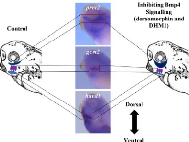
Figure 18. Inhibiting Bmp4 signalling resulted in a ventral shift of the hand1
expression domain. ... 65
Figure 19. Inhibiting Bmp4 signalling resulted in a ventral shift of the gcm2
and pax1 expression domain in the developing pharyngeal region. .. 67-68
Figure 20. Inhibiting Bmp4 signalling resulted in a ventral shift of the dorsal
marker, hoxa3. ... 70-71
Figure 21. T7E1 assay results demonstrating that CRISPR/Cas9 genome
xii
Figure 23. Mutations in hand1 resulted in no change of the dorsal marker, prrx2, expression domain within the developing pharyngeal
region ... 78
Supp. Fig. 1. Images demonstrating that inhibiting Shh signalling resulted in a
dorsal shift of hand1 expression within the pharyngeal region. ... 91
Supp. Fig. 2. Images showing inhibition of Shh signalling resulted in a dorsal
shift in the expression domain of the intermediate marker gcm2
within the developing pharyngeal region. ... 92
Supp. Fig. 3. Images demonstrating inhibiting Shh signalling resulted in a
dorsal shift of pou3f3 expression within the developing
pharyngeal region. ... 93
Supp. Fig. 4. Images demonstrating that inhibiting Bmp4 signalling resulted in
a ventral shift of hand1 expression within the pharyngeal region. .... 94
Supp. Fig. 5. Images showing that inhibiting Bmp4 signalling resulted in a
ventral shift of gcm2 expression within the pharyngeal region. ... 95
Supp. Fig. 6. Images demonstrating that inhibiting Bmp4 signalling resulted in
a ventral shift of pax1 expression within the pharyngeal region. ... 96
Supp. Fig. 7. Images demonstrating that inhibiting Bmp4 signalling resulted in
a ventral shift of hoxa3 expression within the pharyngeal region. ... 97
Supp. Fig. 8. Images of embryos that had hand1 mutated using CRISPR/Cas9 showed a dorsal shift in the expression domain of gcm2. ... 98
Supp. Fig. 9. Mutations in hand1 resulted in a trending dorsal shift of the hoxa3
xiii µg – Micrograms
µL – Microlitres
µM – Micromolar
bHLH – basic helix-loop-helix
Bmp4 – Bone morphogenetic factor 4
cDNA – complementary deoxyribonucleic acid
CRISPR/Cas9 – clustered regularly interspaced short palindromic repeats/
caspase 9
CY – cyclopamine
dH2O – distilled water
DIG – digoxigenin
DMH1 – dorsomorphin homolog 1
DMSO – dimethyl sulfide
DNA - deoxyribonucleic acid
RNA - ribonucleic acid
EtOH – ethanol
g - grams
gcm2 – Glial Cells Missing Homolog 2
h - hour
hand1 – Heart and Neural Crest Derivatives Expressed 1
hoxa3 – Homeobox A3
xiv mg - milligram
mL - millilitre
mM – millimolar
mn - minute
mRNA – messenger ribonucleic acid
ºC – Degrees Celsius
pax1 – Paired Box 1
PCR – polymerase chain reaction
PMA – purmorphamine
prrx2 – Paired Related Homeobox 2
RA – retinoic acid
rpm – revolutions per minute
sgDNA – single guide deoxyribonucleic acid
Shh – Sonic Hedgehog
T7E1 – T7 endonuclease 1
TGF-β - Transforming growth factor beta 1
UV – ultraviolet
Wnt - Wingless-related integration site
Chapter 1: Introduction
Craniofacial morphogenesis is a complex developmental process that requires the
precise orchestration of many molecular and morphogenetic events (Ataliotis et al., 2005;
Ferguson and Graham, 2004; Graham and Smith, 2001; Ho et al., 1994; Noden and
Trainor, 2005; Rinon et al., 2007). Structures such as the muscles of mastication, nerves
needed for facial expression and the thyroid and parathyroid glands are a few of the
important adult structures of the head and neck region that require proper early
embryonic development of the pharyngeal region (Frisdal and Trainor, 2014). Since
many adult structures and features of the head and neck region originate from the
developing pharyngeal region, this area has been the focus of many studies to understand
how patterning occurs and potential developmental origins of disorders and diseases
(Escriva et al., 2002; Jones and Trainor, 2004; Scambler, 2000; Stewart et al., 2013). The
purpose of this thesis is to advance the knowledge of how the pharyngeal region develops
and to determine the factors that are regulating the dorsoventral patterning of the
developing pharyngeal complex.
1.1 Xenopus laevisas a model for development
The South African clawed frog, Xenopus laevis, is a well-characterized model of development that has been used extensively over the past century to investigate many
aspects of early embryogenesis. Some of the numerous advantages it has for studying early
development include that many early developmental signalling pathways, morphological
processes, and genes are all conserved between X. laevis and mammalian development. These conserved characteristics make Xenopus an appropriate model of development when investigating developmental events and diseases that occur in humans (Dickinson, 2016;
Haremaki et al., 2015; Nie and Bronner, 2015).
One of the significant advantages of using X. laevis as a model of development is the ability of the embryos to tolerate extensive manipulation. Embryos can be exposed to
reagents, have essential tissue extirpated, or DNA, mRNA or proteins can be injected to
test specific hypotheses about the roles of signalling pathways or specific genes during
early embryonic development (Blum and Ott, 2018; Tandon et al., 2017; Wheeler and
occurs externally in very simple culture conditions. Xenopus embryos can be treated directly by adding the reagents to the media in which the embryos are developing. (Gordon
et al., 2010; Tabler et al., 2014).
Pertinent to this thesis is that the conserved biological characteristics of Xenopus also enables the use of CRISPR/Cas9 genome editing technology. The ability to rapidly inject
large numbers of embryos with CRISPR/Cas9 reagents allows one to generate large
numbers of embryos with mutations in specific genes that are required in conserved
developmental events (Bhattacharya et al., 2015; Tandon et al., 2017).
X. laevis also has a well-documented fate map of the early blastomeres allowing for physical or chemical manipulation of specific embryonic regions (Dale and Slack, 1987).
The ease of manipulation coupled with the well-defined fate map allows for the
investigation into the exact developmental stages when specific tissues or signalling
pathways are required. Xenopus embryos all develop at the same rate that is strictly dependent on temperature and this allows one to manipulate the rate of development by
simply changing temperature. Lastly, perhaps the most beneficial property of Xenopus as a model of development is that the females can produce hundreds of eggs which
synchronously develop once fertilized. This is advantageous to studies comparing
experimental and control embryos, often using whole-mount in situ hybridization to study the localization of RNA, because large cohorts of embryos can be easily obtained. This
allows for hundreds of observations and hence a large N-value when statistically analyzing
those images.
One of the few complexities of using X. laevis as a model system is that these frogs are tetraploid. Specifically, X. laevis is an allelotetraploid species because it is a result of a hybridization event between two parental species. Two diploid progenitors, one closely
related to Xenopus tropicalis and another ancient diploid Xenopus are the sources of the two diploid sets of chromosomes. Those two progenitor species had 20 chromosomes and
the reason that X. laevis has 38, rather than 40 chromosomes, is that chromosome 9 and 10 have fused (Matsuda et al., 2015; Session et al., 2016). The two sets of chromosomes are
referred to as the long chromosome and the short chromosome and individual genes have
hand1s) (Session et al., 2016). Therefore, genetic studies, specifically ones dealing with mutating genes, can be more complex. For example, X. laevis has four potential targets in the one cell embryo for a particular gene product if using CRISPR/Cas9 gene editing. In
order to circumvent this complexity, multiple guide RNAs for CRISPR/Cas9 studies may
be required to target both the long and short chromosome version of a particular gene in
order to ensure that all gene copies are mutated (Tandon et al., 2017).
As mentioned earlier the vast majority of cellular pathways and developmental events
are conserved between X. laevis and their mammalian counterparts. Two of the signalling pathways that are conserved are the Sonic hedgehog (Shh) and Bone morphogenetic
protein (Bmp) signalling pathways which regulate morphological and patterning events
throughout X. laevis development (Briscoe et al., 1999; Liem et al., 2000; Liem Jr. et al., 1997; Timmer et al., 2002). Not only are signalling pathways conserved but embryonic
structures are conserved as well. One such structure is the developing pharyngeal region.
In fact, segmentation and the creation of slits in the developing pharyngeal region is a
defining characteristic of all chordates. The pharyngeal region and its patterning are the
focus of this thesis.
1.2 Early embryonic development of the pharynx
The fully developed pharynx is located in the neck region and is situated between the
oral and nasal cavity and the esophagus and larynx. The pharynx is crucial to the survival
of all vertebrates given that the pharynx facilitates respiration by connecting the nasal and
oral cavity to the larynx, while, also connecting the oral cavity to the esophagus allowing
for digestive functions. One of the key reasons why the developing pharyngeal region is
the focus of many developmental studies is that this area later gives rise to bones, cartilages,
tissues, arteries, veins, and nerves of the head and neck region (Frisdal and Trainor, 2014).
Pharyngeal endoderm also gives rise to the thymus, parathyroid, thyroid, and
ultimobranchial bodies and disruptions in patterning can give rise to defects in these organs
(Graham and Smith, 2001). Developmental studies of the pharyngeal region are also
important because one in three congenital disorders affects the head and neck region and
Pierre Robin sequence and DiGeorge syndrome (Jones and Trainor, 2004; Scambler, 2000;
Stewart et al., 2013).
As mentioned earlier, the basic pharyngeal region structure is conserved across all
vertebrates (Square et al., 2015). This allows one to study the development of the pharynx
and facial malformations associated with human disorders in model organisms that can be
easily manipulated, either pharmacologically or genetically, including mice, zebrafish, and
X. laevis (Ataliotis et al., 2005; Stewart et al., 2013).
During early embryonic development, the developing pharyngeal region can be
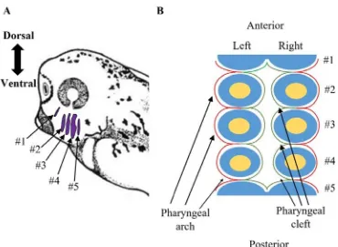
identified as the reiterated series of outgrowths called pharyngeal arches on the lateral side
of the embryo towards the ventral side of the developing head (Fig. 1A). The number of
pharyngeal arches is species-dependent and can range from four to nine. X. laevis
possesses seven pharyngeal arches which develop in sequential order. The first pharyngeal
arch first develops at stage 23 and by stage 35 (Appendix) the first five pharyngeal arches
can be identified on the lateral sides of the embryo (Fig. 1A). The development of the
pharyngeal region is complex since it requires interaction between all three germ layers
(endoderm, mesoderm and ectoderm) along with migrating neural crest cells (Ataliotis et
al., 2005; Ferguson and Graham, 2004; Graham and Smith, 2001; Ho et al., 1994; Noden
and Trainor, 2005; Rinon et al., 2007). The ectoderm surrounds the exterior of the
pharyngeal arches which is defined as the pharyngeal cleft and groove. The interior portion
of the pharyngeal arches, which is referred to as the pharyngeal pouch, is laminated with
the endoderm (Fig. 1B). The ectoderm and endoderm come in close proximity with one
another in the pharyngeal clefts, therefore, it is suspected that this close interaction between
the two layers may be required for the complete perforation and opening of the pharyngeal
gill slits in X. laevis embryos. This hypothesis is based on previous research showing that proximity of ectoderm and endoderm control the opening of the mouth in X. laevis embryos (Dickinson and Sive, 2006; Tabler et al., 2014). In the arch region between the clefts where
the ectoderm and endoderm are adjacent, the space found between the ectoderm and
endoderm is made up of mesoderm and neural crest cells that have delaminated from the
the development of the pharyngeal region, each of the pharyngeal arches will give rise to
specific skeletal, vascular, and muscle derivatives (Frisdal and Trainor, 2014).
In frogs the first pharyngeal arch will give rise to structures including but not limited to
gill primordium, maxilla, and mandible, while the second pharyngeal arch later develops
into the second aortic arch, upper part of the hyoid bone and stapes (Frisdal and Trainor,
2014). In the later stages of development the lower hyoid bone and common carotid artery
originate from the third pharyngeal arch, whereas the thyroid and thymus derive from the
fourth pharyngeal arch (Frisdal and Trainor, 2014). The fifth pharyngeal arches later gives
Figure 1. Diagrams displaying where the pharyngeal arches are located in a stage 35
X. laevis embryos and the organization of the germ layers and neural crest cells within
the pharyngeal region. (A) The pharyngeal arches (purple) of a stage 35 X. laevis
embryos can be identified by the reiterated series of outgrowths on lateral side of the head
region of the embryo. (B) A schematic through the transverse section of the pharyngeal
region of X. laevis. The exterior of the pharyngeal arches is covered by ectoderm (red), while, the interior of the pharyngeal arches is covered by a layer of endodermal cells
(green). Within the pharyngeal arches the ectoderm and endoderm cover a layer of neural
crests cells (blue) and at the centre of the group of neural crest cells is a mesodermal core
During development of the arches, there is a clear anteroposterior pattern to the
developing arches as evident by the different tissues formed from pharyngeal arches
(Escriva et al., 2002; Frisdal and Trainor, 2014). This anteroposterior pattern is in part,
regulated by retinoic acid (RA) signalling (Escriva et al., 2002). In Amphioxus, addition of exogenous RA results in an anterior shift in the expression of AmphiTR2/4 which is found within the second pharyngeal arch while blocking RA signalling by exposing
Amphioxus to BMS009, an RA inhibitor, was able to shift the expression of AmphiTR2/4
to the posterior region of the developing pharyngeal region (Escriva et al., 2002).
Furthermore, it has been established that the Hox genes are key elements of the anteroposterior pattern in the developing pharyngeal region (Hunt et al., 1991; Maconochie
et al., 1999). More specifically, different Hox genes are expressed in different neural crest cell subpopulations and the presence or absence of certain Hox genes will give neural crest cells positional identity. The identity of neural crest cells found in specific pouches along
the anteroposterior axis of the developing pharyngeal region are co-linear with the Hox
gene numbers in the region of the neural tube where the neural crest cells are derived (Hunt
et al., 1991a, 1991b; Maconochie et al., 1999; Trainor and Krumlauf, 2000; Tümpel et al.,
2002, 2008). If the Hox genes are misregulated, such as by silencing Hoxa3, skeletal elements, normally found in the first pharyngeal arch, shift into the second pharyngeal arch
and structures such as Meckel’s cartilage, incus, and malleus which are normally found in
the second pharyngeal arch are shifted into the first pharyngeal arch region
(Gendron-Maguire et al., 1993; Kontges and Lumsden, 1996; Rijli et al., 1993). This anteroposterior
patterning has been extensively studied due to the role of individual arches in forming
specific structures such as a the thyroid, thymus and bones of the face (Frisdal and Trainor,
2014; Minoux and Rijli, 2010).
Not only does the developing pharyngeal region display anteroposterior patterning but
it also displays patterning along the dorsoventral axis (Square et al., 2015). In order to
understand the evolution of skeletal components derived from the arches during
development, Square et al. (2015) demonstrated that several specific genes are
differentially expressed along the dorsoventral axis of the developing pharyngeal region in
In both studies, expression of genes such as hand2, dlx2/4 and pou3f3 were restricted to the ventral, intermediate or dorsal regions of the developing pharyngeal region (Jeong et
al., 2008; Talbot et al., 2010). Also dlx genes were demonstrated to play important roles in patterning the developing pharyngeal region and in regulating proper formation of
skeletal structures arising from this area (Jeong et al., 2008; Talbot et al., 2010). Although
these studies have shown that the developing pharyngeal region displays dorsoventral
patterning, to my knowledge the signalling molecules that are responsible for establishing
that pattern remain unknown (Jeong et al., 2008; Square et al., 2015; Talbot et al., 2010).
Perhaps the best understood example of dorsoventral patterning of a structure in the
early embryo is in the neural tube where opposing gradients of Shh and Bmp4 signalling
are necessary to establish a pattern of neuronal identities along that axis (Briscoe et al.,
1999; Liem et al., 2000; Liem Jr. et al., 1997; Timmer et al., 2002). Expression profiles of
Shh and Bmp4 around the pharyngeal region suggest that they may be available to help
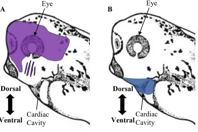
generate the dorsoventral pattern in the developing pharyngeal region (Fig. 2). If so, it
should be noted that the gradients would be inverted as compared to the neural tube (Fig.
3) with Shh found on the dorsal side and Bmp4 found on the ventral side of the developing
Figure 2. Diagrams depicting the localization of Shh and Bmp4 mRNA in the head
and neck region of stage 35 X. laevis embryos. (A) Shh expression (purple) is observed anterior to the pharyngeal region including and up to the first pharyngeal arch, and dorsal
to the five pharyngeal arches. (B) Bmp4 expression (blue) is observed ventral to the pharyngeal region. Data is based on previous expression studies (Koide et al., 2006;
Rankin et al., 2012).
Eye Eye
Cardiac Cavity
1.3 Axial Patterning in the Embryo
The anteroposterior, dorsoventral, and left-right axes are three axes along which an
embryo develops. X. laevis has been extensively used to study how these three axes are generated and the possible consequences of their malformation due to aberrant molecular
signalling or incorrect cellular divisions (Altaba and Melton, 1989; Campione et al., 1999;
Suzuki et al., 1994). In particular, the molecular mechanisms that establish the
dorsoventral axis of the embryo is particularly well understood in Xenopus. A microtubule-based mechanism initiated by entry of the sperm centriole drives beta-catenin to the dorsal
side of the just fertilized embryo causing a canonical wingless (wnt) signal that defines that
side of the embryo as being dorsal (Weaver and Kimelman, 2004). Later in the developing
Xenopus embryo the neural plate forms from ectoderm on the dorsal side of the embryo. That plate then rolls to form the neural tube and the subsequent tube then establishes its
own dorsoventral pattern (Christen and Slack, 1997; McMahon et al., 1998; Yost, 1990).
A neural tube is found in all vertebrate embryos and is the precursor to the brain and
spinal cord, thereby making correct patterning of the neural tube crucial to the embryo’s
survival. One characteristic of dorsoventral patterning in the neural tube is that specific
subtypes of neurons differentiate according to their position in the dorsoventral axis and
this is crucial for proper motor, sensory and interneuron neuron development (Barth et al.,
1999; Basler et al., 1993; Ericson et al., 1996; McMahon et al., 1998; Nguyen et al., 2000;
Sander et al., 2000; Vallstedt et al., 2001; Yamada et al., 1993).
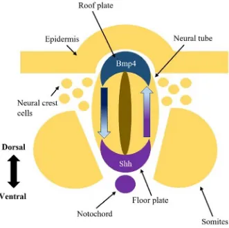
A key element in establishing the dorsoventral axis in the neural tube is secretion of the
Shh ligand (Ericson et al., 1996; Roelink et al., 1995). The secretion of the Shh ligand
from the notochord initiates the secretion of Shh in the floor plate. The further from this
ventral source of the ligand within the neural tube, the lower the concentration of the Shh
ligand resulting in the formation of a Shh gradient (Ericson et al., 1996; Roelink et al.,
1995). Concurrently, Bmp4 is secreted from the epidermis dorsal to the neural tube,
prompting the cells found in the roof plate of the neural tube to also secrete Bmp4 (Liem
Jr. et al., 1995, 1997). The Bmp4 concentration is high at the dorsal pole of the neural tube
with levels of Bmp4 decreasing ventrally along the dorsoventral axis (Liem Jr. et al., 1995,
(Briscoe et al., 1999; Liem et al., 2000; Liem Jr. et al., 1997; Timmer et al., 2002). The
levels of signalling ligand that the cells in the neural tube receive provides cells with
positional information along the dorsoventral axis so that they can express and/or repress
specific transcription factors (Nguyen et al., 2000; Sander et al., 2000). A combinatorial
code based on the expression of these transcription factors specifies the different subtypes
of neurons according to their position along the dorsoventral axis. For example, sensory
neurons differentiate near the dorsal side of the neural tube and motor neurons are clustered
near the ventral pole of the neural tube (Briscoe et al., 1999; Ericson et al., 1995; Liem et
al., 2000; Liem Jr. et al., 1995, 1997; Roelink et al., 1995; Timmer et al., 2002).
The dorsoventral patterning of the neural tube is just one case of many processes
where proper expression of Shh and Bmp4 are required for correct embryonic
development. Another such developmental event that requires these signalling pathways
is during the development of the kidneys (Nishinakamura and Sakaguchi, 2014; Yu et al.,
2002). More specifically Shh signalling is required to promote mesenchymal cell
proliferation, and regulates the time point at which differentiation of smooth muscle
progenitor cells occurs in the ureteral mesenchyme (Yu et al., 2002). Mice in which Shh
was mutated displayed phenotypes of renal hypoplasia, hydronephrosis and hydroureter
demonstrating the necessity of expressing Shh at the correct time points and locations
during the development of the kidney (Yu et al., 2002). Whereas Bmp4 is required to
prevent ureteric bud attraction and combined with the Bmp antagonist, Gremlin, are
required to initiate ureteric budding (Nishinakamura and Sakaguchi, 2014). Mice in
which Bmp4 is mutated exhibit characteristics similar to human congenital anomalies of
the kidney and the urinary tract including hypoplastic kidneys and hydroureter
(Nishinakamura and Sakaguchi, 2014). A third region of the developing embryo where
these signalling ligands displayed a similar expression profiles to the neural tube is
around the developing pharyngeal region (Fig. 2) (Barnett et al., 2012; Rankin et al.,
2012). The location of Shh and Bmp4 expression within the developing pharyngeal
region is the inverse of their positioning within the neural tube with respect to the
dorsoventral axis. Shh is expressed dorsal to the developing pharyngeal region and
Bmp4 is expressed ventral to the developing pharyngeal region (Fig. 2) (Barnett et al.,
Figure 3. Diagram demonstrating the expression localization of Shh and Bmp4 in the
neural tube. In the neural tube, the initial secretion of the Shh ligand (purple) originates
from the notochord at the ventral side of the neural tube and causes cells located in the
floor plate to also secrete the Shh ligand. Along the dorsoventral axis, there is a gradient
of Shh protein from a high concentration found around the ventral pole and decreasing
levels moving towards the dorsal side of the neural tube. A similar pattern is observed with
the Bmp4 (blue) protein levels along the dorsoventral axis. A higher concentration of
Bmp4 ligand is observed at the dorsal pole and decreases moving towards the ventral pole.
Together these signalling molecules create opposing gradients which pattern the neural
tube along the dorsoventral axis.
1.4 Sonic hedgehog (Shh) signalling
Sonic hedgehog (Shh) is one of the three Hedgehog signalling proteins (the other two
are Indian and Desert hedgehog) and has been implicated in many biological processes
throughout an organism’s lifespan including events in early development, tissue
regeneration, stem cell renewal and cancer (Bailey et al., 2008; Ericson et al., 1995;
Machold et al., 2003). In addition to its role in patterning the neural tube, Shh is required
for many critical patterning events in invertebrates and vertebrates (Ericson et al., 1995;
Laufer et al., 1994; Rankin et al., 2016). One such place that Shh plays a critical role is
during the development of the limb (Laufer et al., 1994; Tickle and Towers, 2017). Here
Shh not only provides positional information for cells along the anteroposterior axis (thumb
to little finger) but also stimulates mesenchymal cell proliferation to control the width of
the limb and regulates the anteroposterior length of the apical ectodermal ridge which is
important for developing the correct structures along the proximo-distal axis of the
developing limb (Laufer et al., 1994; Tickle and Towers, 2017). With respect to Xenopus,
Shh is expressed in defined locations during specific stages of development such as ventral
to the neural tube, in the floor plate, in the limb bud, and dorsal to the pharyngeal region
(Ericson et al., 1995; Koide et al., 2006; Laufer et al., 1994).
The Shh ligand is a protein that requires multiple modifications for the ligand to become
a functional signalling protein. The Shh protein precursor is initially proteolytically
cleaved into an amino terminus (Shh-N) and a carboxy terminus (Shh-C) peptides
(Choudhry et al., 2014). Shh-N has been demonstrated to be the peptide which is crucial
for Shh signalling (Choudhry et al., 2014). Following cleavage, auto-proteolysis occurs
which then allows for a cholesterol moiety to be added to the C-terminus of Shh-N peptide
(Choudhry et al., 2014). Next, Skinny hedgehog acyltransferase attaches a palmitoyl group
on the N-terminal of Shh which is vital for regulating the secretion of Shh and the ability
for Shh to signal at longer-ranges (Chamoun et al., 2001; Lewis et al., 2001; Liu et al.,
2016; Zeng et al., 2001). Once it has undergone these post-translational modifications it
can be used in intercellular signalling.
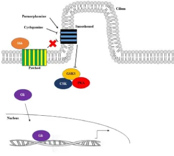
When there is an absence of the fully post-translationally modified Shh ligand, the
inactive form by the transmembrane receptor protein, Patched, and regulating the activity
of the downstream transcription factors Gli1, Gli2, and Gli3. With no ligand present, Gli1
will be phosphorylated, targeting it for degradation, and Gli2 and Gli3 will be proteolyzed
to produce their repressor forms by the glycogen synthase kinase (GSK3), tyrosine-protein
kinase (CSK) and protein kinase A (PKA) complex preventing upregulation of Shh target
genes.
However, when the fully post-translationally modified Shh ligand is present and bound
to Patched, Smoothened is no longer negatively regulated by Patched. This allows
Smoothened to locate to the cilium thereby allowing Gli1 and Gli2 to be processed into
their activator conformations. Therefore, the Gli1 and Gli2, in their transcriptional
activator forms, are then able to translocate to the nucleus where they up regulate
Gli-responsive target genes by outcompeting the Gli3 repressor (Fig. 4).
Two of the most effective small molecular reagents that are used to either
pharmacologically activate or inhibit the Shh signalling pathway are purmorphamine and
cyclopamine, respectively (Chen et al., 2002; Sinha and Chen, 2006). Purmorphamine, the
Shh signalling activator, has been shown to activate Shh signalling by directly targeting
the heptahelical bundle of Smoothened causing a conformational change that results in
Smoothened remaining in its active form even in the absence of the Shh ligand (Sinha and
Chen, 2006). Consequently, Smoothened retains its active conformation and Gli3
repressor is marked for degradation while, Gli1 and Gli2 are no longer marked for
degradation allowing them to translocate to the nucleus where they upregulate Shh target
genes leading to the pathway being constitutively active.
Cyclopamine is a well-established Shh signalling inhibitor (Chen et al., 2002).
Cyclopamine inhibits the Shh signalling pathway by directly binding to the heptahelical
bundle of the co-receptor, Smoothened, consequentially causing a conformational change
of Smoothened (Chen et al., 2002). This conformational change results in Smoothened
remaining in its inactive form even when the Shh ligand is bound to Patched (Chen et al.,
2002). Since Smoothened is restricted to its inactive conformation, the Gli1 activators are
phosphorylated, priming them for degradation, while, Gli2 and Gli3 transcription factors
where the transcription factors repress Shh target genes leading to the inhibition of the Shh
Figure 4. The Shh signalling pathway. The Shh signalling pathway is initiated when
the Shh ligand binds to the transmembrane receptor, Patched. As a result of this
interaction, Patched which is localized near the base of the cilium exits and Smoothened
now migrates into the cilium. Since Smoothened is now present in the cilium,
Smoothened’s activity is no longer inhibited and it can now prevent the Gli proteins from
being degraded or converted into repressors by the GSK3, CSK and PKA complex.
Therefore, the Gli proteins can act as activators and then translocate to the nucleus where
they upregulate Gli-responsive target genes. Purmorphamine is a small molecule
activator of the Shh signalling pathway which results in the constrictive upregulation of
Shh target genes. Conversely, cyclopamine is used as a small molecule inhibitor of the
Shh signalling pathway and when administered it causes inhibition of the Shh signalling
1.5 Bmp4 signalling
Bone morphogenetic proteins (Bmps) are a group of signalling molecules that are a part
of the Transforming Growth Factor-β (TGF- β) superfamily of secreted proteins. Bone
morphogenetic protein 4 (Bmp4) is highly conserved among vertebrates and plays crucial
roles in embryogenesis and maintenance of adult tissue homeostasis (Bei and Maas, 1998;
Qian et al., 2013; Timmer et al., 2002). Bmp4 is particularly well known for its role as the
dorsal signal during the dorsoventral patterning of the neural tube in vertebrates (Timmer
et al., 2002). Bmp4 also regulates dorsoventral patterning of somite derivatives and
anteroposterior patterning of the limbs (Beck et al., 2001; Drossopoulou et al., 2000;
Schmidt et al., 1995).
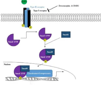
Bmp4 signalling is first initiated when the Bmp4 ligand binds to the Bmp type I
receptors ALK2, ALK3, and ALK6 and type II serine/threonine kinase heterodimeric
receptors causing the type II receptor to transphosphorylate the type I receptor. This in
turn leads to the phosphorylation of the receptor-regulated Smad 1/5/8 intracellular proteins
which then form a complex with Smad 4 protein, a common-mediator. The Smad 1/5/8 –
Smad 4 complex then translocates to the nucleus where the complex upregulates Bmp4
target genes (Fig. 5).
Several small molecular reagents have been identified that can broadly and selectively
inhibit the Bmp pathways. One common broad inhibitor of the Bmp pathways is
dorsomorphin although it is also known to inhibit 5' AMP-activated protein kinase
signalling (Gao et al., 2008; Yu et al., 2008). A more commonly used specific inhibitor of
the Bmp4 pathway is Dorsomorphin Homolog 1 (DMH1) (Hao et al., 2010, 2014).
Dorsomorphin blocks the Bmp signalling pathway by preventing phosphorylation of the
BMP type I receptors by the Bmp type II receptor while DHM1 inhibits the Bmp4
signalling pathway by binding to the intracellular kinase domain of the Bmp type I receptor
(Hao et al., 2010, 2014; Yu et al., 2008). Therefore, following treatment with
Dorsomorphin or DMH1, Smad 1/5/8 proteins are no longer able to be phosphorylated and
thus cannot form a complex with Smad 4 protein in order to translocate to the nucleus to
Figure 5. Bmp4 signalling pathway. The Bmp4 signalling pathway is initiated when a
Bmp4 ligand binds to the Bmp type I and II heterodimeric receptors resulting in the
phosphorylation of the Bmp type I receptor. This phosphorylation event leads to the
phosphorylation of Smad 1/5/8 which then is able to form a complex with a Smad 4 protein.
This Smad complex then translocates to the nucleus where it upregulates Bmp4 target
genes. Two small molecular reagents that are used to inhibit the Bmp signalling pathway
1.6 Regional dorsoventral pattern within the developing pharyngeal complex
In order to understand dorsoventral patterning, I have examined the expression of genes
that have regional expression within the developing pharyngeal complex. I have roughly
divided the expression domains into three regions based on their expression along the
dorsoventral axis: ventral, intermediate and dorsal expression domains. The expression
domains are visualized using whole-mount in situ hybridization. All of the genes used as regional markers encode transcription factors that are critical in the development of
structures originating from the pharyngeal region such as the thyroid, bones of the ear, and
cartilage of the head and pharynx (Berge et al., 1998; Firulli et al., 2014; Günther et al.,
2000; Jeong et al., 2008; Liu et al., 2007; Manley and Capecchi, 1998; Su et al., 2001).
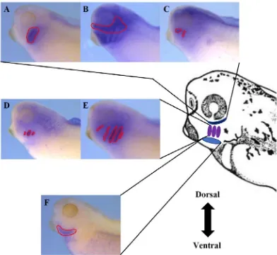
1.6.1 Ventral pharyngeal region marker
The transcription factor whose expression profile was chosen as the ventral developing
pharyngeal marker was hand1. Expression of hand1 is initiated following gastrulation at the end of stage 12 (Session et al., 2016). Following stage 12, hand1 is expressed in the cardiac progenitor cells, lateral plate mesoderm, and the developing pharyngeal region
(Angelo et al., 2000; Deimling and Drysdale, 2009, 2011). Specifically, with respect to
the pharyngeal region at stage 35, hand1 is expressed in the ventral region of the developing pharyngeal complex between the posterior of the cement gland and the posterior of the
developing pharyngeal complex (Fig. 6F). Along the dorsoventral axis hand1 expression begins at the most ventral edge of the developing pharyngeal complex and extends roughly
1.4 µm ventrally. The expression profile of hand1 has also been used to investigate whether retinoic acid (RA) signalling regulates the anteroposterior patterning of the lateral
plate mesoderm (Deimling and Drysdale, 2009). More specifically, hand1 expression was used as a marker of the anterior-middle domain of the lateral plate mesoderm (Collop et
al., 2006; Deimling and Drysdale, 2009).
Hand1 is not only important as a marker of the developing ventral pharyngeal region but also could be a possible downstream regulator of the dorsoventral patterning of the
placentation and the linear heart tube fails to loop in the correct direction resulting in either
a linear heart tube at the midline or slight looping to the right then left (Riley et al., 1998).
If the placentation defects are rescued, the embryos still die from cardiovascular defects
(Riley et al., 1998). Not only is hand1 a vital transcription factor in the initial morphogenesis of the heart, but it also plays an essential role in ventricular myocyte
differentiation and expression of a subset of cardiac genes (Smart et al., 2002).
Hand1 also plays a role in the development of other lineages including the lung and trachea along the anteroposterior axis of the developing embryo (Fernandez‐Teran et al.,
2003; Hoyos et al., 2016; Rankin et al., 2016). Here hand1 expression is restricted to the heart, pharyngeal mesenchyme, and the posterior lateral plate mesoderm, but lacking
expression in the foregut lateral plate mesoderm region (Rankin et al., 2016). This hand1
expression which outlines the presumptive lung field is crucial for later development of the
lung demonstrating one of many key roles the hand1 transcription factor plays during embryonic development (Rankin et al., 2016).
The hand1 gene is of particular interest to this study because hand1 is expressed in the developing ventral pharyngeal region and has been demonstrated to be necessary for proper
morphogenesis (Firulli et al., 2014). I found that not only is hand1 expressionregulated by Shh and Bmp4 signalling during the dorsoventral patterning of the developing
pharyngeal region, but I hypothesize that hand1 is itself is a regulator of the pattern.
1.6.2 Intermediate pharyngeal region markers
The expression domain of the transcription factors gcm2 and pax1 were chosen as markers of the intermediate developing pharyngeal region. This is the region where the
gill slits will eventually open to the pharyngeal cavity. Gcm2 is a master regulator of the parathyroid which is derived from endoderm of the pharyngeal region (Correa et al., 2002;
Kebebew et al., 2004). Gcm2 has been demonstrated to be necessary for proper parathyroid gland development, expression of the parathyroid hormone, and proper expression prevents
conditions like hyperparathyroidism (Correa et al., 2002; Günther et al., 2000; Liu et al.,
2007). Detection of gcm2 mRNAbegins in the oocyte and is observed until stage 12 at which point the expression becomes barely detectable until stages 29-30 when its
of the second, third, and fourth pharyngeal arches of the developing pharyngeal region at
stage 35 (Lee et al., 2013) (Fig. 6D).
The second RNA chosen as a marker for the intermediate region of the developing
pharyngeal complex was pax1. The pax1 gene encodes a transcription factor that plays important roles during embryonic development. With respect to the developing pharyngeal
complex, it is crucial for the proper development of the parathyroid glands and for complete
separation of the pharyngeal pouch (Su et al., 2001). During X. laevis development pax1
is expressed between stages 22 and 38 where it is constrained to the pharyngeal and the
perinotochordal regions of the embryo (Gray et al., 2009; Sánchez and Sánchez, 2013,
2015). Within the developing pharyngeal region at stage 35, pax1 expression is restricted to the first five pharyngeal arches (Fig. 6E).
1.6.3 Dorsal pharyngeal region markers
The expression domains that were used to mark the developing dorsal pharyngeal region
were those of hoxa3, and prrx2, and pou3f3. The hoxa3 gene encodes a transcription factor and is part of the A cluster of homeobox genes on chromosome 7 which are known to be
regulators of patterning during embryonic development (Chojnowski et al., 2016; Manley
and Capecchi, 1998). Proper expression of hoxa3 is crucial for many craniofacial derivatives of the developing pharyngeal region such as cranial nerves, throat cartilage,
thyroid and the parathyroid glands (Chojnowski et al., 2016; Manley and Capecchi, 1998).
The expression of hoxa3 is detectable from the end of gastrulation at stage 12 and continues beyond stage 40 (Session et al., 2016). Expression of hoxa3 is observed in several regions including the hindbrain, spinal cord, and developing pharyngeal region (McNulty et al.,
2005; Square et al., 2015). At stage 35 when the developing pharyngeal region can be
observed on either side of the head region of the embryo, the hoxa3 expression is restricted to the pharyngeal tissue which surrounds the third and fourth pharyngeal arches (Fig. 6A).
The transcription factor, prrx2, is crucial for proper development of select facial bones and if misexpressed, can lead to severe craniofacial malformations (Berge et al., 1998).
Within the developing pharyngeal region its expression is necessary for the proper
development of the mandibular processes, dentaries and the nasal cavity (Balic et al., 2009;
is low and prrx2 expression does not become prominent until stage 20 and declines to low levels again by stage 40 (Session et al., 2016). Throughout, these developmental stages
prrx2 expression can be detected in the head region, mouth primordium and the developing pharyngeal region (Square et al., 2015). With respect to the developing pharyngeal region
and this study at stage 35, prrx2 expression is found just posterior to the cement gland extending to the posterior of the developing pharyngeal region. Though the prrx2
expression can be observed in the ventral and dorsal regions of the developing pharyngeal
complex, no expression is present in the intermediate region (Fig. 6B). The dorsal
expression domain of prrx2 is the focus of this thesis when comparing the expression domain between control embryosand embryos in which signaling was altered or hand1
expression knocked-down.
The last gene whose expression is used in this thesis as a marker for the dorsal region
of the developing pharyngeal complex was pou3f3.The transcription factor, pou3f3, is a member of the POU domain family of genes which have been implicated in many
development processes, however, pou3f3 expression has been specifically demonstrated to be crucial for proper development of pharyngeal derivatives such as squamosal bone, jugal
bone, and if it is lost, there is failure of the stapes to detach from the styloid process
(Cosse-Etchepare et al., 2018; Jeong et al., 2008; Ryan and Rosenfeld, 1997). In Xenopus, its mRNA is first detected at stage 8. After stage 12, pou3f3 is once again expressed until stage 40 where the expression returns to levels similar to those at stage 8 (Session et al.,
2016). Throughout stages 12 to 40 pou3f3 expression is mainly localized to the anterior region of the embryo including the anterior neural fold, fore- and hindbrain, and the
developing pharyngeal region (Cosse-Etchepare et al., 2018; Square et al., 2015).
Figure 6. Diagram displaying the expression domains of the markers of ventral,
intermediate and dorsal regions of the developing pharyngeal complex. The mRNA
expression profiles used as markers for the dorsal region of the developing pharyngeal
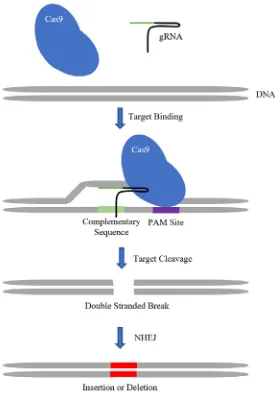
1.7 CRISPR/Cas9 Genome Editing
Over the past ten years one approach to modifying the genome that has gained
tremendous popularity is CRISPR/Cas9 genome editing technology. Instead of using
morpholinos to knock-down gene expression, many laboratories are now using
CRISPR/Cas9 to cause mutations in genes which in turn result in the knocking-down of
their expression (Tandon et al., 2017). The two key molecules that cause the mutations in
the gene of interest are the Cas9 enzyme, and a portion of RNA called the guide RNA
(gRNA). The enzyme and gRNA work together as a complex inside the cell’s nucleus
where the complex searches along the DNA for small sequences called protospacer
adjacent motifs (PAM) sites (Pickar-Oliver and Gersbach, 2019). These PAM sites allow
the Cas-9 enzyme to grip the DNA resulting in its destabilization leading to the unzipping
of the double-stranded helix (Pickar-Oliver and Gersbach, 2019). Following, the
opening of the DNA, the gRNA moves along the DNA searching for the complementary
sequence (Pickar-Oliver and Gersbach, 2019). Once the complementary sequence is
found the gRNA activates the Cas-9 enzyme which in turn cleaves the DNA into two
separate pieces (Pickar-Oliver and Gersbach, 2019). The double-stranded break is
repaired by non-homologous end-joining which is an error-prone repair mechanism that
introduces insertions or deletions at the site of the break (Pickar-Oliver and Gersbach,
2019). One of the possible outcomes of the insertions or deletions is a frameshift
mutation leading to a premature stop codon resulting in a non-functional gene
(Pickar-Oliver and Gersbach, 2019).
Using CRISPR-Cas9 genome editing technology in the model system, X. laevis offers a few more challenges than to researchers working with other models of development
such as mice or human cells. This is because X. laevis are tetraploid meaning that they carry two complete genomes. The two sets can be differentiated based on small
differences in chromosome size and so one set has been designated long and the other
short (Session et al., 2016). Although there has been some evolutionary loss of one copy
of many genes, for the majority of the genes there are essentially four functional copies
(Session et al., 2016). Therefore, when attempting to knock-down genes in X. laevis
(Yang et al., 2013). Nevertheless, CRISPR/Cas9 technology is able to efficiently mutate
both sets of genes without any clear toxic effects (Bhattacharya et al., 2015; Blitz et al.,
2013; DeLay et al., 2018). CRISPR/Cas9 genome editing will be the method used to
Figure 7. Diagram depicting how CRISPR/Cas9 genome editing technology
introduces an insertion or deletion into the gene of interest. The enzyme and gRNA
work together as a complex searching along the DNA for small sequences called
protospacer adjacent motifs (PAM) sites. These PAM sites allow the Cas9 enzyme to
grip the DNA resulting in its destabilization leading to the unzipping of the
double-stranded helix. The gRNA then searches for the complementary sequence. Once the
complementary sequence is found the gRNA activates the Cas9 enzyme which in turn
creates a double stranded break. The break is repaired by non-homologous end-joining
which is an error-prone repair mechanism that introduces an insertion or deletion at the
site of the break. The insertion or deletions may cause a coding frameshift leading to the
1.8 Purpose of the research
The purpose of this study is to discover underlying signalling mechanisms that regulate
the dorsoventral patterning of the developing pharyngeal region in X. laevis. I utilized information from previous studies that have demonstrated clear patterns of gene expression
along the dorsoventral axis in the developing pharyngeal region. Based on the observation
that Shh is located dorsal to the developing pharyngeal region, while Bmp4 is located
ventral to the developing pharyngeal region, there is the potential for a counter gradient
role for these molecules in the dorsoventral patterning of the pharyngeal complex, similar
to the known patterning described for the neural tube (Barnett et al., 2012; Le Dréau and
Martí, 2012; Rankin et al., 2012; Square et al., 2015). Therefore, I hypothesize that the correct dorsoventral gene expression pattern observed in the developing pharynx is regulated by opposing gradients of Shh and Bmp4 signalling. This thesis will investigate whether Shh and Bmp4 provide positional information to cells found along the
dorsoventral axis so that the cells can activate and/or repress specific transcription factors
known to be involved in craniofacial development. Small molecule reagents were used to
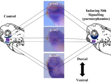
alter the signalling pathways in embryos prior to the pharyngeal patterning. I predict that
activating Shh signalling by exposing the embryos to purmorphamine will result in a
ventral shift of the mRNA expression profiles (Fig. 8). In contrast, I predict that exposing
embryos to cyclopamine, thereby inhibiting Shh signalling, will cause a dorsal shift in the
mRNA expression domains in the three regions of the developing pharyngeal complex
(Fig. 9). Similarly, in purmorphamine treated embryos, I predict a ventral shift of the
mRNA expression domains in the three regions of the developing pharyngeal complex
when exposed to the Bmp inhibitors dorsomorphin and DMH1 (Fig. 10). Finally, a second
hypothesis is that hand1 regulates the dorsoventral patterning of the developing pharyngeal region. This is due to the ventrally restricted localization of hand1 mRNA in the developing pharyngeal region and Shh signaling and the hand genes have been demonstrated to cooperatively regulate embryonic morphogenetic events (Firulli et al.,
2017; Rankin et al., 2016; Riley et al., 1998). I predict that when the hand1 gene is mutated, the mRNA expression profiles in the intermediate and dorsal regions will be shifted
pharyngeal region is patterned and could be later used to predict the origin of defects in
Figure 8. Diagram depicting the prediction of the change in the mRNA localization
when inducing Shh signalling by exposing the X. laevis embryos to purmorphamine.
On the left side of the diagram is a X. laevis head and neck region with the light blue, purple and dark blue coloured areas marking the ventral, intermediate and dorsal regions of the
developing pharyngeal complex, respectively. The three centre images are X. laevis
embryos stained for hand1, gcm2, and prrx2 which are markers for the ventral, intermediate and dorsal regions, respectively. On the right side is a diagram predicting the
ventral shift of the markers of the three regions when inducing Shh signalling through the
Figure 9. Diagram depicting the predictions of the change in the mRNA localization
following cyclopamine inhibition of Shh. On the left side of the diagram is a X. laevis
head and neck region with the light blue, purple and dark blue coloured areas marking the
ventral, intermediate and dorsal regions of the developing pharyngeal complex,
respectively. The three centre images are X. laevis embryos stained for hand1, gcm2, and
prrx2 which are markers for the ventral, intermediate and dorsal regions, respectively. On the right side is a diagram predicting the dorsal shift of the markers of the three regions
Figure 10. Diagram depicting the predictions of the change in the mRNA
localization when inhibiting Bmp4 signalling by exposing the X. laevis embryos to
dorsomorphin and DMH1. On the left side of the diagram is a X. laevis head and neck region with the light blue, purple and dark blue coloured areas marking the ventral,
intermediate and dorsal regions of the developing pharyngeal complex, respectively. The
three centre images are X. laevis embryos stained for hand1, gcm2, and prrx2 which are markers for the ventral, intermediate and dorsal regions, respectively. On the right side is
a diagram predicting the ventral shift of the markers of the three regions when inhibiting
Figure 11. Diagram depicting the predictions of the change in the mRNA
localization after hand1 mutation by CRISPR/Cas9 technology. On the left side of
the diagram is a X. laevis head and neck region with the purple, and dark blue coloured areas marking the intermediate, and dorsal regions of the developing pharyngeal
complex, respectively. The two centre images are X. laevis embryos stained for gcm2, and prrx2 which are markers for the intermediate, and dorsal regions, respectively. On the right side is a diagram predicting the dorsal shift of the markers of the intermediate
Chapter 2: Materials and Methods
2.1 Generation of X. laevis embryos
In order to induce ovulation, female X. laevis were injected with 800 IU of human chorionic gonadotrophin (Intravet) the night before eggs were to be fertilized. Male X. laevis were sacrificed, and testes were removed and stored in 200% Steinberg’s solution. All Steinberg’s solutions were prepared from Steinberg’s stock solution A (68g NaCl, 1g
KCl, 4.09g MgSO4-7H2O, 1.58g Ca(NO3)2-4H2O) and Steinberg’s stock solution B (11.2g
Tris-HCl pH 7.4). Eggs were squeezed from the females into 80% Steinberg’s solution
and in vitro fertilization was accomplished using minced testes. Embryos were de-jellied at stage 4 with 2.5% cysteine (pH 8.0) and washed several times with 20% Steinberg’s
solution to remove excess 2.5% cysteine. Embryos were staged according to the
Nieuwkoop and Faber staging (Appendix) (P.D. Nieuwkoop and J. Faber, 1994). All
embryos were cultured in 3 mL of 20% Steinberg’s solution at 18ºC.
2.2 Shh activator and inhibitor
Cyclopamine and purmorphamine (Toronto Research Chemicals) were prepared as 20
mM and 10 mM stock solutions in 95% ethanol and DMSO, respectively, and stored at
-20ºC. Based on research by Lewis and Krieg (2014) that determined effective
concentrations for Xenopus embryos, I treated stage 13 embryos with 100 μM
cyclopamine or 20 μM purmorphamine. Control embryos were treated with either 15 μL
of 95% ethanol or 3 μL of DMSO in 3 mL of 20% Steinberg’s solution. Embryos were
continuously exposed to cyclopamine or purmorphamine from stage 13 until stage 35
when they were fixed for whole-mount in situ hybridization. The embryos were treated starting at stage 13 (Appendix) to avoid interfering with gastrulation and were fixed at
stage 35 (Appendix) since this is the optimal stage to view the developing pharyngeal
region.
2.3 Bmp4 inhibitors
In order to inhibit Bmp signalling, embryos were treated with 40 μM DMH1 (Adooq
Bioscience) (Rankin et al., 2015) or 10 μM dorsomorphin (Fisher Scientific) (Pieper et