MreB cytoskeleton dynamics and its
role in cell wall synthesis in
Bacillus subtilis
Julia Domínguez Escobar
der Fakultät für Biologie
der Ludwig-Maximilians-Universität München
Characterization of the actin-like MreB
cytoskeleton dynamics and its role in cell wall
synthesis in Bacillus subtilis
vorgelegt von
Julia Domínguez-Escobar
aus Mexiko
Erstgutachter: Prof. Dr. Thorsten Mascher Zweitgutachter: Prof. Dr. Marc Bramkamp
1
The peptidoglycan cell wall (CW) and the actin-like MreB cytoskeleton are the major determinants of cell morphology in non-spherical bacteria. Bacillus subtilis is a rod-shaped Gram-positive bacterium that has three MreB isoforms: MreB, Mbl (MreB–like) and MreBH
(MreB-Homologue). Over the last decade, all three proteins were reported to localize in dynamic
2
that it is still associated to the CW elongation machinery but might be defective in an interaction important for MreB morphogenetic function. Thus, this mutant appears as as a good candidate to start characterizing the interactions between the three MreB isoforms and components involved in CW elongation. It might also help to understand the function of components of theCW-synthetic complexes, and how they are coordinated to achieve efficient CW synthesis. Finally, to investigate how the integrity of the CW is maintained, we studied the localization and dynamics of the LiaIH-system, which is the target of LiaRS, a two-component system involved in cell envelope stress response. We found that under stress conditions, when liaI and LiaH genes are expressed, the proteins form static complexes that coat the cell membrane. LiaI is required for the even distribution of the LiaH in the membrane. Taken together, these data suggest that LiaIH complexes may protect the cell from CW damage.
3
1. Dominguez-Escobar, J., Arnaud Chastanet, A., Crevenna, A. H., Vincent Fromion, V.,
Wedlich-Söldner, R., and Carballido-López R. (2011) Processive Movement of MreB-Associated Cell Wall Biosynthetic Complexes in Bacteria. Science 333:225-228.
2. Spira, F., Dominguez-Escobar, J., Müller, N., and Wedlich-Söldner, R. (2012) Visualization of Cortex Organization and Dynamics in Microorganisms, using Total Internal Reflection Fluorescence Microscopy. Journalof Visualized Experiments 63:e3982.
3. Rueff, A.S., Chastanet, A., Domínguez-Escobar, J., Jao, Z., Yates, J., Prejean, M.V., Delumeau, O., Noirot, P., Roland Wedlich-Söldner, R., Filipe,S. R.,and Carballido-López, R. (in press) An early cytoplasmatic step of peptidoglycan synthesis is associated to MreB in Bacillus subtlis. Molecular Microbiology.
4. Domínguez-Escobar, J*., Wolf, D*., Fritz, G., Höfler, C., Wedlich-Söldner, R., and
4
aa amino acid
ATP adenosine triphosphate
CW cell wall
EDTA ethlylene diaminetetraacetic acid
F-actin filamentous actin
GFP green fluorescent protein
GTP guanosine triphosphate
Hsp heat shock protein
IFs intermedia filaments
LPS lipopolysaccharide
LTA lipoteichoic acid
OD600nm optical density measured at a wavelength of 600 nm
ON over night
PBP penicillin binding protein
PG peptidoglycan
RT room temperature
TCS two component system
TIRFM total internal reflection fluorescence microscopy
ts temperature sensitive
WACA Walker A cytoskeletal ATPases
wt wild-type
5
Publications ... 3
Abbreviations ... 4
Contents ... 5
List of figures ... 8
List of appendices ... 10
1. Introduction ... 1
1.1. Bacterial cell wall ... 1
1.1.1. Cell wall architecture ... 2
1.1.2. Peptidoglycan synthesis ... 4
1.1.2.1. Bacillus subtilis PBPs and autolysins ... 7
1.1.2.2. Cell wall synthesis in Bacillus subtilis ... 9
1.2. Bacterial cytoskeleton ... 10
1.2.1. Actin homologues ... 11
1.2.2. Tubulin homologues ... 13
1.2.3. Intermediate filaments homologues... 14
1.2.4. Additional bacterial cytoskeletal proteins ... 15
1.3. MreB proteins ... 16
1.3.1. Biochemical properties of MreB ... 17
1.3.2. Subcellular localization of MreB proteins ... 18
1.3.3. Role of MreB proteins in cell shape determination in Bacilus subtilis ... 20
1.3.4. Role of MreB proteins in cell wall synthesis in Bacilus subtilis ... 20
1.4.Stress-inducible cell envelope proteins LiaI and LiaH of Bacillus subtilis ... 23
1.5. Total Internal Reflexion Microscopy ... 26
1.6. Aims of the thesis ... 28
2. Material and Methods ... 29
2.1. Solutions and media ... 29
2.2. Strains and plasmids ... 29
2.3 Oligonucleotides ... 34
2.4 Media supplements ... 37
2.5. Experimental procedures ... 38
2.5.1. DNA Methods ... 38
2.5.1.1. Oligonucleotides ... 38
2.5.1.2. Polymerase chain reaction (PCR) ... 38
2.5.1.3. Elution of DNA fragments from an agarose gel ... 38
2.5.1.4. Plasmid purification ... 39
2.5.1.5. Agarose gel electrophoresis of DNA fragments ... 39
2.5.1.6. Restrictions endonuclease digestion ... 39
2.5.1.7. Ligation of DNA fragments ... 40
6
2.5.2.2. SDS-polyacrylamide gel electrophoresis... 42
2.5.2.3. Coomassie staining ... 42
2.5.2.4. Western Blot ... 43
2.5.3. Manipulation in E. coli ... 43
2.5.3.1. Preparation of competent cells ... 43
2.5.3.2. Transformation of competent E. coli cells ... 44
2.5.4. Manipulation in B. subtilis ... 44
2.5.4.1. Preparation of B. subtilis competent cells ... 44
2.5.4.2. Transformation of B. subtilis ... 44
2.5.4.3. Preparation of chromosomal DNA (phenol/chloroform method) ... 45
2.5.4.4. Preparation of chromosomal DNA (Wizard® Genomic DNA Purification Kit) ... 46
2.5.4.5. B. subtilis colony PCR ... 46
2.5.4.6. Construction of mreBH and mbl deletion strains ... 47
2.5.4.7. Construction of RWB2 and RWB3 plasmids ... 47
2.5.4.8. Construction of point mutations ... 48
2.5.4.9 Construction of inducible GFP/mRFPruby fusion proteins at the ectopic AmyE locus ... 48
2.5.4.10. Construction of fusion proteins in native locus ... 49
2.5.5. Vancomycin staining ... 50
2.5.6. Cell wall drug or enzyme treatments ... 50
2.5.7. Growth curves ... 51
2.5.8 Light Microscopy ... 51
2.5.8.1. Sample preparation for microscopy ... 51
2.5.8.2 Epifluorescence microscopy ... 52
2.5.8.3 Total internal fluorescence microscopy... 52
2.5.8.5. Fluorescence recovery after photobleaching analysis ... 53
2.5.9. Data presentation and statistical analysis ... 54
3. Results ... 55
3. 1. MreB proteins localize to dynamic patches in growing cells ... 55
3.2. Processive motility of CW elongation complexes ... 61
3.3 Patch motility is not treadmilling-driven ... 64
3.4. Patch motility is driven by CW synthesis ... 67
3.5. MreB restricts motility of CW elongation complexes ... 70
3.6. Identification of an MreB mutant with growth and cell-shape defects ... 72
3.7. GFP-MreBEERmut3 is not a temperature sensitive mutant ... 77
3.8. GFP-MreBEERmut3 forms patches and displays motility similar to wild-type GFP-MreBpatches ... 79
3.9. The equivalent Mbl mutant displays no growth and cell shape defects ... 81
3.10. LiaI and LiaH form discrete foci coating the inner membrane in live Bacillus subtilis ... 83
7
4.1. Processive movement of MreB-associated cell wall biosynthetic complexes in bacteria ... 90
4.2. The molecular basis for a morphogenetic role of MreB ... 93
4.3. Localization, interactions and dynamics of the cell envelope stress inducible proteins LiaI and LiaH in Bacillus subtilis ... 96
5. Summary... 98
Appendices ... 113
Acknowledgments ... 125
8
Figure 1.1. Scheme of gram-positive and gram-negative bacterial cell envelope ... 2
Figure 1.2. Three models of cell wall architecture ... 4
Figure 1.3. Schematic view of the PG synthesis pathway in gram-positive bacteria ... 5
Figure 1.4. Mur biosynthetic pathway of lipid-linked PG intermediates ... 6
Figure 1.5. Schematic models of the two cell wall synthesis machineries ... 10
Figure 1.6. Ribbon representation of the three-dimensional monomer structures Actin, MreB and ParM ... 12
Figure 1.7. The superfamily of bacterial actin homologs ... 13
Figure 1.8. Subcelullar localization of MreB proteins ... 19
Figure 1.9. Lia stress response in Bacillus subtilis ... 24
Figure 1.10. Scheme of total internal reflection microscopy ... 27
Figure 3.1. Expression levels of GFP-MreB, GFP-Mbl, GFP-MreBH and MreBH-GFP fusions. ... 56
Figure 3.2. Motile MreB patches ... 56
Figure 3.3. Patch speed distributions by cell ... 58
Figure 3.4. Characterization of MreB proteins patch motility. ... 59
Figure 3.5. Intensity distributions and correlation curves for MreB patches of each isoform... 60
Figure 3.6. Colocalization of the three MreB isoforms ... 61
Figure 3.7. Processive motility of CW elongation complexes ... 63
Figure 3.8. Diffusive movement of PBPs and the autolysin LytE ... 64
Figure 3.9. Patch motility is not driven by treadmilling ... 66
Figure 3.10. Patch motility is driven by PG synthesis ... 68
Figure 3.11. PG concentration modulate patch motility rate ... 70
Figure 3.12. MreB deletion generates faster motion and a loss of directionality of morphogenetic factors ... 71
Figure 3.13. Cartoon of the Termotoga maritima MreB monomer structure ... 73
Figure 3.14. Sequence alignment of MreB-like proteins and actin... 74
Figure 3.15. A three amino acids substitution in MreB causes defects in growth and morphology in Bacillussubtilis ... 76
Figure 3.16. GFP-MreBEERmut3 growth at different temperatures... 78
Figure 3.17. GFP-MreBEERmut3 forms dynamic patches ... 80
Figure 3.18. The MblEQKmut mutant has not defects ... 82
Figure 3.19. Subcellular localization of LiaI-GFP and LiaH-GFP in Bacillus subtilis ... 84
9
10
Appendix 1. Table A1. Solutions and buffers ... 113
Appendix 2. Table A2. Growth media ... 115
Appendix 3. Table A3. Patch speeds ... 117
Appendix 4. Table A4. Trace angles ... 119
Appendix 5. Table A5. Colocalization values ... 120
Appendix 6. Movie legends ... 121
1
1. Introduction
1.1. Bacterial cell wall
2
Figure 1.1. Scheme of gram-positive and gram-negative bacterial cell envelope (Silhavy et al., 2010).
Gram-negativebacteriahave inner and outer membrane and the peptidoglycan is located in the periplasm. In contrast, Gram-positive bacteria have one membrane and a ticker peptidoglycan that contains teichoic acids. Inner membrane proteins (IMP), outer membrane proteins (OMP), lipoproteins (LP), covalently attached proteins (CAP), lipoteichoic acid (LTA), wall teichoic acid (WTA).
1.1.1. Cell wall architecture
3
glycan strands are rather rigid (Vollmer and Bertsche, 2008). The length distribution of the glycan chains is very variable and the number of disaccharides (DS) units depends on the strain and growth conditions. Escherichia coli and most of the other gram-negativespecies studied have a mean of 20-40 DS units and each unit has length of 1.03 nm. The glycan strands are longer (50-60 DS units) in newly synthetisized strands and shorter in PG of stationary phase cells (Vollmer and Seligman, 2010).The thickness of E. coli murein is between 1.5 and 10 nm (Vollmer and Holtje, 2004). An AFM study revealed that B. subtilis glycan strands contain up to 5000 DS units (~5 µm), with an average of 1300 DS units (1.3 µm) (Hayhurst et al., 2008). Cryo-TEM revealed that the thickness of the B. subtilis cell wall is 33 nm and consists of an inner wall zone which has been proposed to be an equivalent of the periplasm, and an outer wall zone (Matias and Beveridge, 2005).
4
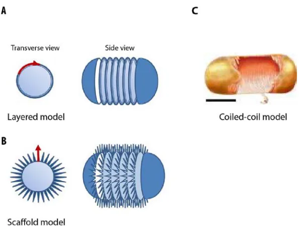
Figure 1.2. Three models of cell wall architecture (modified from (Hayhurst et al., 2008b; Young, 2011)). (A)
In the layered (hoop) model, the peptidoglycan chains lie parallel to the plasma membrane. (B) In the scaffold model, the peptidoglycan chains extend perpendicular to the plasma membrane. (C) In the coiled-coil model the peptidoglycan chains form a rope coiled around the cell. Arrows: direction of glycan strands.
1.1.2. Peptidoglycan synthesis
5
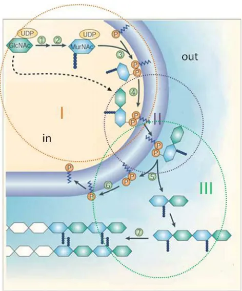
Figure 1.3. Schematic view of the PG synthesis pathway in gram-positive bacteria (modified from (Walsh,
2003)). Synthesis of cytoplasmic PG precursor, flipping of the precursor across the membrane and incorporation into the pre-existing cell wall are indicated as I, II and III surrounded by circles. Numbers inside green circles indicated steps of biosynthesis catalysed by the enzymes: MurA (1), MurB, C, D, E, F, Alr, D-Ala– D-Ala ligase (2), MraY (3), MurG (4), phosphates are added by transglycosylation and pyrophosphorylation (5 and 6), and finally, a peptide bond between the peptide chains is formed (step 7).
6
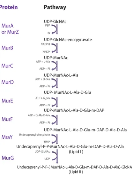
Figure 1.4. Mur biosynthetic pathway of lipid-linked PG intermediates (modified from (Scheffers, 2012)).
7
of Lipid II into the growing PG structure by PBPs (Figure 1.6, step III). PBPs catalyze transglycosylation and transpeptidation reactions responsible for the formation of glycosidic and peptide bonds of the PG (Archibald et al., 1993). Elongation of glycan chains occurs by the formation of glycosidic bonds between a Lipid II and a Lipid II PG–linked strand (Scheffers, 2012). After the transglycosylation reaction, the undecaprenyl-phosphate is released and dephosphorylated to yield the lipid carried bactoprenol, which becomes available for a second round of synthesis (Fraipont et al., 2006). After elongation of the glycan chain, there is a transpeptidation reaction in wich the terminal D-Alanyl-D-Alanyl bond of a lipid-linked murein precursor is cleaved by a transpeptidase. Next, a peptaide bond is formed between the carboxyl group of the penultime D-Ala of the precursor molecule and the diaminopimelic acid (m-A2pm) in a peptide sidechain of the growing sacculus (Scheffers, 2012). The incorporation of Lipid II into the existing PG strand requires cleavage of old glycan strands by autolysins (see below), a process that must be tightly controlled to allow insertion without disrupting the structural integrity of the PG.
1.1.2.1. Bacillus subtilis PBPs and autolysins
The CW is subject to continuos turnover, with PG being synthesized and hydrolysed at the same time. PBPs are involved in PG elongation and maturation; and cell wall hydrolases allow insertion of a new PG strand in the meshwork (Scheffers, 2012).
8
membrane where PG synthesis takes place. HMW PBPs are subdivided into class A and B, depending of their primary structure and the catalytic activity of the N-terminal domain. In class A, the terminal domain possesses glycosyltransferase activity, where as in class B, the N-terminal domain has no known catalytic activity and is believed to interact with other proteins involved in the cell cycle or important for protein folding and stability. The C-terminal domain of both classes has a transpeptidase activity (for review see (Sauvage et al., 2008)). B. subtilis
contains four genes encoding class A PBPs. One of them, ponA, gives rise to two proteins, PBP1 and PBP1b that are different due to C-terminal processing of the original protein (Popham and Setlow, 1995). However, deletion of all four genes does not stop PG synthesis, indicating that there should be more proteins capable of performing transglycosylation (Scheffers, 2012). There are six genes that encode class B HMW PBPs in B. subtilis: pbpA, pbpH, pbpB, pbpC, spoDV and pbpI. PBP2b (homologue of PBP3 of E. coli) is the only essential PBP in B. subtilis. PbpH and PBP2a are expressed during vegetative growth and have redundant roles in cell wall elongation (Scheffers, 2012). Finally, there are two classes of LMW PBPs: endopeptidases and carboxypeptidases. The two endopeptidases known in B. subtilis, PBP4 and PBPX, can be deleted without any phenotypic effects. The four carboxypeptidases, PBP5, PBP5* (which is a different PBP than PBP5), DacF and PBP4a, play different roles during PG maturation during vegetative growth or sporulation (Scheffers, 2012). In a study where the localizationof 13 B. subtilis PBPs was investigated, three different patterns were described: (i) homogenous localization along the cell envelope and at the septum, (ii) only at the septum and (iii) spotty localization, which was originally interpreted as helical-like pattern (Scheffers et al., 2004b). Nothing was known about the dynamics of the different PBPs.
9
endopeptidases according to their hydrolytic bond specificity (for a review see (Smith et al., 2000)). The B. subtilis genome contains 35 confirmed or predicted autolysins, which are clustered in 11 families on the basis of amino acid sequence similarities (Smith et al., 2000). Proteins from each family have been characterized, implicating them in processes such as cell wall turnover, cell wall modification, cell separation, competence, cell motility or germination (for a review see (Vollmer et al., 2008; Yamaguchi et al., 2004). However, it has proven difficult to assign specific functions to individual PG hydrolases because of their apparent redundancy. LytE, a member of the DL-endopeptidase II family has been shown to be involved in maturation of the lateral cell wall during vegetative cell growth (Carballido-Lopez et al., 2006; Yamaguchi et al., 2004). DL-endopeptidases hydrolyse the peptide bond between d-glutamate and A2pm (Smith et al., 2000). More recently, it was shown that the cell wall hydrolase YvcE also plays a role in cell wall elongation and that a yvcE lytE double mutant strain is not viable (Bisicchia et al., 2007).
1.1.2.2. Cell wall synthesis in Bacillus subtilis
10
(Lutkenhaus et al., 2012)). DivIB, DivIC, FtsL, PBP2b, PBP1, FtsW and GpsB/YpsB control synthesis of septal peptidoglycan (Scheffers, 2012). While FtsZ recruits the septal wall-synthetizing machinery, it is thought that in similar way the MreB cytoskeleton could direct PG synthesis by localizing cell wall elongation machineries (see below in section 1.3.4). MreC, MreD, RodA, PbpH, Pbp2A and LytE are members of these complexes (Carballido-Lopez and Formstone, 2007).
Figure 1.5. Schematic models of the two cell wall synthesis machineries (Carballido-Lopez and Formstone,
2007). The Z-ring (made by FtsZ) functions as a scaffold for a group of proteins that synthesize the division septum. The MreB cytoskeleton functions as a scaffold for the elongation complexes that synthesize lateral cell wall. The division machinery localizes at the mid cell and the elongation complexes are organized in a helical-like pattern. Question marks indicate unknown or putative players associated with the two machinaries.
1.2. Bacterial cytoskeleton
11
proteins (molecules that convert the energy of ATP hydrolysis into mechanical force). Each type of filaments has distintic mechanical properties, dynamics, and biological roles, but all three certain fundamental principles, the ability of self-assembly and to form dynamic structures (Alberts, 2008). The cytoskeleton was previously thought to be a feature only of but in organization and cell shape without counterparts in eukaryotes have been found (see below) (Ingerson-Mahar and Gitai, 2012), indicating that there are probably more to be discovered and characterized.
1.2.1. Actin homologues
Actin is the most abundant intracellular protein in eukaryotic cells, being a key cytoskeleton protein involved in essential processes like cell polarity, cell division, DNA segregation or cell migration (Alberts, 2008). Eukatyotic actin is a protein of 42 kDa with a polypeptide chain of 375 aa folded in two subdomains that are stabilized by an adenine nucleotide lying in between (Pollard et al., 2008). A four-subdomain nomenclature has been traditionally adopted (Figure 1.6). Actin polymerizes in presence of ATP and is found in two different conformations: globular actin (G-actin) and filamentous actin (F-actin).
12
similar to actin polymerisation (van den Ent et al., 2001) (Figure 1.6). Over the last decade it has been shown that, in addition to MreB, the family of bacterial actin homologues comprises the chromosomally encoded FtsA and MamK proteins and a variety of proteins encoded on extra-chromosomal plasmids like ParM (Figure 1.6), Alfa (actin like filament) and Alps (actin-like proteins highly divergent) (Figure 1.7), involved in different cellular processes such as cell shape determination, cell division, magnetosome organization and plasmid segregation (for reviews see (Carballido-Lopez, 2006; Shaevitz and Gitai, 2010).
Figure 1.6. Ribbon representation of the three-dimensional monomer structures Actin, MreB and ParM (modified from (Roeben et al., 2006)). The ADP molecule bound to actin and ParM is shown in ball-and-stick
13
Figure 1.7. The superfamily of bacterial actin homologs (Shaevitz and Gitai, 2010). The bacterial actins have
been identified based on sequence homology. The subfamilies that have been experimentally shown to polymerize are colored.
1.2.2. Tubulin homologues
Tubulin is a major component of the eukaryotic cytoskeleton. It forms stiff structures named microtubules, cylindrical polymers of α- and ß- tubulin, which provide support for a variety of cellular components. Microtubules serve as tracks for directed movement via a large number of motor proteins (Pollard and Earnshaw, 2002). The first tubulin-like protein identified in bacteria was FtsZ, which exhibits structural homology (Lowe and Amos, 1998) and similarities relative to eukaryotic tubulin such as the presence of a tubulin signature motif and the ability to bind and hydrolyse GTP, and to undergo GTP-dependent polymerization into protofilaments and tubules (Erickson, 1995).
14
cryotomographic and photo-activation localization microscopy (PALM), suggest that the Z-ring is a discontinuous structure (Fu et al., 2010; Strauss et al., 2012). The prevailing model is that the Z-ring works as a scaffold that localizes a cascade of proteins (divisome) coordinating proper cell division (see Fig. 1.7). It is also widely established that the Z-ring helps to generate the motive force for cell constriction at the sites of division (Li et al., 2007).
In addition to FtsZ, two tubulin-like genes, btubA and btubB, were identified in Prosthecobacter dejongeii (Schlieper et al., 2005). BtubA and BtubB have higher sequence homology to eukaryotic tubuin than to FtsZ and in some species the three proteins coexist (Pilhofer et al., 2011). The function of BtubA and BtubB proteins remains to be elucidated. Finally, two additional FtsZ-like proteins, TubZ and PhuZ, involved in plasmid and phage segregation respectively, were recently identified (Kraemer et al., 2012; Makarova and Koonin, 2010).
1.2.3. Intermediate filaments homologues
Intermediate filaments (IFs) are a family of related proteins that share a common sequence of a rod-like domain with variable head and tail domains at the two ends. IFs self-assemble into apolar filaments of 10 nm diameter. IFs are flexible but strong polymers that provide mechanical support for cells (Pollard et al., 2008). Crescentin, a protein of 430 aa with a coiled-coil-rich structure, was the first bacterial IF-like protein to be identified. Crescentin is required for the crescent shape of
15
(filament-forming protein) in Streptomyces coelicolor, revealed that it also forms filaments in live cells and deletion of the protein generates growth and morphological defects (Bagchi et al., 2008).
1.2.4. Additional bacterial cytoskeletal proteins
More proteins able to polymerize into linear filaments that mediate cellular organization, without eukaryotic homologues have been identified as part of the bacterial cytoskeleton (for a recent review see (Ingerson-Mahar and Gitai, 2012)). These proteins play a structural or regulatory role in promoting cellular organization acting as scaffolds or exerting forces on targets such as membranes or chromosomes. Walker A cytoskeletal ATPases proteins (WACAs) are the best characterized filament-forming proteins without counterparts in eukaryotic cells (Lowe and Amos, 2009). ParA and MinD are the two most-studied WACAs. ParA is involved in plasmid/chroosome segregation (Gerdes et al., 2000) and MinD in the determination of the septum placement (Lutkenhaus, 2007). ESCRT (endosomal sorting complex required for transport) and bactofilins homologues has been also found in archea and some bacteria (Ingerson-Mahar and Gitai, 2012). The Ctp synthase CtpS, an enzyme responsible for the synthesis of CTP (cytidine triphosphate) from UTP (uridine triphosphate), ATP (adenosine triphosphate) and glutamine, regulates crescentin assembly preventing excessive C. crecescentus
16 1.3. MreB proteins
Among bacterial actin-like proteins, the chromosomally encoded MreB proteins are the most widely conserved and remain the best characterized so far. MreB is almost always present in rod-shaped bacteria and absent in spherical (coccoid) bacteria. Gram-negative bacteria, e. g, E. coli
have one mreB gene encoded in an operon together with mreC and mreD. Gram-positive bacteria often have the mreBCD operon plus extra copies of mreB. The gram-positive, rod-shaped bacterium B. subtilis has three mreB homologues: mreB, mbl (mreB-like) and mreBH (mreB
-Homologue). MreB is also present in cell wall-less bacteria, like Chlamydophila pneumoniae,
where it might play a role in cell division by spatially organizing lipid biosynthesis (Gaballah et al., 2011). MreB was originally described to localize underneath the cytoplasmic membrane, in a filamentous helix-like structure (Figge et al., 2004; Gitai et al., 2005; Jones et al., 2001; Kruse et al., 2003). The finding of CW synthesis in a helical pattern (Daniel and Errington, 2003; Kawai et al., 2009a; Tiyanont et al., 2006b) and that several morphogenetic factors (PBPs, MreC, MreD, rodA and autolysins) also localize in a helical pattern in an MreB-dependent fashion led to a model where MreB proteins play a role in cell shape determination by organizing PG synthesis (Carballido-Lopez and Errington, 2003) (for more detail see section 1.4.4). It was also proposed that MreB contributes to cell shape maintenance via a mechanical function of the MreB filaments (Soufo and Graumann, 2010; Wang et al., 2010). MreB is involved in Streptomyces sporulation,
17
isoforms (Schirner and Errington, 2009). Therefore and because of more discrepancies between different groups, the MreB role in chromosome segregation has been highly debated (for a recent review see (Chastanet and Carballido-Lopez, 2012).
1.3.1. Biochemical properties of MreB
Several difficulties in the expression (e.g. gen mutations) and purification (e.g. protein aggregation) of the MreB proteins have precluded their biochemical characterization. Therefore there are just a few studies on MreB1 from the thermophile Termotoga maritime and one study on the assembly propeties of B. subtilis. The MreB monomer consist of two domains also subdivide in two: IA-IB and IIA-IIB (Figure 1.6). The two larger subdomains (IA-IIA) have a common conserved fold that comprises a five stranded beta-sheet surrounded by three α-helices. The smaller domains (IB-IIB) are more diverse within the superfamily, probably having specific functions (Carballido-Lopez, 2012). The conservation of the structures raised the possibility of conserved biochemical properties and/or functions between actins and MreB proteins (Chastanet and Carballido-Lopez, 2012). Actin polymerization includes nucleation (formation of dimers and trimers), elongation, and stady state (Alberts, 2008). MreB assembles into filaments with a subunit repeat similar to that of F-actin (van den Ent et al., 2001). However, is not well understood whether MreB nucleation is highly favourable and fast, or there is not nucleation phase like in actin (Esue et al., 2005). In contrast to actin, the mechanism of annealing (join of filaments trough direct association of filaments ends) does not contribute to MreB polymerization (Esue et al., 2005). MreB hydrolyses ATP and GTP, indicating that the protein is an ATPase as well as a GTPase. The critical concentration (Cc) is the concentration of actin monomers in equilibrium
18
concentrations below Cc, there is net depolymerization of filaments (Lodish et al. 2000). MreB exhibits a critical concentration (~ 3 nM) of two orders of magnitude lower than that of actin (0.1 µM) (Carballido-Lopez, 2006; Pollard et al., 2008). Also in contrast to actin that forms helical filaments, it was observed that MreB forms straight protofilaments. However, a recent study showed that MreB forms both, linear and helical protofilaments, probably reflecting the bound state of nucleotides, where linear filaments are bound to NDP and helical filaments to NTP (Esue et al., 2005). MreB polymerizes without accessory proteins into bundles that display different morphologies and sometimes into closed rings (Esue et al., 2005). Because the phosphate release is almost simultaneous to polymer assembly and the absence of dynamic instability, it has been suggested that ADP/GDP–bound MreB exist in the filament (Chastanet and Carballido-Lopez, 2012). Recently, the assembly properties of B. subtilis MreB were also investigated. Interestingly it was found that B. subtilis MreB polymerization occurs in absence of nucleotide (Mayer and Amann, 2009). Further studies will clarify whether aggregation of MreB was observed or that B. subtilis MreB actually has a different polymerization mechanism. Altought MreB and actin share a structural homology (van den Ent et al., 2001) it might be that the biochemical properties of both proteins are not that similar.
1.3.2. Subcellular localization of MreB proteins
19
helix is observed, leading in a ring at the future division site (Figge et al., 2004; Gitai et al., 2004a; Vats and Rothfield, 2007). Time-lapse and FRAP experiments showed that MreB (and Mbl) filaments are highly dynamic structures that are continuously remodeling during growth and division (Carballido-Lopez, 2012). In B. subtilis cells, Mbl filaments turnover was reported to occur along their length, with a half-time of recovery of about 8 minutes (Carballido-Lopez and Errington, 2003) and filaments of MreB (or Mbl) were observed to perform a full turn around the diameter of the cell with a estimated average speed of 0.07 µm.s-1 (Defeu Soufo and Graumann, 2004). Direct motion of MreB-YFP in C. crescentus was also observed in a quantitative single molecule study, in which treadmilling of MreB monomers within the filaments was proposed to drive such motion (Kim et al., 2006b).
Figure 1.8. Subcelullar localization of MreB proteins (modified from (Carballido-Lopez and Errington, 2003;
20
1.3.3. Role of MreB proteins in cell shape determination in Bacilus subtilis
In B. subtilis the mreB and mbl genes are essential for cell viability under normal growth conditions, while deletion of the third homologue mreBH has no effects, but prevents growth in a minimal medium with low concentrations of magnesium (Mg2+) or under stress conditions (Carballido-Lopez et al., 2006; Jones et al., 2001; Kawai et al., 2009a). Before lysis, cells lacking MreB become swollen and bulge at the cell poles (Formstone and Errington, 2005), and cells lacking Mbl bend and twist, adopting a coiled helical shape (Jones et al., 2001; Schirner and Errington, 2009). Absence of MreBH in cells under stress conditions generates curved and bended cells, especially in stationary phase (Kawai et al., 2009a). Interestingly, high concentrations of Mg2+ recue the lethal phenotypes of mreB and mbl null mutants and restored wild-type morphology of a mreBH null mutant (Carballido-Lopez et al., 2006; Formstone and Errington, 2005). The mechanism behind the Mg2+ complementation is not known, but since Mg2+ also rescues growth and morphological defects of other proteins involved in several aspects of cell wall synthesis (e.g. MreC, MreD, RodA, PBP1, etc. (Leaver and Errington, 2005a; Murray et al., 1998b; Rogers et al., 1976) it might be an unspecific mechanism.
1.3.4. Role of MreB proteins in cell wall synthesis in Bacilus subtilis
21
and Mbl complexes; and PBP1 and PBP2a in the MreBH complex (Kawai et al., 2009a). Indicating that MreB (and Mbl/MreBH) filaments are directly associated with PG synthesizing complexes. In addition, White and co-workers showed that C. crescentus MreB filaments are required for the organization of several cytosolic murein biosynthetic enzymes (MraY, MurB, MurC, MurE and MurF), suggesting the existence of cytoplasmic complexes involved in PG precursor synthesis (White et al., 2010). However, nothing is known in B. subtilis about this aspect.
22
The current model of cell wall elongation machineries includes the transmembrane proteins MreD and MreC as linkers to couple MreB proteins to the extracellular cell wall synthetic machinery (Carballido-Lopez and Formstone, 2007). MreC and mreD are immediately downstream of mreB, encoded in the same operon (Formstone and Errington, 2005). MreC is predicted to have a single transmembrane span with its major C-terminal domains outside the cytoplasmic membrane and MreD is predominantly hydrophobic with four or six transmebrane spans and both N- and C-terminal inside in the cell (Leaver and Errington, 2005a). In B. subtilis
23
1.4.Stress-inducible cell envelope proteins LiaI and LiaH of Bacillus subtilis
24
PliaI is completely LiaR-dependent (Jordan et al., 2006). In addition to bacitracin, the PliaIpromoter
is also induced by nisin, ramoplanin and vancomycin, i. e. by antibiotics that interfere with the lipid II cycle (Mascher et al., 2004). The lia operon is also weakly induced by detergents, ethanol, alkaline shock and secretion stress (Hyyrylainen et al., 2005; Mascher et al., 2004; Pietiainen et al., 2005; Wiegert et al., 2001). LiaG is a putative membrane-anchored protein of unkown function, LiaI is a small hydrophobic protein of unknown function too with two putative transmembrane helices, and LiaH is a cytoplasmic protein member of the phage-shock protein family (Jordan et al., 2006; Wolf et al., 2010) (Figure 1.9.).
Figure 1.9. Lia stress response in Bacillus subtilis (modified from (Wolf et al., 2010)). Proteins surrounded by
circles are drawn at their known or predicted subcellular localization. LiaH is shown in its oligomeric form. Single ended arrows indicate activation, double-ended arrows interactions and T-shaped lines inhibitions. Abbreviations as follows: CM - cytoplasmic membrane; CW - cell wall; OM - outer membrane.
In E. coli the phage-shock protein A (PspA) is part of the PspF regulon, which includes
25
26 1.5. Total Internal Reflexion Microscopy
27
work to study the localization and dynamics of the MreB proteins and components of the CW machinery.
Figure 1.10. Scheme of total internal reflection microscopy (Alberts, 2008). TIRF microscopy uses an
28 1.6. Aims of the thesis
In bacterial cell biology PG synthesis and chemical composition are well understood, but the
structure of the sacculus and the mechanisms controlling its growth remain elusive. The current
model is that the helical localization of actin-like MreB proteins spatially organizes the cell wall biosynthetic complexes that mediate sidewall elongation.
TIRFM is a sensitive technique for studying events at cell surfaces and its known that MreB
filaments localize underneath the membrane spanning the cell cylinder. The first aim of my thesis
was to do a comprehensive characterization of MreB isorforms (MreB, Mbl and MreBH) dynamics in B. subtilis. The elongation complexes are thought to contain the essential transmembrane proteins MreC and MreD, RodA and RodZ, PG hydrolases, and PBPs, the enzymes that catalyze PG elongation and cross-linking. The second aim was to study the spatial organization and dynamics of the components of the cell wall elongation complexes, as well as the
interactions with the MreB cytoskeleton. Based on structural homology between MreB and actin,
and on single study treadmilling was previously suggested to explain the MreB motility. The third aim was to study the molecular basis of the MreB filaments motility.
Because of its many essential functions, the cell envelope is a prime target for numerous antibiotics. Therefore it is crucial for cell survival to continuously monitor and maintain envelope integrity. To investigate how the integrity of the cell wall is maintained, the fourth aim was to study the localization and dynamics of the LiaIH-system, the target of LiaRS, a two component system involved in cell envelope stress response.
Genetics, TIRFM live cell imaging, fluorescence recovery after photobleaching and drug/enzyme
29
2. Material and Methods
2.1. Solutions and media
Tables of solutions and growth medium details are given in appendices 1 and 2.
2.2. Strains and plasmids
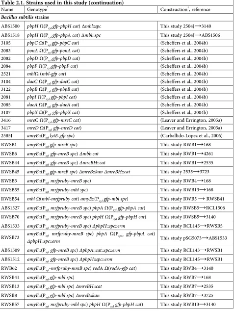
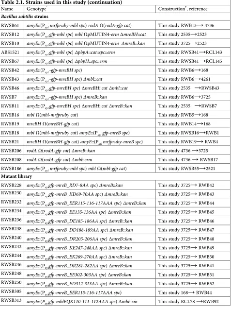
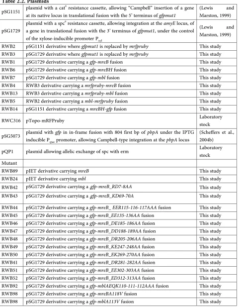
Bacterial strains and plasmids used in this study are given in tables 2.1 and 2.2 respectively.
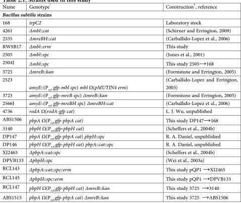
Table 2.1. Strains used in this study
Name Genotype* Construction†, reference Bacillus subtilis strains
168 trpC2 Laboratory stock
4261 ∆mbl::cat (Schirner and Errington, 2009) 2535 ∆mreBH::cat (Carballido-Lopez et al., 2006) RWSB17 ∆mbl::erm This study
2505 ∆mbl::spc (Jones et al., 2001) 2504J ∆mbl::spc This study 2505→168
3725 ∆mreB::kan (Formstone and Errington, 2005) 2523
amyE::(Pxyl gfp-mbl spc) mbl Ω(pMUTIN4 erm)
(Carballido-Lopez and Errington, 2003)
3723 amyE::(Pxyl gfp-mreB spc) ∆mreB::kan (Formstone and Errington, 2005)
2566J amyE::(Pxyl gfp-mreBH spc) ∆mreBH::cat (Carballido-Lopez et al., 2006)
4736 rodA Ω(rodA-gfp cat) L. J. Wu, unpublished ABS1506 pbpA Ω(Pxyl gfp-pbpA cat) This study DP147→168 3140 pbpH Ω(Pxyl gfp-pbpH cat) (Scheffers et al., 2004b)
DP147 pbpA Ω(Pxyl gfp-pbpA cat) pbpH::spc R. A. Daniel, unpublished
DP146 pbpH Ω(Pxyl gfp-pbpH cat) pbpA::cat::spc R. A. Daniel, unpublished
XI2465 ∆pbpA::cat::spc (Scheffers et al., 2004b) DPVB133 ∆pbpH::spc (Wei et al., 2003a)
30 Table 2.1. Strains used in this study (continuation)
Name Genotype* Construction†, reference Bacillus subtilis strains
ABS1500 pbpH Ω(Pxyl gfp-pbpH cat)∆mbl::spc This study 2504J→3140
ABS1518 pbpH Ω(Pxyl gfp-pbpA cat)∆mbl::spc This study 2504J→ABS1506
3105 pbpC Ω(Pxyl gfp-pbpC cat) (Scheffers et al., 2004b)
2083 ponA Ω(Pxyl gfp-ponA cat) (Scheffers et al., 2004b)
2082 pbpD Ω(Pxyl gfp-pbpD cat) (Scheffers et al., 2004b)
2084 pbpF Ω(Pxyl gfp-pbpF cat) (Scheffers et al., 2004b)
2521 mblΩ (mbl-gfp cat) (Scheffers et al., 2004b) 3104 dacC Ω(Pxylgfp-dacC cat) (Scheffers et al., 2004b)
3122 pbpB Ω(Pxylgfp-pbpB cat) (Scheffers et al., 2004b)
2081 pbpI Ω(Pxylgfp-pbpI cat) (Scheffers et al., 2004b)
2085 dacA Ω(Pxylgfp-dacA cat) (Scheffers et al., 2004b)
3107 pbpX Ω(Pxyl gfp-pbpX cat) (Scheffers et al., 2004b)
3416 mreC Ω(Pxyl gfp-mreC cat) (Leaver and Errington, 2005a)
3417 mreD Ω(Pxyl gfp-mreD cat) (Leaver and Errington, 2005a)
2585J amyE::(Pxyl lytE-gfp spc) (Carballido-Lopez et al., 2006)
RWSB1 amyE::(Pxyl gfp-mreB spc) This study RWB1→168
RWSB6 amyE::(Pxyl gfp-mreB spc) ∆mbl::cat This study RWB1→4261
RWSB44 amyE::(Pxyl gfp-mreB spc) ∆mreBH::cat This study RWB1→2535
RWSB45 amyE::(Pxyl gfp-mreB spc) ∆mreB::kan ∆mreBH::cat This study 2535→3723
RWSB5 amyE::(Pxyl mrfpruby-mreB spc) This study RWB4→168
RWSB55 amyE::(Pxyl mrfpruby-mbl spc) This study RWB13→168
RWSB54 mbl Ω(mbl-mrfpruby cat) amyE::(Pxyl gfp-mbl spc) This study RWB5 → RWSB41
ABS1527 amyE::(Pxyl mrfpruby-mreB spc) pbpA Ω(Pxyl gfp-pbpA cat) This study RWSB5→RCL1506
RWSB70 amyE::(Pxyl mrfpruby-mreB spc) pbpH Ω(Pxyl gfp-pbpH cat) This study RWSB5→3140
ABS1533 amyE::(Pxyl mrfpruby-mreB spc) ∆pbpH::spc::erm This study RCL145→RWSB5
RWSB73 amyE::(Pxyl mrfpruby-mreB spc) pbpA Ω(Pspac gfp-pbpA cat)
∆pbpH::spc::erm This study pSG5073→ABS1533
ABS1509 amyE::(Pxyl gfp-mreB spc) ∆pbpA::cat::spc::erm This study RCL143→RWSB1
ABS1512 amyE::(Pxyl gfp-mreB spc) ∆pbpH::spc::erm This study RCL145→RWSB1
RWB62 amyE::(Pxyl-mrfpruby-mreB spc) rodA Ω(rodA-gfp cat) This study RWB4→3140
RWSB41 amyE::(Pxyl gfp-mbl spc) This study RWB7→168
RWSB13 amyE::(Pxyl gfp-mbl spc) ∆mreBH::cat This study RWB7→2535
RWSB8 amyE::(Pxyl gfp-mbl spc) ∆mreB::kan This study RWB7→3725
31 Table 2.1. Strains used in this study (continuation)
Name Genotype* Construction†, reference Bacillus subtilis strains
RWSB61 amyE::(Pxyl mrfpruby-mbl spc) rodA Ω(rodA-gfp cat) This study RWB13→ 4736
RWSB12 amyE::(Pxyl gfp-mbl spc) mbl ΩpMUTIN4-erm ∆mreBH::cat This study 2535→2523
RWSB10 amyE::(Pxyl gfp-mbl spc) mbl ΩpMUTIN4-erm ∆mreB::kan This study 3725→2523
ABS1521 amyE::(Pxyl gfp-mbl spc) ∆pbpA::cat::spc::erm This study RWSB41→RCL143
RWSB67 amyE::(Pxyl gfp-mbl spc) ∆pbpH::spc::erm This study RWSB41→RCL145
RWSB42 amyE::(Pxyl -gfp-mreBH spc) This study RWB6→168
RWSB43 amyE::(Pxyl -gfp-mreBH spc) ∆mbl::cat This study RWB6→4261
RWSB46 amyE::(Pxyl gfp-mreBH spc)∆mreBH::cat ∆mbl::cat This study 2535 →RWSB43
RWSB7 amyE::(Pxyl -gfp-mreBH spc) ∆mreB::kan This study RWB6→3725
RWSB11 amyE::(Pxyl gfp-mreBH spc)∆mreBH::cat ∆mreB::kan This study 2535 →RWSB7
RWSB16 mbl Ω(mbl-mrfpruby cat) This study RWB5→168 RWSB19 mreBH Ω(mreBH-gfp cat) This study RWB14→168 RWSB18 mbl Ω(mbl-mrfpruby cat) amyE::(Pxyl gfp-mreB spc) This study RWSB16→RWB1
RWSB21 mreBH Ω(mreBH-gfp cat) amyE::(Pxyl mrfpruby-mreB spc) This study RWB19→ RWB4
RWSB206 rodA Ω(rodA-gfp cat) ∆mreB::kan This study 4736 →3725 RWSB208 rodA Ω(rodA-gfp cat) ∆mbl::erm This study 4736 → RWSB17 RWSB186 amyE::(Pxyl mrfpruby-mbl spc) mbl Ω(mbl-gfp cat) This study RWSB55→2521 Mutant library
RWSB228 amyE::(Pxylgfp-mreB_RD7-8AA spc) ∆mreB::kan This study 3725→ RWB42
32 Table 2.1. Strains used in this study (continuation)
Name Genotype* Construction†, reference Bacillus subtilis strains
RWSB309 amyE::(Pxylgfp-mreB_A118V spc) ∆mreB::kan This study 3725→ RWB44 RWSB311 amyE::(Pxylgfp-mblA113V spc) ∆mbl::cm This study RCL78 →RWB92 TMB321 amyE::(PxylLiaH-gfp spc) D. Wolf, unpublished
TMB322 amyE::(PxylLiaI-gfp spc) D. Wolf, unpublished
TMB1328 LiaH Ω(LiaH-gfp cat) D. Wolf, unpublished TMB1394 ∆liaI (clean deletion) D. Wolf, unpublished TMB1407 LiaH Ω(LiaH-gfp cat) ∆liaI (clean deletion) D. Wolf, unpublished TMB1421 LiaI Ω(LiaI-gfp cat) D. Wolf, unpublished TMB1441 amyE::(PxylLiaI-gfp cm) LiaI Ω(LiaI-mrfpruby cat) D. Wolf, unpublished
Escherichia coli strains
FB72 DY329, mreB’-rfp-‘mreB (Bendezu et al., 2009b)
Caulobacter crescentus strains
LS3814 xylΩPxyl gfp-mreB neo (Gitai et al., 2004a)
*. Resistance gene abbreviations: kan, kanamycin; spc, spectinomycin; cat, chloramphenicol; erm, erythromycin. Other abbreviations: ∆, deletion; Ω, insertion.
33
pSG1151 plasmid with a catat its native locus in translational fusion with the 5’ terminus of resistance cassette, allowing “Campbell” insertion of a gene gfpmut1 (Lewis Marston, 1999) and
pSG1729 plasmid with a spc
a resistance cassette, allowing integration at the amyE locus, of
a gene in translational fusion with the 3’ terminus of gfpmut1, under the control of the xylose-inducible promoter Pxyl
(Lewis and Marston, 1999) RWB2 pSG1151 derivative where gfpmut1 is replaced by mrfpruby This study RWB3 pSG1729 derivative where gfpmut1 is replaced by mrfpruby This study RWB1 pSG1729 derivative carrying a gfp-mreB fusion This study RWB6 pSG1729 derivative carrying a gfp-mreBH fusion This study RWB7 pSG1729 derivative carrying a gfp-mbl fusion This study RWB4 RWB3 derivative carrying a mrfpruby-mreB fusion This study RWB13 RWB3 derivative carrying a mrfpruby-mbl fusion This study RWB5 RWB2 derivative carrying a mbl-mrfpruby fusion This study RWB14 pSG1151 derivative carrying a mreBH-gfp fusion This study RWC316 pTopo-mRFPruby Laboratory stock
pSG5073 plasmid with inducible P gfp in in-frame fusion with 804 first bp of pbpA under the IPTG
spac promoter, allowing Campbell-type integration at the pbpA locus
(Scheffers et al., 2004b)
pQP1 plasmid allowing allelic exchange of spc with erm Laboratory stock Mutant
RWB89 pJET derivative carrying mreB This study RWB24 pJET derivative carrying mbl This study RWB42 pSG1729 derivative carrying a lgfp-mreB_RD7-8AA This study
RWB43 pSG1729 derivative carrying a lgfp-mreB_KD69-70A This study
34
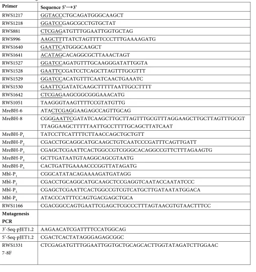
A list of oligonucleotides is given in table 2.3
Table 2.3. Oligonucleotides
Primer Sequence 5’→3’
RWS1217 GGTACCCTGCAGATGGGCAAGCT RWS1218 GGATCCGAGCGCCTGTGCTAT
RWS881 CTCGAGATGTTTGGAATTGGTGCTAG
RWS996 AAGCTTTTATCTAGTTTTCCCTTTGAAAAGATG RWS1640 GAATTCATGGGCAAGCT
RWS1641 ACATAGCACAGGCGCTTAAACTAGT
RWS1527 GGATCCAGATGTTTGCAAGGGATATTGGTA RWS1528 GAATTCCGATCCTCAGCTTAGTTTGCGTTT RWS1529 GGATCCACATGTTTCAATCAACTGAAATC RWS1530 GAATTCGATATCAAGCTTTTTAATTGCCTTTT RWS1642 CTCGAGAAGCGGCGGGAAACATG
RWS1051 TAAGGGTAAGTTTTCCGTATGTTG MreBH-6 ATACTCGAGGAAGAGCCAGTTGCAG
MreBH-8 CGGGAATTCGATATCAAGCTTGCTTAGTTTGCGTTTAGGAAGCTTGCTTAGTTTGCGT TTAGGAAGCTTTTTAATTGCCTTTTGCAGCTTATCAAT
MreBH-P1 TATCCTTCATTTTCTTAACCAGCTGCTGTT
MreBH-P2 CGACCTGCAGGCATGCAAGCTGTCAATCCCGATTTCAGTTGATT
MreBH-P3 CGAGCTCGAATTCACTGGCCGTCGGGCACAGGCCGTTCTTTAGAAGTG
MreBH-P4 GCTTGATAATGTAAGGCAGCGTAATG
MreBH-P5 CACTGATTGAAAACCCGGTTATAGATG
Mbl-P1 CGGCATATACAGAAAAGATGATAGG
Mbl-P2 CGACCTGCAGGCATGCAAGCTCCGAGGTCAATACCAATATCCC
Mbl-P3 CGAGCTCGAATTCACTGGCCGTCGTCATGCTTGATAATATGGACA
Mbl-P4 ATACCCATTTCCAGTGACGAGCTGCA
RWS1166 CGACGGCCAGTGAATTCGAGCTCGCCCTTTAGTAACGTGTAACTTTCC
Mutagenesis PCR
3’-Seq-pJET1.2 AAGAACATCGATTTTCCATGGCAG 5’-Seq-pJET1.2 CGACTCACTATAGGGAGAGCGGC RWS1331
35
RWS1306
69-70F TGGCTCTTCGCCCGATGGCAGCAGGCGTTATCGCTGATTA RWS1305
69-70R TAATCAGCGATAACGCCTGCTGCCATCGGGCGAAGAGCCA RWS1530
MreB115-116-117F
GGCATTACAGCTGTTGCAGCAGCGGCTGTTATCGATGCGACA
RWS1308
135-136F CGCGTGACGCGTATCCGATTGCAGCACCTTTTGCCGCAGCAATCGG RWS1307
135-136F CCGATTGCTGCGGCAAAAGGTGCTGCAATCGGATACGCGTCACGCG RWS1309
185-186F CAGTCAATCCGTGTAGCCGGTGCTGCAATGGATGACGCGATTATC RWS1310
185-186R GATAATCGCGTCATCCATTGCAGCACCGGCTACACGGATTGACTG RWS1311
188-189F GTAGCCGGTGATGAGATGGCTGCAGCGATTATCAACTACATC RWS1312
188-189R GATGTAGTTGATAATCGCTGCAGCCATCTCATCACCGGCTAC RWS1313
205-206F CGTACAATCTGATGATCGGTGCAGCAACGGCTGAAGCGATTAAAAT RWS1314
205-206R ATTTTAATCGCTTCAGCCGTTGCTGCACCGATCATCAGATTGTACG RWS1315
247-248F AAACAATTGAAATTACAGGAGCAGCAATTTCTAACGCTCTACGCGA RWS1316
247-248R TCGCGTAGAGCGTTAGAAATTGCTGCTCCTGTAATTTCAATTGTTT RWS1317
269-270F AAGCAGTGAAGAGCACACTCGCAGCAACACCGCCTGAGCTTGCAGC RWS1318
269-270R GCTGCAAGCTCAGGCGGTGTTGCTGCGAGTGTGCTCTTCACTGCTT RWS1319
281-282F CCGCCGGTTAACACTATACCTGCTGCCATGATATCTGCTGCAAGCT RWS1320
281-282R AGCTTGCAGCAGATATCATGGCAGCAGGTATAGTGTTAACCGGCGG RWS1321
302-303F ATTTGGACAAAGTCATCAGCGCAGCAACAAAAATGCCGGTCCTTAT RWS1322
36
RWS1323
312-313F AAATGCCGGTCCTTATCGCCGCAGCACCGCTTGATTGTG RWS1324
312-313R ATCGCTACACAATCAAGCGGTGCTGCGGCGATAAGGACCGGCATTT RWS1996
115-116-117_MblF
CTGCCCGACGAATATTACATCCGTTGCAGCAGCAGCAATTAAAGAAGCTGCAGA
37
Supplements added to growth medium and the concentration they were used in, are listed in table 2.4.
Table 2.4. Media supplements
Supplement Stock concentration Final concentration
B. subtilis E. coli
Antibiotics
ampicillin 100 mg·ml-1 - 100 µg·ml-1
chloramphenicol 10 mg·ml-1 5 µg·ml-1 -
erythromycin 10 mg·ml-1 1-5 µg·ml-1 -
kanamycin 25 mg·ml-1 5 µg·ml-1 -
phleomycin 5 mg·ml-1 0.2 µg·ml-1 -
spectinomycin 100 mg·ml-1 200 µg·ml-1 -
tetracyclin 20 mg·ml-1 10 µg·ml-1 -
phosphomycin 25 mg·ml-1 700 µg·ml-1 -
vancomycin 20 mg·ml-1 100 µg·ml-1 -
Amino acids
tryptophan 1% 0.01% -
Other supplements
IPTG 100mM 1 mM 1 mM
xylose 20% 0.05-0.5% -
MgSO4 1 M 10-25 mM -
glucose 20% 0.5% -
38 2.5.1. DNA Methods
2.5.1.1. Oligonucleotides
Oligonucleotides were purchased from Metabion (International AG, Germany) and aliquots were stored at -20 °C in a concentration of 100 µM.
2.5.1.2. Polymerase chain reaction (PCR)
PCR reactions were performed according to the manufacturer’s recommendations. Taq polymerase (MPI facility) was used to confirm insertions or deletions; Phusion (NEB) polymerase was used for creating genetic constructs.
2.5.1.3. Elution of DNA fragments from an agarose gel
DNA fragments were cut out of an ethidium bromide stained agarose gel and the DNA purified using the Wizard®SV Gel and PCR Clean-Up System kit (Promega) according to the manufacturer’s recommendations.
39
Plasmids were purified using E.Z.N.A.TM Plasmid Miniprep Kit (Omega Bio-tek) according to the
manufacturer’s recommendations.
2.5.1.5. Agarose gel electrophoresis of DNA fragments
Agarose gel electrophoresis was performed using 1% agarose gels containing 1% ethidium bromide (Carl Roth) in 1xTBE buffer. Samples were mixing with loading dye prior to loading. The voltage used for electrophoresis was 90-120 volts.
2.5.1.6. Restrictions endonuclease digestion
DNA was digested for usually 1-3 in the conditions recommended by of the suppliers of the enzyme, using the following mix:
Reaction mixture:
3 µl DNA fragments
3 µl 10 x BSA
3 µl X NEBuffer
1 µl Enzyme I
1 µl Enzyme II
19 µl ddH2O
40
DNA fragments were ligated together using the following mix and the reaction was incubated for 1 h at RT.
Reaction mixture:
2 µl Plasmid (backbone)
6 µl DNA fragment or ddH2O (negative control) 1 µl 10 X T4 DNA Ligase buffer
1 µl T4 DNA Ligase
2.5.1.8. DNA sequencing
Sequencing of PCR products or plasmids was performed by the sequence facility in the Max Planck Institute of Biochemistry (Germany). Using the following mix:
3 µl Plasmid or DNA fragment
4 µl ddH2O
0.5 µl Primer (forward or reverse each)
2.5.1.9 Mutagenesis PCR
41
5 µl 10 x Pfu polymerase buffer (inc Mg) 1 µl dNTP mix (10 mM)
0.5 µl Primer 1 (100 pmol/ml) 0.5 µl Primer 2 (100 pmol/ml) 1 µl plasmid template (100 ng) 2 µl Pfu polymerase*
40 µl H2O ∑= 50 µl
PCR Program
Duration Temperature cycles
60 seconds 94 °C 1
30 seconds 94°C 12
30 seconds 55°C 12 minutes 68°C
10 minutes 72°C 1
∞ 4°C
42 2.5.2.1. Preparation of protein samples
Cultures were grown in 10 ml LB with the desired xylose concentration at 37 °C to OD600nm 1.0. Cells were harvest by centrifugation and resuspended in a mix of 100 µl of Z buffer, 1μl lysozyme (10mg/mL stock solution) and 0.1 µl dtt (1M stock solution), and incubated 30 min at 37 °C. Then 100µl of loading buffer were added and cells were boil 10 min at ~ 95 °C.
2.5.2.2. SDS-polyacrylamide gel electrophoresis
SDS-polyacrylamide gels at 10% were used and electrophoresis was performed in Laemmili 1x buffer at 200V. Samples were incubated for 10 min at 95 °C before loading.
2.5.2.3. Coomassie staining
For Coomassie staining of protein bands, the gel was washed with ddH2O. Next, the gel was covered with coomassie stain, microwaved on high power from 40 seconds to 1 minute
43
SDS-polyacrylamide gels wererinsed with H2O and then soaked in transfer buffer. Transfer on a PVDF membrane was performed for 1.5 h at 140 mA. After protein transfer, the membrane was incubated for 1 h at RT in blocking buffer, then for 3 h in blocking solution at RT or over night at 4 ˚C containing the primary antibody. Polyclonal anti-MreB, anti-Mbl and anti-GFP, were added at 1:10,000, 1:7,500 and 1:5,000, respectively. After washing the membrane 3 times for 30 min in blocking buffer, the secondary antibody (anti-rabbit-HRP conjugate, Sigma) was added at 1:5,000 and incubated 1h at RT. The membrane was washed three times for 15 min with blocking buffer. Detection was performed with an ECL kit (Amersham).
2.5.3. Manipulation in E. coli
2.5.3.1. Preparation of competent cells
44
Frozen competent cells were thawed on ice. Fifty µl were transferred to a 1.5 ml tube and plasmid DNA or a ligation reaction was added. After incubation on ice for 30 min, cells were heat-schocked for 2 min at 42°C, and placed back on ice for 2 min. Upon addition of 150 µl of YT medium, cells were plated on plates containing the appropriate antibiotic.
2.5.4. Manipulation in B. subtilis
2.5.4.1. Preparation of B. subtilis competent cells
To make B. subtilis competent for the uptake of DNA, 10 ml of MD medium (supplemented with 100μl of 10% CAA) were inoculated with a colony of a freshly streaked strain and incubated at 37°C until OD600 1-1.5. Then, an equal volume of warm MD medium was added and further incubated for 1 h at 37°C. Aliquotes of 800 µl of the now competent cells were either used for transformation immediately or mixed with glycerol (10%) and glucose (1%) and rapidly frozen in liquid nitrogen, then stored at -80 °C, for a later use.
2.5.4.2. Transformation of B. subtilis
45
were checked by colony PCR.
2.5.4.3. Preparation of chromosomal DNA (phenol/chloroform method)
46
A second method was used to avoid using phenol/chloroform. Three ml of LB containing the appropriate antibiotics were inoculated with a single colony from a freshly streaked strain and incubated ON at 37°C. One and a half ml of the cell culture were pelleted and resuspended in 450 µl of 50 mM EDTA with 10 µl of Lysozyme (100 µg/ µl stock). The sample was mixed and incubated for 20 min at 37°C. Then 700 µl of Nuclei Lysis Solution (Promega) were added and samples were gently mixed. Two hundred µl of Protein Precipitation Solution (Promega) were added, vortexed vigorously for 20 s and incubated on ice for 5 min. Cells were centrifuged at 13 000 rpm and 800 µl of supernatant were added into a tube containing 700 µl isopropanol and gently mixed. DNA was pelleted, washed with 600 µl of 70% EtOH and rehydrated with 200 µl of TE buffer.
2.5.4.5. B. subtilis colony PCR
A small fraction of a colony from a fresh streaked strain was resuspended in 100 µl ddH2O and treated with two cycles of boiled-frezzing (10 min, 95 ˚C and freeze in liquid N2) to break cells. For a 50 µl PCR reaction, 35 µl of resuspended colony in H2O were mixed with 15 µl of the mix below.
5 µl 10 x Taq polymerase buffer 1 µl dNTP mix (10 mM) 0.5 µl Primer 1 (100 pmol/ml) 0.5 µl Primer 2 (100 pmol/ml) 0.5 µl Taq polymerase
47
loaded).
Duration Temperature cycles
5 min 94 °C 1
30 seconds 94 °C 29
30 seconds annealing T°C- 5 °C 1 min/kb 72 °C
10 minutes 72 °C 1
∞ 4 °C
2.5.4.6. Construction of mreBH and mbl deletion strains
Genes were deleted by replacing the coding sequence with the erythromycin antibiotic cassette (pMUTIN (Vagner et al., 1998)). Approximately 1,500 bp up- and downstream the target gene were amplified using primer pairs 1/2 and 3/4 (MreBH-P1/MreBH-P2 and MreBH-P3/MreBH-P4 for the mreBH deletion and Mbl-P1/Mbl-P2 and Mbl-P3/Mbl-P4 for the mbl deletion) (see table 2.3). Then a joining PCR was performed using primer pairs 1/4 to join the two flanking fragments with the erythromycin antibiotic cassette, which contained homology regions (~20 bp) with the two flanking regions (flanking region – erm - flanking region). Finally, B. subtilis was
transformed with joining PCR product previously purified. Transformants were selected on erythromycin plates and verified by colony PCR.
2.5.4.7. Construction of RWB2 and RWB3 plasmids
48
mRFPruby (laboratory collection), using primers RWS1640/RWS1641 and RWS1217/RWS1218 respectively, and inserted into pSG1151 and pSG1729 previously digested with EcoRI/SpeI and
KpnI/BamHI enzymes, respectively.
2.5.4.8. Construction of point mutations
Point mutations into the mreB, mbl and mreBH genes were constructed based on the protocol of QuickChange® Site Directed Mutagenesis Kit (Stratagen). Oligonucleotides encoding the desired point mutation were designed (see table 2.3) and the PCR was performed on pJET templates carring mreB, mbl and mreBH genes (RWB89, RWB24 and RWB15). The resulting PCR products were digested with DpnI, and then transformed into E. coli DH5-alfa. Plasmids were purified and the point mutations verified by sequencing (see mutagenesis PCR section 2.5.1.9).
2.5.4.9 Construction of inducible GFP/mRFPruby fusion proteins at the ectopic AmyE locus
The coding sequence of the mreB genewas inserted into plasmids pSG1729 (Lewis and Marston, 1999) and RWB3 previously digested with XhoI/HindIII and XhoI/EcoRI enzymes respectively, giving rise to plasmids RWB1 and RWB4. Transformation of B. subtilis 168 with these plasmids resulted in strains RWSB1 and RWSB5 carrying the gfp-mreB or mrfpruby-mreB fusion respectively under control of the xylose-inducible promoter Pxyl at the ectopic amyE locus. Using
the same strategy, the mbl gene was inserted into pSG1729 and RWB3 previously digested with
EcoRI/BamHI, giving rise to plasmids RWB7 and RWB13, which were transformed into B. subtilis 168 to generate strains RWSB41 and RWSB55 carrying gfp-mbl and mrfpruby-mbl fusions,
49
generate strain RWSB42.
To introduce the gfp fusions in the different deletion backgrounds, strains carrying a fusion protein were transformed with chromosomal DNA from the null mutant strains (see table 2.3). To generate GFP fusions carrying mutanted genes, pJET derived plasmids (see Table 2.2) were digested by XhoI/HindIII (for mreB mutated alleles) and EcoRI/BamHI (for mbl and mreBH
mutated alleles) and inserted into a previously restricted (with XhoI/HindIII or EcoRI/BamHI) pSG1729 plasmid. Each plasmid was transformed into strains 3725 (for MreB mutants), RCL78 (Mbl mutants) and 2535 (MreBH mutants), generating the mutant strains listened in table 2.1. All pJET derivated plasmids carrying a mreB, mbl or mreBH mutated gene were generated using the mutagenesis PCR previously described (see above, section 2.5.1.9), except plasmids RWB42 and RWB44. Plasmid RWB42 was generated by amplifying the mreB gene from B. subtilis
chromosomal DNA using primers RWS1331/ RWS996 and ligated into pJET. Plasmid RWB44 was generated by a triple ligation of the following components: pJET backbone digested with
XhoI/HindIII, a fragment (~680 bp) from RWB89 digested with enzymes PvuII/HindII and a PCR product (~340 bp) amplified using primers RWS881/RWSB1996 and later digested with XhoI/PvuII.
2.5.4.10. Construction of fusion proteins in native locus
50
subtilis 168 with RWB5 and RWB14 plasmids resulted in strains RWSB16 and RWSB19 carrying
mbl-mRFPruby or mreBH-gfp respectively.
2.5.5. Vancomycin staining
Vancomycin staining was performed as previously described (Daniel and Errington, 2007). A mixture of equal amounts of vancomycin (Sigma) and a BODIPY-FL conjugated vancomycin (Van-FL, MolecularProbes) was added for 5 minutes to a final concentration of 1 µg/ml.
2.5.6. Cell wall drug or enzyme treatments
For inhibition of cell wall biosynthesis, stock solutions were added to exponentially growing B. subtilis cultures to reach the following final concentrations: 1 µg/ml (low drug treatment) and 700 µg/ml (high drug treatment) phosphomycin, 100 µg/ml vancomycin and 2 mg/ml lysozyme. Eight to ten min after addition of vancomycin and 20-30 min after addition of phosphomycin cells were washed extensively with LB to observe recovery of MreB dynamics. Aliquots of non-treated cultures were grown in parallel as controls. Control cultures treated with antibiotics lysed after 2-3 hours. For disruption of cell wall integrity, 5 µl of a lysozyme stock solution was added to 100 µl
51
A single colony of a freshly streaked strain was inoculated in 5 ml of LB mediumu (with appropriate supplements) and incubated overnight at 30 °C. Overnight pre-cultures were diluted in fresh LB (with appropriate supplements) to an OD600nm 0.05 and 150 µl of each sample was place in a 96 well plate.OD600 nm was measured every 15 min for 24 hours in a Bioscreen C shaker (Oy Growth Curves Ab). OD600 nm for manual growth curves was measured every hour for more than eight hours in a Genesys 10 UV spektralphotometer (Thermo Scientific).
2.5.8 Light Microscopy
2.5.8.1. Sample preparation for microscopy
For sample preparation, pre-cultures of B. subtilis were grown overnight in LB medium supplemented with 20 mM MgSO4 (LB-Mg) and appropriate antibiotic selection, from freshly isolated colonies on plates. Day cultures were performed by diluting the ON pre-culture to an OD600 of 0.01-0.05 in LB-Mg and grown at 30°C. Expression of fluorescent xylose-inducible fusions was induced by addition of xylose at 0.5% (for MreB, Mbl and MreBH fusions), 0.3% (for MreC/D fusions), 0.05% (for PBP fusions) or 0.05% (for LiaH and LiaI fusions). E. coli strain FB72 was grown at 37°C in LB as described (Bendezu et al., 2009a). Caulobacter crescentus strain LS3814 was grown at 30°C in PYE supplemented with 0.03% xylose for induction of the gfp-mreB
fusion as described (Gitai et al., 2004b). Samples for microscopic observation were taken at mid-exponential phase (OD600 of 0.5–0.7) and immobilized on 1.2 % agarose-coated microscope slides