Original article
Expression pattern of PAX3 and PAX6 genes during
human embryogenesis
JANOS TERZIC
1and MIRNA SARAGA-BABIC
2*
1Department of Physiology and 2Department of Histology and Embryology, Medical School, University of Split, Split, Croatia
ABSTRACT The expression of human PAX6 and PAX3 genes was investigated in 6 human 6-9 week old conceptuses by in situ hybridization. During human embryonic development (6-8 developmental weeks), PAX6 was expressed in the ventricular zone of telencephalon and diencephalon, and in the ventricular and ventral intermediate zones of medulla oblongata and spinal cord. PAX6 expression was detected in both layers of the optic cup, optic stalk and prospective corneal epithelium. Infundibulum and Rathke’s pouch of the pituitary gland showed hybridization signal as well. In the early fetal period (9 developmental weeks) PAX6 expression increased in the spinal cord. In the eye, hybridization signal characterized the corneal and lens epithelium, pigmented and neural retina, while it was missing in the optic disc and nerve. In a 6-week human embryo, transcripts of PAX3 gene were observed in the ventricular zone at the mesencephalic-rhombencephalic border, and in the dorsal part of the ventricular zone and the roof plate of the medulla oblongata and the spinal cord. In the 8-9-week fetus, PAX3 expression increased in dorsal parts of the spinal cord. PAX3 characterized ectomesenchyme of the upper and lower jaw, and tongue. During early human development, PAX6 and PAX3 genes seem to be involved in the brain regionalization and establishment of dorso-ventral polarity of the spinal cord. Additionally, PAX6 participates in organogenesis of the eye and the pituitary gland, and PAX3 in the development of face and neck mesenchyme.
KEY WORDS:
human embryos, PAX3 gene, PAX6 gene, central nervous system
0214-6282/99/$15.00 © UBC Press
Printed in Spain
www.ehu.es/ijdb
*Address for reprints: Department of Histology and Embryology, Medical School, University of Split, PAK, KB Split, Spinciceva 1, 21000 Split, Croatia. FAX: (385 21) 365 738. e-mail: [email protected]
Introduction
Pax genes encode transcription factors which are involved in development and differentiation of the central nervous system both in vertebrates and invertebrates. The murine Pax gene family consists of nine members (Walther et al., 1991; Wallin et al., 1993) which are grouped into six different classes (Callaerts et al., 1997). Classification of Pax genes is based on the presence of gene products containing the obligatory paired domain, additional content of octapeptide, and complete or partial homeodomain (Callaerts et al., 1997). Except for Pax1 and Pax9, all Pax genes are spatially and temporally expressed during development of the central nervous system (Deutsch and Gruss, 1991; Chalepakis et al., 1994; Wallin et al., 1993). At early developmental stages, Pax3, Pax6 and Pax7 are expressed in the entire developing neural tube, except for the Pax6 gene which is excluded from the mesencephalic roof (Jostes et al., 1991; Walther and Gruss, 1991; Goulding et al., 1993). At midgestation, the expression of Pax3/ Pax7 is retracted with a rostral limit at the level of posterior commissure, while the Pax6 expression in the forebrain persists
during embryogenesis and in certain structures in adulthood (Stoykova et al., 1996). The midbrain-hindbrain boundary is the rostral limit of expression of the late Pax genes –Pax2, Pax5, Pax8 (Nornes et al., 1990; Asano and Gruss, 1992).
Genetic lesions in Pax3 and Pax6 genes lead to developmental abnormalities. In Pax3 mutants, alterations such as deletions and point mutations affect the DNA-binding properties of the product of Pax3 (Epstain et al., 1991, 1993; Chalepakis et al., 1994). The heterozygous mice Splotch mutants have pigmentation distur-bances of the abdomen, tail and feet, while homozygous mice mutants die with severe defects of neural tube and neural crest derivatives (Mansouri et al., 1994, 1996). Mutations in the human PAX3 gene have been described in patients with Waardenburg syndrome (Tassabehji et al., 1993), displaying abnormalities of the eye and nose formation, deafness and pigmentation distur-bances. The removal of the notochord or implantation of an additional notochord can alter the dorsoventral patterning of Pax3 and Pax6 gene (Goulding et al., 1993). Expression of Pax3, Pax7 and Pax6 genes can be also regulated by the roof plate and overlaying ectoderm possibly via secretion of bone
morphoge-´
´
v
netic proteins (BMPs) (Liem et al., 1995). In human dysraphic disorders non closure of vertebral arches and neural tube, and defects of surface ectoderm are associated with abnormal noto-chord (Saraga-Babic and Saraga, 1993; Saraga-Babic et al., 1993; 1996a,b). This suggests a possible role for PAX3 or PAX6 genes in genesis of such disorders.However, until now, in human patients with neural tube defects no significant association with altered PAX3 gene has been found (Hol et al., 1996).
Among Pax genes, Pax6 is the earliest one expressed in the developing central nervous system, eyes and nose. During normal mouse development, early restriction of Pax6 into the histogenetic fields correlates with formation of distinct forebrain structures and nuclei. In Small eye mouse mutants, severe defects of the forebrain are found in regions of Pax6 expression (Stoykova et al., 1996). Complete loss of the eyes and nasal cavities are seen in homozy-gous mice mutants, while heterozyhomozy-gous mutants characterize only smaller eyes (Hill et al., 1991). Mutations in the human PAX6 gene
were identified in patients with aniridia syndrome (Glaser et al., 1994) and Peter’s anomaly (Hanson et al., 1994).
Preliminary investigations on the early 3-6-week human embryos disclosed importance of several PAX genes in the development of the central nervous system, but in the development of other organs as well. PAX3 is found in the neural grove and later in the closed neural tube in the region of mesencephalon, rhombencepha-lon and spinal cord (Gerard et al., 1995). PAX6 is expressed very early, just after the closure of the neural tube. Afterwards, it is expressed in the prosencephalon, rhombencephalon, spinal cord, somites and optic vesicle (Gerard et al., 1995). Recent analyses of congenital human dis-eases, and transgenic and spontaneous mice mutants revealed the importance of the Pax genes in organogenesis of the brain and spinal cord, kidney, pancreas, eye, nose and limb muscles (Dahl et al., 1997). The marked correla-tion exists between the expression of Pax genes in developing organs and their roles in normal and abnormal formation of particular organs in animal mutants and human syndromes. For better understanding of the mechanisms under-lying human congenital disorders, a thorough analysis of expression pattern of PAX genes during human development is of essential impor-tance. Preliminary study on early human em-bryos gave data about organs in which PAX genes appear during development, but detailed description of their distribution and expression pattern is still missing. In this study we investi-gated the expression pattern of PAX3 and PAX6 genes during embryonic and early fetal period.
Results
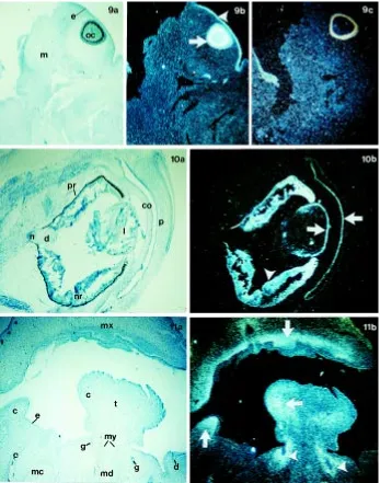
The expression pattern of PAX3 and PAX6 genes was investigated in human embryos between the 6th and 9th developmental week Fig. 1. Transversal section through the head region of the 6-week human embryo.(a)
Under bright-field illumination the telencephalon (t), diencephalon (d), and optic cup (c), as well as the developing pituitary gland –infundibulum (i) and Rathke’s pouch (rp) close to the junction with pharynx are seen. The brain tissue consists of the ventricular zone (vz), the intermediate zone (iz) and the marginal zone (mz). (b) Under dark-field illumination PAX6 hybridization is detected in the ventricular zone of the telencephalon and diencephalon, in the optic cup, infundibulum and Rathke’s pouch (arrows).
Fig. 2. Transversal section through the head region of the 6-week human embryo.(a)
Under bright-field illumination mesencephalon (me) and rhombencephalon (ro) are seen. (b)
Under dark-field illumination PAX3 hybridization is observed in the ventricular zone at the mesencephalon-rhombencephalon border (arrows).
(during the period of the most intense organogenesis). Both genes were expressed in the developing central nervous system –brain and spinal cord. PAX6 was additionally expressed in the developing eye, while PAX3 in the derivatives of the neural crest– the spinal ganglia and the ectomesenchyme of the face. Sense and antisense probes for PAX3 and PAX6 were used as control probes.
Brain
In the 6th week of development the human brain already shows signs of division into five main parts: telencephalon, diencephalon, mesencephalon, metencephalon and myelencephalon.
Expression of PAX6 gene is seen in the ventricular zone of telencephalon and diencephalon. It is also expressed in the optic cup and stalk. PAX6 hybridization signal characterizes the in-fundibulum, the evagination of floor of diencephalon (prospective neurohypophysis) and Rathke’s pouch, an outpocketing of
deum (prospective adenohypophysis) (Fig. 1a,b). Transcripts of PAX3 gene are observed in the ventricular zone at the mesencephalic-rhomben-cephalic border in embryo of the same develop-mental age. (Fig. 2a,b).
At the same developmental age, expression of PAX6 gene is seen in the middle part of ventricular zone of medulla oblongata and within bilateral clusters of cells in the ventral intermediate zone (Fig. 3a.b). Expression of PAX3 gene is seen only in the dorsal part of the ventricular zone of medulla oblongata, and not in any part of the intermediate zone (Fig. 4a,b).
Spinal cord
The spinal cord of a 6-week human embryo shows three characteristic zones: the ventricular zone (prospective ependymal cells), the interme-diate zone (prospective gray matter) and the mar-ginal zone (prospective white matter).
PAX6 gene has a very weak hybridization signal in the middle part of the ventricular zone of a 6-week spinal cord (Fig. 5a,b). Very weak expression of PAX3 gene is observed in the dorsal part of the ventricular zone in embryo of the same develop-mental age (not shown). The same embryo displays clear expression of both PAX6 and PAX3 genes in specific areas of the brain (see Figs. 1,2,3, and 4). Subsequent differentiation of the spinal cord is associated with increase of hybridization signal of both PAX6 and PAX3 genes. At later developmen-tal stages, in the 8-9- week fetus, strong expression of PAX6 gene is seen in the ventral part of the ventricular zone, while weaker expression charac-terizes its middle part. The cluster of cells in the ventral intermediate zone (basal plate) also shows
and the pigmented and neural epithelium of developing retina and iris. Hybridization signal is not present in the optic disc and optic nerve (Fig. 10a,b).
Derivatives of pharyngeal arches (jaws, tongue)
Mesoderm which forms the embryonic connective tissue of the face is ectomesenchyme derived from the neural crest cells. The first pharyngeal arch is involved in the development of the face (including jaws), while the tongue develops from parts of all four pharyngeal arches (Sadler, 1995).
In the 8-9 week human fetus, PAX3 hybridization characterizes connective tissue (lamina propria) of the developing upper and lower jaw, and tongue. Expression is particularly strong in the connective tissue immediately below the epithelium and around the excretory ducts of salivary glands. Hybridization signal is not present in the surface epithelium, myoblasts and Meckel’s carti-lage (Fig. 11a,b).
Discussion
Investigations on the Pax genes during development of different animal species (Bopp et al., 1986; Dressler et al., 1988; Krauss et al., 1991; Goulding et al., 1993) and humans (Gerard et al., 1995; Terzic et al., 1998) disclosed important roles of those genes in body Fig. 3. Transversal section at the level of medulla oblongata (mo) of the 6-week human embryo.(a) Under bright-field illumination the medulla oblongata consists of the ventricular zone (vz), the intermediate zone (iz) and the marginal zone (mz). (b) Under dark-field illumination, expression of PAX6 gene is seen in the middle part of the ventricular zone (arrow) and in part of the ventral intermediate zone (arrowheads).
Fig. 4. Transversal section at the level of medulla oblongata (mo) of the 6-week human embryo. (a) Under bright-field illumination the medulla oblongata consists of the three characteristic zones (see Fig. 3). (b) Under dark-field illumination expression of PAX3 gene is seen in the dorsal part of the ventricular zone (arrows).
a strong hybridization signal. At the cervico-thoracic region, the hybridization signal in the ventricular zone is thinner (Fig. 6a,b) than at the thoraco-lumbar region (Fig. 7a,b) as differentiation of the spinal cord is more advanced in the cranial part of the embryo. Hybridization signal is excluded from the floor plate and roof plate areas.
In the 9-week fetus, the strong PAX3 expression is detected in the dorsal 2/3 of the ventricular zone and in the roof plate, as well as in the spinal ganglia and spinal nerves (Fig. 8a,b).
Eye
In the 4th week of development, the optic vesicle develops as an outgrowth of the diencephalon, the stem of the vesicle being the optic stalk. Subsequently, under inductive influence of the optic vesicle, the lens placode invaginates to form the lens vesicle. The optic vesicle becomes the double walled optic cup, giving rise to pig-mented (outer wall) and neural part (inner wall) of the retina (Sadler, 1995).
In the 6-week embryo, hybridization signal of PAX6 gene is seen in the surface ectoderm (prospective corneal epithelium) and in the neural and pigmented retinal layers of the optic cup (Fig. 9a,b,c). It is also seen in the optic stalk (prospective optic nerve)(not shown). Later on, the cells in the posterior wall of the lens vesicle elongate to form the lens fibers. In a 8-9-week fetus, PAX6
patterning. They were found to be involved in organogenesis of the central nervous system, sense organs, kidneys, pancreas, nose and limb muscles (Mansouri et al., 1994; Dahl et al., 1997; Terzic et al., 1998). Pax genes were also associated with appearance of different syndromes and oncogenesis in humans and with animal mutants (Wehr and Gruss, 1996). They are evolutionary highly conserved in structure and function as they have similar expres-sion patterns in different species, including man.
Due to numerous investigations in experimental animals, par-ticularly mouse, knowledge on Pax genes has significantly im-proved. During normal mouse development expression of Pax6 gene is seen in the developing spinal cord and brain. In the spinal cord it is found in the ventricular zone of the intermediate and basal plates, and in the part of ventral intermediate zone. Early Pax6 expression characterizes prosencephalon, mesencephalon and
rhombencephalon. Later on, it is seen in all parts of the brain except in the roof of mesencephalon (Callaerts et al., 1997).All structures of developing eye and the olfactory epithelium express Pax6 gene (Walther and Gruss, 1991). Recent analyses disclose the impor-tance of Pax6 for the formation of the forebrain (Stoykova et al., 1996) and pancreas (St Onge et al., 1997). In the homozygous mouse Pax6 mutant Small eye, the development of the eye and nose are affected. In human mutation aniridia, patients are suffer-ing from iris defects, but other eye structures are affected as well (Hill et al., 1991; Ton et al., 1991; Glaser et al., 1994).
Investigations on 3-6-week human embryos (Gerard et al., 1995) gave only general information on early expression of PAX genes in certain organs, but without specific details on their distribution within cell layers. Our study was done on later develop-mental stages (6-9 postovulatory weeks), describing changes in Fig. 5. Transversal section through the thoracic part of axial organs in the 6-week human embryo. (a)
Under bright-field illumination, three zones can be distinguished in the spinal cord (sc): the ventricular zone (vz), the intermediate zone (iz) and the marginal zone (mz). Spinal ganglia (sg), vertebral column (vc). (b)
Under dark-field illumination, a very weak hybridization signal of PAX6 gene is seen in the middle part of the ventricular zone (arrow).
Fig. 6. Transversal section through the cervico-thoracic level of axial organs in the 8-9 week human fetus. (a) Under bright-field illumination the spinal cord (sc) consists of a thin ventricular zone (vz) and the well developed intermediate (iz) and marginal zones (mz), roof plate (r) and floor plate (f). Spinal ganglia (sg), vertebral column (vc) and vertebral arches (va) are seen as well. (b) Under dark-field illumination PAX6 hybrid-ization signal is seen in the middle ventricular zone (arrow) except the floor plate area, and bilaterally in the ventral intermediate zone (arrowhead).
Fig. 7. Transversal section through the thoraco-lumbar level of axial organs in the 8-9 week human fetus. (a) Under bright-field illumination the ventricular zone (vz) of the spinal cord (sc) is thinner ventrally than dorsally. Ventral gray horns (vh) of the intermediate zone (iz) are better developed than dorsal horns (dh). Marginal zone (mz), spinal ganglia (sg), vertebral column (vc), vertebral arches (va). (b)
Under dark-field illumination PAX6 is expressed in the middle part of the ventricular zone (arrow) and bilaterally in part of the ventral gray horns (arrow-head).
Fig. 8. Transversal section through the thoracic level of axial organs in the 9-week fetus. (a) Under bright-field illumination the ventricular zone (vz) of the spinal cord (sc) is thin. The intermediate zone (iz) with well developed ventral (vh) and dorsal gray horns (dh), and the marginal zone (mz) are seen. Floor plate (f), roof plate (r), spinal ganglia (sg), vertebral column (vc), vertebral arches (va). (b) Under dark-field illumination PAX3 hybridization characterizes dorsal 2/3 of ventricu-lar zone (arrow) including the roof plate, and spinal ganglia (arrowheads).
expression pattern of PAX3 and PAX6 genes during later embryonic and early fetal period. In embryonic stage of human development, PAX6 was expressed in the mitotically active ventricular zone of telencephalon and dien-cephalon, as well as in the ventricular zone and part of ventral intermediate zone of myelencephalon and spinal cord. In mouse development, Pax6 respected anatomical borders of neuromeres, having a segment-like expression pattern in diencephalon (Stoykova and Gruss, 1994; Stoykova et al., 1996). The caudal limit of Pax6 expression in the human brain was at the diencephalon-mesencephalon border.
Interestingly, in our study the hybridiza-tion signal was much stronger in the brain than in the spinal cord of the same young human embryo. At later stages, increase of PAX6 expression coincided with the differ-entiation of the human spinal cord. In ani-mals, expression of Pax6 gene could be influenced by neurotrophin (Kioussi and Gruss, 1994) and by removal or implantation of the notochord. The notochord transplan-tation dramatically altered the dorsoventral expression pattern of both Pax6 and Pax3 genes (Goulding et al., 1993), suggesting that those genes participate in the establish-ment of dorso-ventral polarity of the spinal cord (Wehr and Gruss, 1996).
In our study, besides its presence in the nervous structures, PAX6 was also detected in the surface head ectoderm of the prospec-tive corneal and lens epithelium, and in the optic cup and stalk. At the end of the embry-onic period, the expression of PAX6 gene was missing in the optic disc and nerve (derivatives of optic stalk). Investigation on early human embryos (Gerard et al., 1995) could show only expression of PAX6 gene in the stage of eye vesicle. In animals, a de-crease in expression of Pax6 gene in pig-mented retinal epithelium and optic stalk was described at later stages of develop-ment (not investigated in our study). It is believed that Pax6 is not necessary for the formation of the optic cup, but is required at other stages of eye development (Macdonald and Wilson, 1996). Aniridia and Peters’ anomaly in humans displayed phenotypic features that correspond to the developmen-tal expression of PAX6 described in our study (Glaser et al., 1994; Hanson et al., 1994).
Transcripts of PAX6 gene were found in both Rathke’s pouch and infundibulum of the developing human pituitary gland. Until now, Pax6 was described only in the primordium
Fig. 9. Transversal section through the head region of the 6-week human embryo, at the level of the eye. (a) Under bright-field illumination section through the optic cup (oc), head mesenchyme (m) and surface epithelium (e) is seen. (b) Under dark-field illumination PAX6 hybridization charac-terizes optic cup (arrow) and head epithelium overlying the eye –prospective corneal epithelium (arrowhead). (c)Under dark-field illumination, the sense control shows no hybridization signal in the optic cup and prospective corneal epithelium. Remaining hybridization of pigmented epithelium is an artifact as it is visible also in every untreated section.
Fig. 10. Transversal section through the eye of the 8-9- week human fetus. (a) Under bright-field illumination the developing eye consists of the cornea (co), lens (l), neural (nr) and pigmented retina (pr). Optic disc (d), initial part of the optic nerve (n) and palpebrae (p) are seen. (b) Under dark-field illumination PAX6 hybridization is seen in the corneal and lens epithelium (arrows) and in both parts of retina (arrowhead).
of adenohypophysis (Rathke’s pouch) of mouse embryos (Walther and Gruss, 1991; Callaerts et al., 1997). In our study, at earlier developmental stages expression of PAX6 gene extended from diencephalon into the infundibulum as it represents the continua-tion of the brain structure. Interestingly, in Pax6 mouse mutants all tissues expressing Pax6 during development were affected except the pituitary gland (Callaerts et al., 1997). Therefore, other genes rather than Pax6 seem to have a crucial role during pituitary development.
Belonging to the group of early expressed genes in animals, Pax3 is found to be active in the whole brain at early developmen-tal stages, but later its expression domain is restricted rostrally at the level of the diencephalon (Jostes et al., 1991; Goulding et al., 1993; Stoykova and Gruss, 1994). Pax3 is found in the ventricular zone, alar plates and the roof plate of the spinal cord, as well as in the neural crest cells and their derivatives. In the mesoderm, Pax3 is expressed in the primitive streak (Goulding et al., 1993) and in the undifferentiated somites. Later on, it characterizes dermomyotome and limb muscles. The Splotch mouse mutants exhibit spina bifida, exencephaly, neural crest and limb-muscle defects (Epstein et al., 1991; Franz et al., 1993; Tassabehji et al., 1993; Mansouri et al., 1996). Pax3 mutations have been corre-lated with human Waardenburg syndrome, which displays nu-merous defects of neural crest derivatives, deafness, pigmenta-tion deficiency and lateral displacement of the inner corner of the eye (Baldwin et al., 1992; Tassabehji et al., 1993). PAX3 has been found to be mutated in human tumor rhabdomyosarcoma (Barr et al., 1993; Shapiro et al., 1993).
Results of our investigation on human PAX3 gene accord with the described data in mouse embryos. In six-week human embryo, strong expression of PAX3 was found at the mesencephalic-rhombencephalic border and in the dorsal part of ventricular zone of medulla oblongata. These data indicate a role for PAX3 gene, together with other PAX genes in establishment of border between those two parts of the human brain. In mouse development, it is believed that Pax3, together with Pax6 participates in establish-ment of segestablish-mental boundaries in the brain and axonal pathways at the borders of their expression (Stoykova and Gruss, 1994). Similarly to PAX6 gene, hybridization signal of PAX3 was very weak in the dorsal ventricular zone of the spinal cord in early human embryos. With advancing differentiation of the spinal cord, hybrid-ization signal increased in dorsal part of ventricular zone and roof plate area. It also characterized spinal ganglia and spinal nerves (derivatives of the neural crest cells). Expression of PAX3 in dorsal parts of the spinal cord suggests its possible role during proper closure of the neural tube. Experimentally, expression of Pax3 can
be changed by extirpation or implantation of an additional noto-chord (Goulding et al., 1993). Morphological studies on human embryos with spina bifida or anencephaly showed that anomalies of the central nervous system are always combined with vertebral and notochord abnormalities (positional changes, dispersed or supernumerary notochord) indicating a role for the notochord in genesis of axial disorders (Babic and Saraga, 1993; Saraga-Babic et al., 1996a,b). Normal spatial relationship between the axial organs at critical stages of development seems to be of essential importance for expression of genes controlling proper axial development (Saraga-Babic et al., 1993, 1996a,b; Monsoro-Burq et al., 1995). In adult humans with neural tube defects, a PAX3 gene variation was detected. An increased, but insignificant fre-quency of the rare allele of a silent polymorphism in exon 2 was found in such patients (Hol et al., 1996).
As previously reported for Pax3 in mouse development (Stoykova and Gruss, 1994), expression of human PAX3 gene was detected within the derivatives of branchial arches –maxilla, mandible and tongue (the limb development was not in the focus of this study). Previous reports on human embryos did not describe such expres-sion (Gerard et al., 1995). During normal human development, neural crest cells invade the central part of pharyngeal arches giving rise to skeletal structures, connective tissue of the face, teeth, stromal components of the glands etc. The muscles of the head and neck differentiate from somitomeres in the head region, e.g. from the original mesoderm of pharyngeal arches. However, further formation of myoblasts is modified by the surrounding mesenchyme derived from neural crest cells (Fitzgerald and Fitzgerald, 1994; Sadler, 1995). Contrary to muscles in the head, differentiation of limb muscles is influenced by the parietal (meso-dermal) mesenchyme. Different origin of mesenchyme which modifies the formation of limb and face muscles might be one of possible explanations for the absence of face abnormalities in Splotch Pax3 mutants. Another possibility is specific expression of paralogous Pax7 gene in this region (Jostes et al., 1991).
Although highly resembling descriptions in experimental ani-mals, human PAX6 and PAX3 genes displayed small differences in their expression pattern when compared to other species. It refers to the weak expression of both genes in the spinal cord of early human embryos. Unlike in other species, PAX6 was found to be active in the infundibulum of the future pituitary gland. In the eye, it disappeared from the optic stalk earlier than in corresponding stage of mouse development. Expression of PAX3 gene only in the facial connective tissue of pharyngeal arches indicates that other genes rather than PAX3 have an important role during face and neck development. Further studies focusing on the limb develop-ment and analysis of brain structures in humans should give more information on the developmental roles of PAX6 and PAX3 genes.
Materials and Methods
Human material
Normal human embryos and fetuses between 6-9 developmental weeks were collected after spontaneous or legal abortions from the Department of Gynecology and Obstetrics, Clinical Hospital Split, Croatia. The embry-onic tissues were treated as postmortal material with permission of hospital’s Drug and Ethical Committee.
To ensure the use of healthy material in our study, we controlled anamnestic data of women that underwent abortion. In order to eliminate the malformed or poorly preserved embryonic tissue, collected embryos
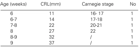
TABLE 1
AGE AND NUMBER OF HUMAN EMBRYOS AND FETUSES USED IN THIS STUDY
Age (weeks) CRL(mm) Carnegie stage No
and fetuses were first examined macroscopically and then microscopically in sections stained with hematoxylin and eosin.
The postovulatory age was estimated from menstrual data, correlated with the crown-rump length (CRL) and Carnegie stages (Moore and Persaud, 1993) (Table 1).
Embryos were dissected into two or three pieces, fixed in 4% paraform-aldehyde in phosphate buffer for several hours and embedded in paraffin. Tissue blocks were serially cut in transversal direction and mounted on glass-slides coated with Chrome alum (Serva) gelatin.
In situ hybridization
The slices were processed through the following steps: dewaxing in xylene, dehydration, washing in PBS, refixing in 4% PFA, washing, protein-ase-K treatment (0,02 mg/ml), washing, 4% PFA, washing, 0,1,M trietha-nolamine treatment, washing and dehydration.
S35labeled probe, specific for PAX3 and PAX6 , was synthesized using T3-polymerase, according to the supplier’s direction (Promega) from corresponding linearized plasmid templates as described in Goulding et al. (1991) for PAX3 and in Walther and Gruss (1991) for PAX6. Probe (1x10⁸ cpm/ml) was dissolved in hybridization buffer (300 mM NaCl, 10 mM dithiothretiol-DDT, 10% dextran sulphate, 50% formamide, 2 mg/ml bovine serum albumine, 2 mg/ml Ficoll, 2 mg/ml polyvinylpyrrolidine).
The hybridization mix was boiled, applied directly onto the section, and covered with siliconized coverslips. After overnight hybridization at 55°C, the following washing procedure was used: 2 x saline-sodium citrate (SSC), 50% formamide, 10 mM beta-mercaptoethanol (15 min. 37°C); 2xSSC, 50% formamide, 10 mM beta-mercaptoethanol (30 min 65°C); 0,5 M NaCl, 10 mM Tris, 5 mM EDTA (15 min 37°C); 0,5 M NaCl 10 mM Tris, 5 mM EDTA (15 min 37°C); 2xSSC, 50% formamide, 10 mM beta-mercaptoethanol (30 min. 37°C); 2xSSC (15 min. room temp.); 0,1xSSC (15 min at room temperature). The sections were dehydrated in ethanol and air dried.
For autoradiography, slices were dipped in Kodak NTB-2 emulsion dilute 1:1 with water. Slices were exposed for up to 21 days and developed in Kodak D-19 solution. For morphological analysis the sections were stained with Giemsa, coverslips were mounted with Eukitt. Photomicro-graphs were taken with bright/dark field microscope.
Acknowledgments
We thank Ms. Asja Miletic and Ms. Christiane Muller for skillful technical assistance. This work is supported by the Ministry of Science and Technol-ogy of the Republic of Croatia (Grant No 108 194). Mirna Saraga-Babic and Janos Terzic were supported by the ESF fellowships. We wish to thank Professor Peter Gruss for his introduction into methods and help in carrying out this investigation, and Dr. Anastassia Stoykova for critical reading of the text and valuable suggestions.
References
ASANO, M. and GRUSS, P. (1992). Pax-5 is expressed at the midbrain-hindbrain boundary during mouse development. Mech. Dev. 33: 27-38.
BALDWIN, C.T., HOTH, C.F., AMOS, J.A., DA-SILVA, E.O. and MILUNSKY, A. (1992). An exonic mutation in the HuP2 paired domain gene causes Waardenburg’s syndrome. Nature 355: 637-638.
BARR, F.G., GALILI, N., HOLICK, J., BIEGEL, J.A., ROVERA, G, and EMANUEL, B.S. (1993). Rearrangement of the PAX3 paired box gene in the pediatric solid tumor alveolar rhabdomyosarcoma. Nature Genet. 3: 113-117.
BOPP, D., BURRI, M., BAUMGARTNER, S., FRIGERIO, G. and NOLL, M. (1986). Conservation of a large protein domain in the segmentation gene paired and in functionally related genes in Drosophila. Cell 47: 1033-1040.
CALLAERTS, P., HALDER, G. and GEHRING, W.J. (1997). Pax-6 in development and evolution. Annu. Rev. Neurosci. 20: 483-532.
CHALEPAKIS, G., GOULDING, M., READ, A., STRACHAN, T. and GRUSS, P. (1994). Molecular basis of splotch and Waardenburg Pax3 mutations. Proc. Natl.
Acad. Sci. USA 91: 3685-3689.
DAHL, E., KOSEKI, H. and BALLING, R. (1997). Pax genes and organogenesis. Bioessays 19: 755-765.
DEUTSCH, U. and GRUSS, P. (1991). Murine paired domain proteins as regulatory factors of embryonic development. Semin. Dev. Biol. 2: 413-424.
DRESSLER, G.R., DEUTSCH, U., BALLING, R., SIMON, D., GUENET, J.L. and GRUSS, P. (1988). Murine genes with homology to Drosophila segmentation genes. Development 104 (Suppl.): 181-186.
EPSTEIN, D.J., VEKEMANS, M. and GROS, P. (1991). Splotch (Sp2h), a mutation affecting development of the mouse neural tube shows a deletion within the paired homeodomain of Pax3. Cell 67: 767-774.
EPSTEIN, D.J., VOGAN, K.J., TRASLER, D.G. and GROS, P. (1993). A mutation within intron 3 of the Pax3 gene products aberrantly splices mRNA transcripts in the Splotch (Sp) mouse mutant. Proc. Natl. Acad. Sci. USA 90: 532-536.
FITZGERALD, M.J.T. and FITZGERALD, M. (1994). Head and neck: pharyngeal arches, pouches and clefts. In Human Embryology (Ed. W.B. Saunders), Balliere Tindall, London, pp. 55-58.
FRANZ, T., KOTHARY, R., SURANI, M.A.H., HALATA, Z. and GRIM, M. (1993). The Splotch mutation interferes with muscle development in the limbs. Anat. Embryol. 187: 153-160.
GERARD, M., ABITBOL, M., DELEZOIDE, A.L., DUFIER, J.L., MALLET, J. and VEKEMANS, M. (1995). PAX-genes expression during human embryonic devel-opment, a preliminary report. C.R. Acad. Sci. Paris 318: 57-66.
GLASER, T., JEPEAL, L., EDWARDS, J.G., YOUNG, R., FAVOR, J. and MAAS, R.L. (1994). Pax6 gene dosage effect in a family with congenital cataracts, aniridia, anophtalmia and central nervous system defects. Nature Genet. 7: 463-471.
GOULDING, M.D., CHALEPAKIS, G., DEUTSCH, U., ERSELIUS, J.R. and GRUSS, P. (1991). Pax3, a novel murine DNA binding protein expressed during early neurogenesis. EMBO J. 10: 1135-1147.
GOULDING, M.D., LUMSDEN, A. and GRUSS, P. (1993). Signals from the notochord and floor plate regulate the region-specific expression of two Pax genes in the developing spinal cord. Development 117: 1001-1016.
HANSON, I.M., FLETCHER, J.M., JORDAN, T., BROWN, A., TAYLOR, D., ADAMS, R.J., PUNNET, H.H. and VAN HEYNINGEN, V. (1994). Mutations at the PAX6 locus are found in heterogeneous anterior segment malformations including Peters’ anomaly. Nature Genet. 6: 168-173.
HILL, R.E., FAVOR, J., HOGAN, B.L.M., TON, C.C.T., SAUNDERS, G.F., HANSON, I.M., PROSSER, J., JODAN, T., HASTIE, N.D. and VAN HEYNINGEN, V. (1991). Mouse Small eye results from mutations in a paired-like homeobox-containing gene. Nature 354: 522-525.
HOL, F.A., GEURDS, M.P., CHATKUPT, S., SHUGART, Y.Y., BALLING, R., SCHRANDER-STUMPEL, C.T., JOHNSON, W.G., HAMEL, B.C. and MARIMAN, E.C. (1996). PAX genes and human neural tube defects: an amino acid substitu-tion in PAX1 is a patient with spina bifida. J. Med. Genet. 33: 655-660.
JOSTES, B., WALTHER, C. and GRUSS, P. (1991). The murine paired box gene, Pax7, is expressed specifically during the development of th nervous and muscular system. Mech. Dev. 33: 27-38.
KIOUSSI, C. and GRUSS, P. (1994). Differential induction of Pax genes by NFG and BDNF in cerebellar primary clusters. J. Cell Biol. 125: 417-425.
KRAUSS, S., JOHANSEN, T., KORZH, V. and FJOSE, A. (1991). Expression pattern of zebrafish pax genes suggests a role in early brain regionalization. Nature 353: 267-270.
LIEM, K.F., REMML, G., ROELINK, H. and JESSEL T.M. (1995). Dorsal differentiation of neural plate cells induced by BMP-mediated signals from epidermal ectoderm. Cell 82: 969-979.
MACDONALD, R. and WILSON, S.W. (1996). Pax proteins and eye development. Curr. Opin. Neurobiol. 6: 49-56.
MANSOURI, A., HALLONET, M. and GRUSS, P. (1996). Pax genes and their roles in cell differentiation and development. Curr. Opin. Cell Biol. 8: 851-857.
MANSOURI, A., STOYKOVA, A. and GRUSS, P. (1994). Pax genes in development. J. Cell Sci. 18 (Suppl.): 35-42.
MONSORO-BURQ, A.H., BONTOUX, M., VINCENT, C. and LE DOUARIN, N.M. (1995). The developmental relationship of the neural tube and the notochord: short and long term effects of the notochord on the dorsal spinal cord. Mech. Dev. 53: 157-170.
´
´
´
MOORE, K.L. and PERSAUD, T.V.N. (1993). Development of tissues, organs and body form. The fourth to eight weeks. In The developing human. Clinically oriented embryology. W.B. Saunders Company, Philadelphia, pp. 70-89.
NORNES, H.O., DRESSLER, G.R., KNAPIK, E.W., DEUTSCH, U. and GRUSS, P. (1990). Spatially and temporally restricted expression of Pax2 during murine neurogenesis. Development 109: 797-809.
SADLER, T.W. (1995). Langman’s Medical Embryology (Ed. J.N. Gardner), Williams and Wilkins, Baltimore.
SARAGA-BABIC, M. and SARAGA, M. (1993). Role of the notochord in the develop-ment of cephalic structures in normal and anencephalic human fetuses. Virchows Arch. (A) 422: 161-168.
SARAGA-BABIC, M., KROLO, M., SAPUNAR, D., TERZIC, J. and BIOCIC, M. (1996a). Differences in origin and fate between the cranial and caudal spinal cord during normal and disturbed human development. Acta Neuropathol. 91: 194-199.
SARAGA-BABIC, M., SAPUNAR, D. and STEFANOVIC, V. (1993). Histological features of axial structurse during embryonic and fetal stages of human craniora-chischisis. Acta. Neuropathol. 86: 289-294.
SARAGA-BABIC, M., STEFANOVIC, V., LEHTONEN, E., SAPUNAR, D., SARAGA, M. and WARTIOVAARA, J. (1996b). Neurulation mechanisms in the human development. Croatian Med. J. 37: 7-14.
SHAPIRO, D.N., SUBLETT, J.E., LI, B., DOWNING, J.R. and NAEVE, C.W. (1993). Fusion of PAX3 to a member of the forkhead family of transcription factors in human alveolar rhabdomyosarcoma. Cancer Res. 53: 5108-5112.
ST-ONGE, L., SOSA-PINEDA, B., CHOWDHURY, K., MANSOURI, A. and GRUSS, P. (1997). Pax6 is required for differentiation of glucagon-producing a-cells in mouse pancreas. Nature 387: 406-409.
STOYKOVA, A. and GRUSS, P. (1994). Roles of Pax-genes in developing and adult brain as suggested by expression patterns. J. Neurosci. 14: 1395-1412.
STOYKOVA, A., FRITSCH, R., WALTHER, C. and GRUSS, P. (1996). Forebrain patterning defects in Small eye mutant mice. Development 122: 3453-3465.
TASSABEHJI, M., READ, A.P., NEWTON, V.E., PATTEON, M., GRUSS, P., HAR-RIS, R. and STRACHAN, T. (1993). Mutations in the PAX3 paired box gene causing Waanderburg syndrome Type 1 and Type 2. Nature Genet. 3: 26-30.
TERZIC, J., MULLER, C., GAJOVIC, S. and SARAGA-BABIC, M. (1998). Expression of PAX2 gene during human development. Int. J. Dev. Biol. 42: 701-707.
TON, C.C.T., HIRVONEN, H., MIWA, H., WEIL, M.M., MONAGHAN, P., JORDAN, T., VAN HEYNINGEN, V., HASTIE, N.D., MEIJERS-HEIJBOER, H., DRECHSLER, M., ROYER-POKORA, B., COLLINS, F., SWAROOP, A., STRONG, L.C. and SAUNDERS, G.F. (1991). Positional cloning of a paired-box and homeobox-containing gene from Aniridia region. Cell 67: 1059-1074.
WALLIN, J., MIZUTANI, Y., IMAI, K., MIYASHITA, N., MORIWAKI, K., TANIGUCHI, M., KOSEKI, H. and BALLING, R. (1993). A new Pax gene, Pax9, maps to mouse chromosome 12. Mammal. Genome 4: 354-358.
WALTHER, C. and GRUSS, P. (1991). Pax6, a murine paired box gene, is expressed in the developing CNS. Development 113: 1435-1449.
WALTHER, C., GUENET, J-L., SIMON, D., DEUTSCH, U., JOSTES, B., GOULDING, M.D., PLACHOV, D., BALLING, R. and GRUSS, P. (1991). Pax: A murine multigene family of paired box containing genes. Genomics 11: 424-434.
WEHR, R. and GRUSS, P. (1996). Pax and vertebrate development. Int. J. Dev. Biol. 40: 369-377.
Received: February 1999 Accepted for publication: September 1999
´
´ ´ v ´
´ ´