Electronic Thesis and Dissertation Repository
7-3-2012 12:00 AM
Individual Differences in White Matter Microstructure Predict
Individual Differences in White Matter Microstructure Predict
Mathematical Achievement
Mathematical Achievement
Anna A. Matejko
The University of Western Ontario
Supervisor Dr. Daniel Ansari
The University of Western Ontario
Graduate Program in Psychology
A thesis submitted in partial fulfillment of the requirements for the degree in Master of Science © Anna A. Matejko 2012
Follow this and additional works at: https://ir.lib.uwo.ca/etd
Part of the Behavior and Behavior Mechanisms Commons Recommended Citation
Recommended Citation
Matejko, Anna A., "Individual Differences in White Matter Microstructure Predict Mathematical Achievement" (2012). Electronic Thesis and Dissertation Repository. 610.
https://ir.lib.uwo.ca/etd/610
This Dissertation/Thesis is brought to you for free and open access by Scholarship@Western. It has been accepted for inclusion in Electronic Thesis and Dissertation Repository by an authorized administrator of
ACHIEVEMENT
(Spine title: Brain Microstructure Predicts Mathematical Achievement) (Thesis format: Monograph)
by
Anna A. Matejko
Graduate Program in Psychology
A thesis submitted in partial fulfillment of the requirements for the degree of
Master of Science
The School of Graduate and Postdoctoral Studies The University of Western Ontario
London, Ontario, Canada
ii
School of Graduate and Postdoctoral Studies
CERTIFICATE OF EXAMINATION
Supervisor
______________________________ Dr. Daniel Ansari
Supervisory Committee
______________________________
Examiners
______________________________ Dr. Rhodri Cusack
______________________________ Dr. Jessica Grahn
______________________________ Dr. Jonathan Fugelsang
The thesis by
Anna Alexandra Matejko
entitled:
Individual Differences in White Matter Microstructure Predict
Mathematical Achievement
is accepted in partial fulfillment of the requirements for the degree of
Master of Science
______________________ _______________________________
Date Chair of the Thesis Examination Board
iii
Abstract
The current study uses diffusion tensor imaging to test whether individual differences in
white matter predict performance on the math subtest of the preliminary Scholastic Aptitude
Test (PSAT). Grade 10 and 11 PSAT scores were obtained from 30 young adults (ages
17-18) with wide-ranging math achievement levels. Tract based spatial statistics was used to
examine the correlation between PSAT math scores, fractional anisotropy (FA), radial
diffusivity (RD) and axial diffusivity (AD). FA in left parietal white matter was positively
correlated with math PSAT scores (specifically in the left superior longitudinal fasciculus,
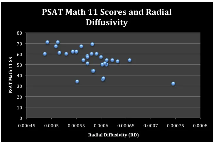
left superior corona radiata, and left corticospinal tract). Furthermore, RD, but not AD, was
correlated with PSAT math scores in these white matter microstructures. The negative
correlation with RD suggests increased myelination in participants with higher PSAT math
scores. Individual differences in FA and RD may reflect variability in experience dependent
plasticity over the course of learning and development.
Keywords
iv
Acknowledgments
First of all, I would like to thank my supervisor Dr. Daniel Ansari. It is impossible to
overstate my gratitude for his endless patience, guidance, and enthusiasm. He has provided
me with numerous opportunities to grow academically for which I will be forever indebted,
and his enthusiasm always inspires me to learn more. Daniel is more than I could have ever
asked for in a supervisor and I will always be grateful for his mentorship and encouragement.
I would also like to extend my sincerest gratitude to Dr. Gavin Price and Dr. Michele
Mazzocco for their feedback and advice throughout this work. I am very grateful to have had
the opportunity to work with both of them.
A special round of thanks goes to my colleagues: Bea Goffin, Christian Battista, Ian
Holloway, Nadia Nosworthy, Stephan Vogel, and Stephanie Bugden, for their stimulating
discussions, advice, and support. Late nights at the lab would not be the same without you.
Most importantly, I would like to give thanks to my family and friends. There are not
enough words to express how grateful I am for their boundless love and support. Thank you
v
Table of Contents
CERTIFICATE OF EXAMINATION... ii
Abstract ... iii
Acknowledgments... iv
Table of Contents... v
List of Tables ... vii
List of Figures ... viii
List of Appendices ... ix
1 Introduction... 1
1.1 The Calculating Brain: Evidence from Neuropsychology and Functional Brain Imaging ... 2
1.1.1 Evidence From Neuropsychological Patients and Early Brain Imaging .... 3
1.1.2 The Fronto-Parietal Network for Calculation ... 4
1.1.3 The Development and Specialization of Left Parietal Circuits for Mathematical Processing ... 9
1.2 Diffusion Tensor Imaging... 11
1.2.1 Properties of Water Diffusion and Anisotropy ... 11
1.2.2 Sources of Anisotropy... 12
1.2.3 Measuring and Calculating the Diffusion Tensor... 13
1.2.4 Diffusion Parameters ... 15
1.2.5 Methods of Analyzing DTI Data ... 17
1.3 Genetic and Environmental Determinants of Individual Differences in White Matter Integrity ... 18
1.4 White Matter Microstructure and Cognitive Function... 19
1.5 White Matter Correlates of Mathematical Cognition ... 22
vi
1.5.3 Summary: Mathematical Cognition and White Matter... 28
1.6 Current Study ... 29
2 Method ... 30
2.1 Participants... 30
2.2 Preliminary Scholastic Aptitude Test ... 31
2.3 MRI Acquisition ... 32
2.4 Analyses... 33
3 Results... 35
3.1 TBSS: Fractional Anisotropy... 35
3.2 TBSS: Radial and Axial Diffusivity ... 38
4 Discussion ... 42
4.1 Limitations & Future Directions... 48
5 Summary and Conclusions... 50
vii
List of Tables
viii
List of Figures
Figure 1: Neural network associated with number and arithmetic processing ... 5
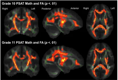
Figure 2: White matter structures showing a positive correlation with FA and PSAT math
scores in Grade 10 and Grade 11... 35
Figure 3: White matter structures showing a positive correlation with FA and PSAT math
scores in Grade 10 and Grade 11, correcting for age and PSAT Critical Reading... 36
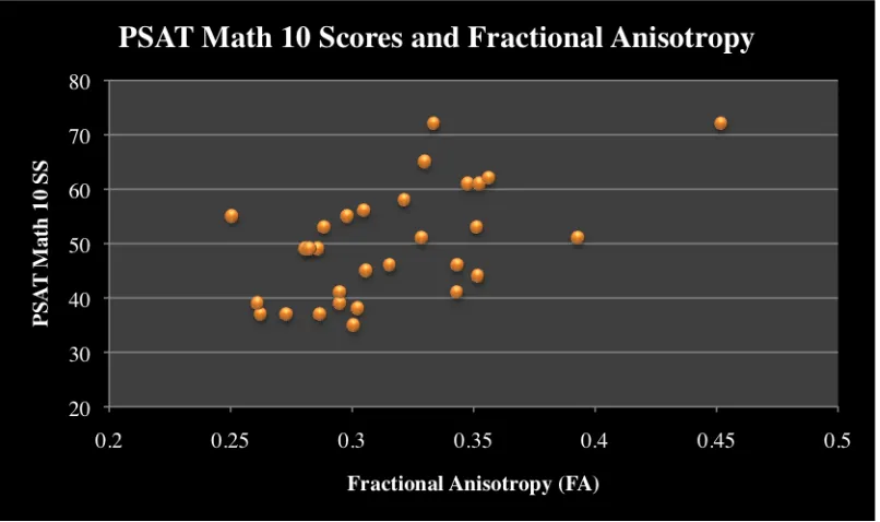
Figure 4: PSAT Math 10 scores and fractional anisotropy... 37
Figure 5: PSAT Math 11 scores and fractional anisotropy... 38
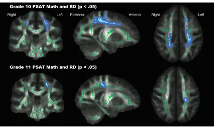
Figure 6: White matter structures showing a negative correlation with RD and PSAT math
scores in Grade 10 and Grade 11, correcting for age and PSAT Critical Reading... 39
Figure 7: White matter structures showing a negative correlation with RD and PSAT math
scores in Grade 10 (p < .03)... 40
Figure 8. PSAT Math 10 and radial diffusivity. ... 40
ix
List of Appendices
1
Introduction
Mathematical skills are of critical importance academically and in everyday life
(Bindler & Bayne, 1991; Bynner & Parsons, 2006; Cartwright, 1996; Parsons & Bynner,
2005). Research has begun to chart the development of mathematical skills and the neural
correlates of complex skills such as arithmetic and calculation. Yet, the neural correlates
of mathematical skills are relatively understudied compared to other cognitive domains.
For example, the functional and anatomical neural underpinnings of reading are
delineated in much more detail (Ben-Shachar, Dougherty, & Wandell, 2007; Price, in
press). Functional and structural neuroimaging techniques are useful to constrain and
inform our understanding of cognitive processes, such as mathematics, and can help
provide neuronally plausible theories of mathematical development. Therefore, a more
complete understanding of the neural circuits involved in mathematical skills will provide
a clearer picture of this complex skill.
Functional neuroimaging has provided converging evidence to suggest that
distinct parietal circuits become increasingly specialized for number and mathematical
processing over development (Ansari, 2008). However, research on the neural
underpinnings of mathematical skills has primarily focused on understanding brain
function during math and number tasks. Comparatively less is known about brain
structure underlying mathematical skills. Only a handful of studies have examined how
white matter microstructures are related to math competency even though connectivity
The study presented in this thesis aims to further elucidate the neural mechanisms
underlying mathematical skills. Specifically, this study focuses on how individual
differences in white matter integrity may be related to mathematical abilities in high
school students. Using Diffusion Tensor Imaging (DTI) to assess white matter integrity,
we aim to determine which white matter tracts are associated with mathematical
achievement. The introduction will begin with a review of the neural and anatomical
correlates of mathematical cognition. This will provide a background and framework for
the present study. First, there will be a brief summary of literature examining the
functional correlates of mathematical skills such as arithmetic and calculation. Second,
the basic principles of diffusion tensor imaging will be reviewed to provide a background
on the methodology used in this study. Third, an overview will be provided on how
diffusion tensor imaging has been used in the past to assess the white matter structures
associated with cognitive functioning. Finally, the available literature on the white matter
correlates of numerical and mathematical cognition will be summarized which will lead
into a description of the present study and how it expands upon previous research on
mathematical cognition and neuroimaging.
1.1 The Calculating Brain: Evidence from Neuropsychology
and Functional Brain Imaging
There is a long history of research exploring the cortical regions supporting
mathematical processing such as calculation (for a review see: Ansari, Holloway, Price,
& van Eimeren, 2008). To date, this research has primarily focused on the functional
combined structural and functional imaging techniques (Kim & Kim, 2005; Olesen,
Nagy, Westerberg, & Klingberg, 2003; van Eimeren et al., 2010), brain structure and
function cannot be thought of as separate entities. The brain acts as an integrated and
interacting system, and studies on brain structure and function should inform each other
on the mechanisms underlying a particular cognitive process. The following section will
provide a brief review of neuropsychological and fMRI literature of mathematical skills
with the intention of providing a framework for the research in this thesis.
1.1.1 Evidence From Neuropsychological Patients and Early Brain
Imaging
It has long been known that the parietal lobe is critical for numeracy. Disorders of
calculation and arithmetic (acalculia) were first systematically studied in the early 1900’s
by Henschen (Grewel, 1952; Henschen, 1919). Since acalculia was observable following
lesions to many regions of the cortex, Henschen concluded that many regions were
essential for calculation, and that calculation was distinguishable from other skills such as
language and music (Henschen, 1919). Later, studies on neuropsychological patients who
had acquired brain damage to the parietal cortex linked these localized lesions to specific
deficits in calculation (Cipolotti, Butterworth, & Denes, 1991; Gerstmann, 1940; Jackson
& Warrington, 1986). Moreover, Gerstmann syndrome is characterized by a collection of
cognitive deficits resulting from a lesion to the angular gyrus in the parietal lobe, usually
in the left hemisphere (Gerstmann, 1940). One of the effects is a particular deficit in the
patient’s ability to calculate.
With the advances of functional neuroimaging, more detailed examinations of
calculation in the human brain could be made. Calculation was one of the first cognitive
regional increases in cerebral blood flow during a calculation task using the intracarotid
133Xe injection technique. They found increased blood flow in the left and right angular
gyrus along with prefrontal regions. These findings have largely been replicated in later
fMRI studies. Similarly, Rueckert et al. (1996) found activation in the angular and
supramarginal gyri during subtraction, with greater activation in the left hemisphere than
the right. Studies on multiplication also reveal predominantly left-hemisphere activation
in the prefrontal cortex, and the inferior and superior parietal cortex (Dehaene et al.,
1996; Rickard et al., 2000). These early studies highlighted the importance of prefrontal
and parietal activation in calculation. Moreover, they primarily reveal a left lateralized
network of brain regions engaged during arithmetic tasks.
1.1.2 The Fronto-Parietal Network for Calculation
In more recent years, a network of regions including the intraparietal sulcus,
superior parietal lobule, angular gyrus, supra-marginal gyrus, and inferior, middle and
superior frontal gyri have been associated with numerical and mathematical processing
(Ansari, 2008; Arsalidou & Taylor, 2011; Cohen Kadosh, Lammertyn, & Izard, 2008;
Dehaene, Piazza, Pinel, & Cohen, 2003). Based on a recent meta-analyses, it is known
that a large fronto-parietal network is involved in calculation regardless of the
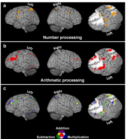
Figure 1. Neural network associated with number and arithmetic processing (adapted from Arsalidou and Taylor (2011)). Activation likelihood estimation maps show brain regions that were consistently activated across multiple studies examining (a) number processing (b) arithmetic processing, combining multiple operations, and (c) arithmetic processing differentiating between addition, subtraction and multiplication.
Calculation is a complex skill that is not process pure (Ansari et al., 2008). It
requires many cognitive processes including attention, working memory, and processing
frontal regions supports domain-general processes such as working memory, attention
and response-selection, whereas parietal activation is calculation specific (Dehaene,
1996). Therefore, calculation needs to be broken down into subcomponents to understand
the role of individual regions in this large fronto-parietal network (Ansari et al., 2008).
To test this hypothesis, Menon, Rivera, White, Glover, and Reiss (2000) modulated
numerically relevant stimuli and task difficulty by varying the presentation rate (task
difficulty) and changing the number of operands in an arithmetic verification task. In
arithmetic verification, the participant is presented with an arithmetic problem and a
solution. The participant must judge whether the solution is correct or incorrect. Rate of
problem presentation modulated activity in the oribitofrontal and insular cortex, whereas
the main effect of manipulating the number of operands was specific to the left and right
angular gyrus and adjoining intraparietal sulcus. These results provide empirical support
to the theory that prefrontal regions are primarily involved in domain general processes
that respond to task difficulty, whereas the parietal cortex responds to calculation specific
processes.
Different regions of the parietal cortex appear to be involved in mathematical
processing depending on the task and the type of strategy being used. Studies have
attempted to disambiguate the role of the above-mentioned regions during mathematical
tasks. For example, the angular gyrus is hypothesized to be involved in the retrieval of
arithmetic facts as opposed to manipulating quantities during calculation. To test how
retrieval versus procedural strategies modulate brain activation, Delazer et al. (2003)
trained participants on a set of multiplication problems. When comparing untrained vs.
activation. In contrast, the left angular gyrus had greater activation for trained vs.
untrained problems, suggesting a shift in activation following training. This shift is likely
a consequence of a greater reliance on automatic fact retrieval. Additional evidence from
Ischebeck et al. (2006) demonstrated that this shift may be specific to the mathematical
operation. Greater activation was found in the left angular gyrus for trained multiplication
problems compared to untrained problems, but not for subtraction problems. While
training multiplication problems increases the likelihood of using retrieval strategies,
subtraction still requires the manipulation of quantities to solve the problem, potentially
explaining differences in activation for these two operations (Ischebeck et al, 2006).
A later fMRI study examined differencess in activation depending on the strategy
being used by using self-reports to assess procedural versus retrieval strategies (Grabner
et al., 2009). Stronger activation in the left angular gyrus was present for trials where
participants reported using fact retrieval, whereas more widespread activation was
observed in the fronto-parietal network for trials for which participants indicated the use
of a procedural strategy, such as calculation. Furthermore, the extent to which individuals
recruit the angular gyrus has been related to individual differences in mathematical
achievement (Grabner et al., 2007). Those who scored higher on a standardized test of
mathematics recruited the angular gyrus less during single- and multi-digit multiplication
(Grabner et al., 2007). Even after accuracy and response time were controlled for, these
effects remained, suggesting that the individual differences in the recruitment of the
angular gyrus do not merely reflect individual differences in performance and are instead
The intraparietal sulcus is also considered an essential part of the network
responsible for mathematical skills (Ansari, 2008). Both human and animal research has
shown that the intraparietal sulcus plays a critical role in numerical magnitude processing
(Holloway & Ansari, 2010; Nieder & Miller, 2004). Several transcranial magnetic
stimulation (TMS) studies have demonstrated the importance of the intraparietal sulcus
by creating virtual lesions to the region and then observing performance changes on
mathematical tasks (Göbel, Walsh, & Rushworth, 2001; Rusconi & Umiltà, 2008;
Sandrini & Rusconi, 2008). To examine the role of the intraparietal sulcus and the
posterior superior parietal lobule in multiplication and subtraction, Andres, Pelgrims,
Michaux, Olivier, and Pesenti (2011) induced virtual lesions to these regions. Inhibiting
activity in the intraparietal sulcus, but not the posterior superior parietal lobule, caused
larger response latencies for multiplication and subtraction problems. Moreover,
participants had increased error rates for multiplication problems. Even though
multiplication often relies heavily on retrieval of arithmetic facts, these results show that
the intraparietal sulcus still plays a critical role in this operation.
The fronto-parietal network for arithmetic has also been found to be important in
advanced mathematical skills. In an fMRI study examining the neural correlates of
integral calculus problem solving, Krueger, Spampinato, and Pardini (2008) found that
solving integrals activated a left-lateralized network that included the intraparietal sulcus,
posterior superior parietal lobe, the posterior cingulate cortex, and the dorsolateral
prefrontal cortex. Thus, higher-level mathematical skills rely on a similar set of regions
arithmetic. Such findings may suggest that higher math skills are scaffolded on the neural
circuitry underlying more basic numerical processing.
1.1.3 The Development and Specialization of Left Parietal Circuits for
Mathematical Processing
Converging evidence suggests that parietal circuits become increasingly
specialized for number magnitude processing and calculation over development (Ansari,
2008). A frontoparietal shift is evident from childhood to adulthood for calculation;
activation in the frontal cortex decreases with age while the parietal cortex becomes more
engaged (Rivera, Reiss, Eckert, & Menon, 2005). Indeed, several of the aforementioned
studies on adults demonstrate that regions in the left parietal cortex are important for
calculation and mathematical skills (Arsalidou & Taylor, 2011; Delazer et al., 2003;
Gerstmann, 1940; Ischebeck et al., 2006; Rueckert et al., 1996).
In order to examine the developmental changes associated with mental arithmetic,
Rivera, Reiss, Eckert, and Menon (2005) used an arithmetic verification task with
addition and subtraction. Participants were presented with arithmetic problems (i.e. 6 – 2
= 4), and were required to respond only if the solution was correct. They found a negative
correlation with age in the anterior cingulate cortex, dorsolateral and ventrolateral
prefrontal cortex, indicating that older participants recruited these regions less than
younger participants during mental arithmetic. In contrast, Rivera et al. (2005) found a
positive correlation with age in the left parietal cortex including the supramarginal gyrus,
anterior intraparietal sulcus, and the left lateral occipital temporal cortex. The authors
suggest younger participants require additional cognitive resources to support their
participants. This is supported by the fact that younger participants also recruited regions
known to be involved in procedural and declarative memory including the hippocampus
and basal ganglia. These findings are particularly convincing since both younger and
older participants had similar accuracy. Furthermore, Rivera et al. (2005) found no
relationship between grey matter density age related changes in activation, ruling out the
possibility that developmental changes in brain activation are a consequence of changes
in brain structure.
The hypothesis of a fronto-parietal shift over development overlaps with the
above-mentioned arithmetic training studies. With training (or development), the
prefrontal cortex is recruited less because task difficulty demands are deceasing, resulting
in a smaller load on domain general processes such as working memory and attention
(Delazer et al., 2003; Ischebeck et al., 2006; Menon et al., 2000). Consistent with this
hypothesis, Rivera et al. (2005) found greater recruitment of the left posterior parietal
cortex with age, and decreasing recruitment of the prefrontal cortex. Overall, these
findings converge to support the notion that there is specialization of the left parietal
cortex for mathematical skills. However, they also highlight the importance of examining
the neural correlates of mathematical processing as a dynamic and changing network.
Changes in this network are evident over development (Rivera et al., 2005) and training
(Delazer et al., 2003; Ischebeck et al., 2006).
Mathematical skills are complex and require knowledge of symbolic numbers,
and the ability to mentally manipulate the quantitative referents of these symbolic
representations in the context of mathematical operations. Therefore it is unlikely that
these regions could constrain our understanding of mathematical processing in the
typically developing brain. Diffusion tensor imaging is a useful tool to examine the
connectivity between cortical regions and may help define the neural networks
underlying mathematical processing.
1.2 Diffusion Tensor Imaging
The present study uses Diffusion Tensor Imaging (DTI) to assess white matter
integrity to determine whether individual differences in white matter predict
mathematical achievement. The purpose of the next section is to provide a brief overview
of the theoretical and methodological background of DTI in order to describe the
methods presented in the present study. The aim is to clarify and explain how DTI
parameters are produced and what the limitations are of this methodology.
1.2.1 Properties of Water Diffusion and Anisotropy
White matter consists of bundles of axons, or tracts, that connect proximal and
distal regions of the brain (Oishi, Faria, Zijl, & Mori, 2011). These white matter tracts
cannot be differentiated in conventional MRI (T1 or T2 scans). DTI isa non-invasive
MRI technique that can be used to measure white matter integrity and provide
information about the axonal organization of the brain (Basser, Mattiello, & LeBihan,
1994; Mori & Zhang, 2006). DTI measures Brownian motion, or water diffusion.
Brownian motion is the principle that water molecules are in constant motion and a given
molecule remains in a particular location for a fixed period of time before moving to a
new random location (Jones, 2008). While it may be impossible to characterize a single
of many molecules from their starting point over a fixed period of time (Jones, 2008).
Diffusion imaging measures the mean displacement of water molecules in a particular
voxel for a fixed period of time (Jones, 2008). By measuring the amount of displacement,
it is possible to make inferences about the surrounding neuroanatomical structures.
Normally diffusion is isotropic when it is unrestricted (in free water or pure liquid),
meaning that water diffuses freely along multiple directions with no primary axis. The
presence of cell membranes, myelin sheath, or a larger density of axons presents
boundaries to diffusion and will influence the total amount and direction of diffusion
(Beaulieu, 2002). Water is more likely to diffuse along a white matter tract than across it.
Consequently, when measuring diffusion over a fixed period of time, the mean
displacement of water is reduced in white matter compared to regions that are more
unrestricted (Jones, 2008). Isotropic diffusion is characteristic of regions such as
cerebrospinal fluid or in grey matter where there are there are multiple fiber orientations
and no aligned fiber structures (Oishi et al., 2011). When the movement of diffusion is
along one primary direction (along one axis but not others), it is called anisotropic
diffusion (Mori & Zhang, 2006).
1.2.2 Sources of Anisotropy
Anisotropy depends on the geometry and structure of surrounding tissues
(Beaulieu, 2002) and typically implies a greater number of axons are oriented along one
direction. In a series of controlled experiments, Beaulieu and Allen demonstrated that the
microtubules, fast axonal transport, and induced gradients do not affect anisotropy in
white matter (Beaulieu & Allen, 1994a, 1994b, 1996). Studies on myelin deficient rats
sufficient for anisotropy (Gulani, Webb, Duncan, & Lauterbur, 2001; Johansen-Berg &
Rushworth, 2009). In another study, Takahashi et al. (2002) determined that diffusion is
more anisotropic in nerve columns with multiple axons and a smaller axonal diameter.
While there is still considerable debate over the neuroanatomical sources of anisotropy,
axonal membranes likely play a primary role and myelination modulates, but is not
sufficient, for anisotropic diffusion (Beaulieu, 2002).
1.2.3 Measuring and Calculating the Diffusion Tensor
In DTI, the MR sequence is sensitized to measure diffusion along multiple
directions. In an MRI, the magnetic field is as homogenous as possible and the precession
rate of protons is relatively homogenous. However, the homogeneity can be disturbed
linearly with a pulse field gradient (Mori & Barker, 1999). The strength, direction, and
time period of the gradient can all be controlled (Mori & Barker, 1999). With the first
dephasing gradient protons begin to precess at different rates along the direction of the
field gradient. A second rephasing gradient is applied in the same direction, but with
opposite magnitude (Mori & Barker, 1999). This rephasing gradient re-phases protons,
however, if the protons have moved in between the gradient pulses, then there is a drop in
signal (Mori & Barker, 1999). Therefore, if there is a drop in signal then diffusion is
present, and the greater the diffusion the greater the signal loss. For example, in
cerebrospinal fluid there is greater amounts of free diffusion, therefore the signal loss is
greater and the resulting image becomes darker (Mori & Barker, 1999). The DTI pulse
sequence is sensitized to multiple directions of diffusion. The resulting images are b
-weighted images, which have an image for every diffusion sensitization gradient.
constant in each direction. It is necessary to obtain at least 6 diffusion gradients in order
to calculate the diffusion tensor (Oishi et al., 2011). The diffusion tensor model is fitted
in each voxel, which approximates the displacement of diffusion with an ellipsoid.
Diffusion that is equal in all directions is represented by spherical ellipsoids, whereas
diffusion along one primary direction (anisotropic diffusion) is represented by a more
elongated ellipsoid. Since the displacement of water is not the same in all directions, the
anisotropy of diffusion (the ellipsoid) can be characterized by the 3 x 3 matrix called the
diffusion tensor ( ) (Mori & Barker, 1999).
The matrix describes the displacements in three directions, and the diagonal
elements in the matrix (
€
DxxDyyDzz) represent three orthogonal axes of diffusion (Jones,
2008). The ellipsoid is defined using the mathematical process of matrix diagonalization.
Eigenvectors (
€
ν1,ν2,ν3) describe the axes of the ellipsoid and eigenvalues (
€
λ1,λ2,λ3)
describe the size of the axes (shape) the ellipsoid. The primary eigenvector (
€
ν1) is
associated with the largest eigenvalue (
€
λ1), and is used as an indicator of fiber
orientation. Once the eigenvalues and eigenvectors have been defined for a particular
voxel, diffusion parameters such as fractional anisotropy (FA), radial diffusivity (RD),
1.2.4 Diffusion Parameters
While there are many additional diffusion parameters that can be informative of
the direction and amount of diffusion, this study focuses on FA, AD, and RD to make
inferences about white matter integrity. Axial diffusivity is the primary direction of
diffusion, and is defined by the primary eigenvalue ( ) whereas radial diffusivity is
defined as the mean of the non-primary directions of diffusion . Fractional
anisotropy (FA) is the diffusion parameter most often used in cognitive neuroscience and
it describes the degree and restrictedness of the diffusion ellipsoid in one single ratio. It is
important to note that FA, RD and AD are not independent since FA is calculated from
all three eigenvalues.
€
FA
=
1
2
(
λ
1−
λ
2)
2+
(
λ
2−
λ
3)
2+
(
λ
3−
λ
1)
2λ
1 2+
λ
2 2+
λ
3 2FA quantifies the fraction of the diffusion tensor that can be attributed to
anisotropic diffusion (Basser, 1995). When diffusion is completely isotropic, FA has a
value of 0, whereas when diffusion is completely anisotropic FA has a value of 1. We
know intact cell membranes, myelin sheath, greater axonal density and axonal diameter
all have an impact on anisotropy (Beaulieu, 2002; Takahashi et al., 2002). However, the
relative contributions of these neuroanatomical sources to anisotropy (higher FA) are still
somewhat unclear, and there does not appear to be a one-to one relationship between a
single white matter component and FA (Johansen-Berg & Rushworth, 2009).
Radial and axial diffusivity are thought to be more sensitive measures of
When comparing dysmyelinated mice to controls, eigenvalues were much more sensitive
to these differences in myelination than FA (Tyszka et al., 2006). Other studies have
further suggested that RD is more sensitive to changes in myelination than AD. In
dysmyelinated mice AD remains unchanged, however radial diffusivity increases (Song
et al., 2002). Furthermore, using a cuprizone model to demyelinate the corpus callosum
in mice, Song et al. (2005) found that the severity of dysmyelination in the corpus
callosum was related to the extent of increased radial diffusivity. With subsequent
remyelination of the corpus callosum, radial diffusivity decreased. In the context of
cognitive neuroscience, Keller and Just (2009) found that training induced changes in FA
were correlated with a decrease in RD, but were not correlated with AD. Therefore, RD
and AD can be very informative in a cognitive neuroscience context by elucidating which
neuroanatomical sources contribute to changes in FA.
Diffusion parameters such as FA change over the course of development.
Specifically, it has consistently been demonstrated that both FA and mean diffusivity
(average diffusion in a voxel) change with age (Bava et al., 2010; Lebel & Beaulieu,
2011; Lebel, Caverhill-Godkewitsch, & Beaulieu, 2010; Lebel, Walker, Leemans,
Phillips, & Beaulieu, 2008). In most white matter tracts, FA shows increases with age
while mean diffusivity decreases with age. These changes may largely be driven by
decreases in radial diffusivity (Qiu, Tan, Zhou, & Khong, 2008). Tracts in different
regions of the brain have different developmental trajectories, namely, tracts connecting
to the frontal lobe, such as the inferior and superior longitudinal fasciculi and the
particularly important to keep in mind when correlating diffusion parameters with
cognitive measures in developmental studies since FA is confounded with age.
1.2.5 Methods of Analyzing DTI Data
There are multiple methods to compare white matter integrity of different
populations or to examine the white matter correlates of particular cognitive functions.
These methods include region of interest (ROI) approaches, voxel-wise correlations, or
obtaining diffusion parameters through tractography. In this study we focus on
whole-brain voxelwise correlations. For a more thorough review of both deterministic and
probabilistic tractography see Jones (2008). Analyses using ROI or tractography
approaches have their merits, especially when examining regions that have previously
been determined to be associated with a particular pathology or cognitive function. For
example, the arcuate fasciculus has been well documented to be associated with language
skills (Rilling et al., 2008), and tractography has been a very useful tool to examine
individual differences in this tract. However, tractography and other ROI approaches do
not allow the whole brain to be studied, therefore some white matter regions may be
missed (Smith et al., 2006). Whole brain voxelwise analyses are especially important
when little research has documented the relationship between a particular cognitive
function and brain microstructure. Since the white matter correlates of mathematical
skills still remain unclear, whole-brain analyses are particularly valuable in charting this
cognitive domain.
In the present study, tract based-spatial statistics (TBSS) is used to examine the
relationship between individual differences in mathematical aptitude and diffusion
brain can be investigated without apriori hypotheses. TBSS is an especially useful
technique since it mitigates many of the spatial alignment problems in other methods for
whole-brain voxelwise correlations (Smith et al., 2007). It is important to note that TBSS
detects local changes in white matter structure and does not provide information about
the integrity of long-range white matter tracts. Using this method, we are able to
determine whether local white matter integrity can predict mathematical competence.
1.3 Genetic and Environmental Determinants of Individual
Differences in White Matter Integrity
Are individuals who are genetically endowed with better-connected brains better
performers on cognitive tests, or is brain anatomy more malleable? Fractional anisotropy
relates to many different neuroanatomical features including intact cell membranes,
myelination, axonal packing or axon diameter (Beaulieu, 2002). Any changes to any of
these neuroanatomical features could have an impact on conduction velocity, the
frequency of impulse firing, length of the refractory period, and the probability of an
axon firing (Fields, 2008; Johansen-Berg, 2010). In turn, transmission of signals from
different cortical regions would be affected, which could have an impact on cognitive or
behavioural outcomes (Johansen-Berg, 2010).
What determines individual differences in these neuroanatomical features is still
unclear. Individual differences in white matter integrity can be interpreted in several
ways: they may be a consequence of genetic factors, environmental influences, or a
to white matter development comes from a DTI study examining 92 identical and
fraternal twins to determine the heritable aspects of white matter integrity (Chiang et al.,
2009). By creating three-dimensional maps of heritability, Chiang et al. (2009)
delineated which regions were more influenced by genetic factors. FA in bilateral frontal,
bilateral parietal and left occipital white matter was found to be more heritable and
75-90% of the variance in FA was explained by genetic factors. While genetic factors likely
play an important role in neuroanatomical development, other research has demonstrated
that myelination is influenced by neuronal activity in mouse models (Demerens et al.,
1996; Ishibashi et al., 2006). By using highly specific neurotoxins that either inhibit or
increase neuronal activity, Demerens et al. (1996) found that increasing and decreasing
activation modulated myelination in neighboring axons. The authors suggest that
neuronal activity likely induces the onset of myelination processes in oligodendrocytes.
Relatedly, Ishibashi et al. (2006) found that astrocytes release myelin promoting factors
in response to ATP from surrounding firing axons. Against this available literature,
genetic influences are important in white matter development, however neuroanatomical
factors such as myelination are still malleable later in development and can be modulated
by axonal activity.
1.4 White Matter Microstructure and Cognitive Function
One way to examine the network of regions involved in a particular cognitive
function is through DTI. White matter tracts connect and integrate distal network nodes,
and transmitting information to and from these regions depends on the integrity of the
to isolate networks underlying cognitive functions (Olesen et al., 2003) and to relate
individual differences in brain structure to cognitive abilities such as learning, memory,
and reading (Johansen-Berg, 2010). Studies examining expert populations, such as
musicians, further support the importance of white matter integrity and brain structure in
high level skills (Bengtsson et al., 2005; Imfeld, Oechslin, Meyer, Loenneker, & Jancke,
2009). Even in non-expert populations, individual differences in cognitive abilities have
been related to higher indexes of white matter integrity. For example, Rudebeck et al.
(2009) found that higher FA in the fornix was related to greater recollection memory (but
not familiarity). Furthermore, DTI has been very useful in determining the white matter
pathways underlying reading and attention in both adults and children (Ben-Shachar,
Dougherty, & Wandell, 2007; Niogi & McCandliss, 2006; Niogi, Mukherjee, Ghajar, &
McCandliss, 2010; Qiu, Tan, Zhou, & Khong, 2008). Examining brain microstructure
with DTI has also been valuable in determining differences in typically and atypically
developing children. FA in left temporo-parietal white matter can account for differences
between children with typical reading scores and children with reading disabilities (Niogi
& McCandliss, 2006). Overall, examining individual differences in white matter integrity
has proven to be very informative in understanding cognitive processes across a wide
range of fields.
Few studies have examined the effects of cognitive development or learning
complex cognitive tasks on diffusion parameters. However, studies training basic motor
skills (whole body balancing, juggling) have demonstrated that there are changes in white
matter following training (Scholz, Klein, Behrens, & Johansen-Berg, 2009; Taubert et al.,
showed greater FA in white matter underlying the right posterior intraparietal sulcus
following the 6-week training period compared to controls (Scholz et al., 2009) .
Importantly, these local increases in white matter integrity remained four weeks later.
Changes in white matter integrity have also been replicated with training on more
cognitively demanding tasks. For example, Hu et al. (2011) trained 25 children to use an
abacus for mental arithmetic. They found higher FA in the corpus callosum, left
occipitotemporal junction, and right pre-motor projection in the trained group compared
to matched controls. The observed differences were largely driven by radial rather than
axial diffusivity and there were no regions that showed lower FA in the trained group. In
another study looking at the effects of learning on DTI parameters, Keller and Just (2009)
examined whether remediation in poor readers would alter their white matter
microstructures to be more characteristic of typically developing children. Following 100
hours of intensive training, poor readers showed significant FA increases in the same
regions that showed reduced FA prior to instruction. Increases in FA were correlated with
decreases in radial diffusivity and unchanged axial diffusivity. Such prolonged periods of
training are not absolutely necessary for observable changes in diffusion parameters. In a
recent study demonstrating rapid changes in DTI measures, Sagi et al. (2012) found
significant reductions in mean diffusivity following only two hours of training on a
spatial learning and memory task. These results demonstrate the importance of
environmental influences, such as training, on white matter. In conclusion, these findings
suggest multiple factors likely play a role in white matter development, and that changes
in white matter integrity are likely a product of the interaction between genetic an
1.5 White Matter Correlates of Mathematical Cognition
The primary focus of this thesis is to better understand the relationship between
white matter microstructures and mathematical cognition. In order to fully elucidate the
aims and motives of the present study, this section will provide a review of previous
research on the white matter correlates of mathematical skills.
Few empirical studies have examined the relationship between white matter
integrity and numerical cognition. Several different approaches have been used to study
this relationship including various kinds of analytical techniques (ROI, tractography and
voxel-wise correlations), and populations (typically and atypically developing children).
Based on a review of the functional neuroimaging literature, it would be a parsimonious
prediction that fronto-parietal connections play a dominant role in skills such as
calculation. However, the sparse available research on the relationship between white
matter and mathematical abilities has yielded mixed results with no consensus on critical
regions.
1.5.1 Typical Development, Brain Microstructure, & Mathematical
Skills
To date, most of the studies examining mathematical skills and brain
microstructure have focused on children. In one of the first studies to examine the
microstructural correlates of mathematical processing, van Eimeren, Niogi, McCandliss,
Holloway, & Ansari (2008) used an anatomical regions of interest approach to relate FA
to two standardized measures of mathematical abilities from the Wechsler Individual
between ages 7-9 (n = 13). Numerical operations is a test of written calculation and
involves simple arithmetic problems including addition, subtraction, multiplication and
division. Mathematical reasoning includes more complex problems including single and
multi-step arithmetic, identifying geometric shapes, interpreting graphs, identifying
mathematical patterns, and solving problems related to statistics and probability. After
correcting for age and reaction time in the regression analysis, van Eimeren et al. (2008)
found a correlation between the numerical operations subtest and FA in the left superior
corona radiata and the left inferior longitudinal fasciculus. The authors speculated that the
inferior longitudinal fasciculus may be related to the efficiency with which the
participants were able to process Arabic numerals since inferior temporal regions have
been related to the visual representations of calculation problems and processing
numerical symbols (Dehaene, 1992). Since the left superior corona radiata has been
related to reading skills (Ben-Shachar et al., 2007), van Eimeren and colleaugues suggest
that these left temporoparietal regions may become co-opted for exact, verbal
mathematical skills. White matter integrity in the left superior corona radiata has also
been correlated with beta values in the left angular gyrus during an arithmetic task with
small and large problem sizes (van Eimeren et al., 2010). This association was
particularly strong for simple arithmetic problems that had a high probability of being
solved by retrieval strategies. In summary, these results highlighted the importance of the
left inferior longitudinal fasciculus and the left superior corona radiata in mathematical
skills.
ROI approaches have also been employed using deterministic tractography in the
2009). The authors assessed mental arithmetic using three measures: simple math facts,
exact two-digit addition, and approximate two digit addition. Simple math facts was a
verification task that included simple multiplication and addition problems, whereas
exact and approximate addition included larger addition problems where the participant
had to select the correct answer from two options. For approximate addition neither
response was numerically correct, and the participant had to select the correct answer
rounded to the nearest decade. FA in the left anterior superior longitudinal fasciculus was
correlated with approximate addition (but not exact addition or simple math facts) in
children ages 10-15 years old (n = 34) (Tsang et al., 2009). Importantly, this correlation
remained after removing variance due to age, intelligence, rapid digit naming, written
calculation, and reading. However, this study was unable to replicate the association
between exact arithmetic and left parietal white matter that van Eimeren et al. (2008)
reported.
Magnitude comparison has shown to be a sensitive measure of math achievement
(Holloway & Ansari, 2009) and is thought to be a foundational skill for later
mathematical skills. Therefore, it may be particularly informative to determine the neural
correlates of such basic number processing. In the first study to relate basic number
processing (magnitude comparison) to white matter microstructure, Cantlon et al. (2011)
examined three regions of the corpus callosum including the genu, isthmus, and the
splenium using deterministic tractography. In a group of 18 children (6 years old) and 14
adults (23 years old), they examined how corpus callosum white matter integrity was
related to accuracy on a magnitude comparison task with digits (Arabic numerals) and
symbolic and non-symbolic versions of the task in both adults and children. This finding
is significant in that the isthmus of the corpus callosum connects the two hemispheres of
the parietal cortex. The authors speculated that weak white matter integrity of the left
isthmus may be related to more right hemisphere activation in children compared to
adults during numerical tasks. They additionally hypothesized that this interhemispheric
connection could play an important role in the maturation of numerical representations.
1.5.2 Atypical Development, Brain Microstructure, & Mathematical
Skills
Several DTI studies have explored the brain-behaviour correlations in populations
with specific deficits in mathematics (i.e. developmental dyscalculia, fetal alcohol
spectrum disorder, velocardiofacial syndrome, etc). Ultimately, it is very hard to interpret
research examining brain-behaviour correlations in atypically developing participants,
and it is even more difficult to make generalizations about neurocognitive processes in
typical development. Any associations between brain microstructure and mathematical
skills in atypically developing populations could be specific to the disorder or a
byproduct of the neuropathology as opposed to a general cognitive process observable in
all individuals. Nevertheless, a review of these studies is useful in order to examine
whether there are any consistent findings across multiple disorders.
Only one study has examined the neuroanatomical correlates of developmental
dyscalculia, a developmental learning disability that is characterized by specific deficits
in mathematics in the absence of any other cognitive disabilities (Shalev, 2004).
Specifically, Rykhlevskaia, Uddin, Kondos, and Menon (2009) revealed FA reductions in
additionally correlated with numerical operations but not mathematical reasoning or word
reading (for a description of these tests see van Eimeren et al. (2008) above). Using this
cluster as a seeding region for probabilistic tractography, Rykhlevskaia et al. (2009)
found a significantly lower probability of connectivity to the right inferior temporal
gyrus. These findings converge with those of van Eimeren et al. (2008) who found
correlations with numerical operations, but not mathematical reasoning, in the inferior
longitudinal fasciculus. Indeed, Rykhlevskaia et al. (2009) suggested that the inferior
longitudinal fasciculus may run through the temporoparietal cluster. One noteworthy
difference between the two studies is that van Eimeren et al. (2008) found a correlation in
the left hemisphere whereas Rykhlevskaia et al. (2009) found one in the right. One might
speculate that these hemispheric differences may be a result of atypical numerical
processing in dyscalculic children, however, further research would need to explore this
possibility, especially since this group of children only had modest mathematical
difficulties. Predominantly right hemisphere correlations have also been found in youths
with multiple sclerosis (Till et al., 2011). Specifically, Till et al. (2011) found that the
corpus callosum, right parietal and right frontal white matter were associated with
performance on the calculation subtest of the Woodcock Johnson, however, these
correlations were not present in the matched control group. Only the corpus callosum was
a significant predictor of calculation skills after age, IQ and working memory were taken
into account. Since FA was averaged across all white matter in each lobe (separately for
each hemisphere) and a ROI analysis was used for the corpus callosum, it is somewhat
unsurprising that only correlations in the corpus callosum remained significant after
matter pathways and mathematical processing likely only uses a portion of these
pathways. Therefore, correlating mathematical skills with FA that has been averaged
across multiple white matter tracts is unlikely to produce significant results since the
mean FA will not be sensitive to individual differences in microstructure. Again, these
results conflict with the literature on typically developing children, which primarily find
left hemisphere correlations.
In the study of white matter integrity in another atypical population,
Barnea-Goraly, Eliez, Menon, Bammer, and Reiss (2005) presented findings in velocardiofacial
syndrome (VCFS) that are more convergent with studies on typically developing
populations. VCFS is a genetic syndrome that has a wide variety of symptoms that
includes an uneven cognitive profile with greater impairments in non-verbal skills such as
visuospatial skills and mathematics. A whole brain regression was used to determine
whether arithmetic skills, measured by subtests in the Wechsler Intelligence Scale for
Children-III and or the Wechsler Adult Intelligence Scale-III, were related to FA after
including IQ and age as nuisance factors (Barnea-Goraly et al., 2005). Results of this
analysis revealed a region of left parietal white matter underlying the intraparietal sulcus,
the supramarginal and angular gyri that was associated with individual differences in
children’s math skills. The authors reasoned that their findings parallel previous
neuroimaging studies (Eliez et al., 2001), and that their results suggest parietal circuits
play an important and critical role in numerical and mathematical processing. However,
these results cannot be generalized to mathematical processing in typical populations.
Indeed, Barnea-Goraly et al. (2005) did not find the same correlation in control subjects.
microstructures and mathematical skills are related to general cognitive processing or
whether they are a product of the neuropathology of the disorder.
The white matter correlates of mathematical proficiency have also been examined
in children with Fetal Alcohol Spectrum Disorder (FASD) (Lebel, Rasmussen, Wyper,
Andrew, & Beaulieu, 2010). Children with FASD have a mixed profile of cognitive
abilities, but have particular deficits on measures of mathematics (Lebel et al., 2010). The
Quantitative Concepts subtest of the Woodcock-Johnson III was used as an assessment of
mathematical skills. This test assesses math concepts, symbols and vocabulary.
Voxel-based analyses revealed several correlations including several positively correlated
clusters: two in left parietal white matter, the left cerebellum, and one negative cluster in
the bilateral brainstem. Tractography from these regions confirmed that the left superior
longitudinal fasciculus, left corticospinal tract, corpus callosum, cerebellar pundicle and
bilateral projection fibers were the contributing tracts in these correlations.
1.5.3 Summary: Mathematical Cognition and White Matter
Together, the available literature on mathematical cognition presents a mixed set
of findings. While we may hypothesize that fronto-parietal may be especially important
for mathematical competence and skills such as arithmetic, several studies have
demonstrated that temporo-occipital, corticospinal and cross-hemispheric connections are
important predictors of math achievement (Cantlon et al., 2011; Rykhlevskaia et al.,
2009; van Eimeren et al., 2008). One possible reason for these diverse findings is that
majority of the aforementioned studies utilize ROI approaches, especially those
examining typically developing individuals. Hence, it remains unclear whether other
most strongly related to mathematical achievement across the whole brain. Moreover,
only one study examining typically developing populations (Tsang et al., 2009) has
inspected tracts specific to mathematical skills by correcting for potentially confounding
variables such as reading skills or IQ. Furthermore, correlations have been run
exclusively with FA while more fine-grained measures, such as RD and AD, have not
been considered. An examination of these measures of white matter can further elucidate
neuroanatomical sources of these correlations (Song et al., 2005). In addition, the
available studies have used measures of mathematical skills focused on basic
mathematical and number skills such as single-digit calculation as opposed to broad,
educational measures of competence. Overall, the white matter correlates of
mathematical processing need to be explored further using whole-brain voxelwise
approaches with adequate corrections for multiple comparisons and appropriate
covariates. This will hopefully help clarify the critical regions involved in mathematical
skills.
1.6 Current Study
It is apparent from the above review of the literature that there currently exists
little consensus on which white matter microstructures are critical for mathematical
processing. Furthermore, the relationship between white matter integrity and broad
mathematical competence is still unclear. This may partly be due to many studies
including wide age ranges (often with small sample sizes), using a variety of methods
(e.g. ROI vs whole-brain analyses; experimental vs standardized assessments of
individuals. To address these outstanding questions and extend our understanding of the
structural neuroanatomy that supports mathematical cognition, the current study uses
diffusion tensor imaging to examine whether individual differences in white matter
integrity predict performance on the math subtest of the preliminary Scholastic Aptitude
Test (PSAT), a broad and widely utilized measure used to predict college achievement.
Importantly we extend previous research on white matter correlates of mathematical
proficiency by examining radial and axial diffusivity in conjunction with FA. The present
study has several aims: (1) Determine whether individual differences in white matter
integrity are associated with broad, complex, and educationally relevant measures of
mathematics; (2) Examine which white matter regions and microstructures predict
mathematical skills above and beyond other measures scholastic aptitude; and (3)
Explore the relationship between mathematical competence and radial and axial
diffusivity to potentially elucidate the neuroanatomical sources of anisotropy.
2
Method
2.1 Participants
Participants were recruited as part of a larger longitudinal study of mathematical
development (Mazzocco & Devlin, 2008) from a large suburban public school district in
the greater Baltimore region of Maryland, USA. Participants were selected from 7
schools with relatively low indices of mobility (to reduce attrition) and were
representative of a broad socioeconomic background. Initial recruitment began in
kindergarten and longitudinal data was collected over a ten-year period. When the cohort
study. They were selected to be representative of individuals with consistently low, low
average, average, high average and consistently high performance on measures of math
achievement from kindergarten to Grade 9. Thirty young adults between ages 17-18 (M=
18.0; SD= 0.4) participated in this study (15 female, 30 right handed). Forty-three
participants were originally scanned, however only 30 had PSAT scores in both Grade 10
and 11.
2.2 Preliminary Scholastic Aptitude Test
We used the math subtest of the PSAT as a measure of mathematical competence.
The PSAT is used to reliably predict college entrance exam scores and is taken by over
3.5 million high school students in the United States every year. This test is also the
qualifying exam for the U.S. Merit-based Scholarship Program. Consequently, this
measure is highly educationally relevant and has been designed to predict future
academic success.
The math subtest of the PSAT has a wide variety of mathematical problems that
range in difficulty; it contains 38 questions including word problems, geometry, algebraic
equations, and complex arithmetic. Importantly, the test, unlike the measures used in
previous studies of the association between individual differences in white matter
structure and mathematical competence, assesses skills beyond basic (single-digit)
arithmetic. The participants in this sample completed the PSAT in Grades 10 and 11. All
participants consented to sharing their official College Board test results which were
obtained from their high school registrar. Since DTI data was collected in Grade 12,
parameters across two different time-points. PSAT subtests have standard scores that
range from 20-80. The national average for the math subtest was 44.3 (SD = 11.1) in
Grade 10 and 48.3 (SD = 11.4) in Grade 11. Participants demonstrated a wide range of
achievement levels on the PSAT math subtest in Grade 10 and Grade 11 (Table 1). As
anticipated, there was a strong correlation between PSAT 10 and 11 math scores, r(28) =
.86, p < .001. To control for non-mathematical academic competence, scores from the
PSAT critical reading subtest were also collected in Grades 10 and 11 (Table 1). The
national average for the critical reading subtest was 41.9 (SD = 11.4) in Grade 10 and
47.0 (SD = 11.5) in Grade 11. This subtest includes sentence completion (deciphering the
logic of a sentence or understanding the meaning of words in context) and reading
comprehension problems (understanding or interpreting written passages). PSAT critical
reading and PSAT math subtests were highly correlated in both Grade 10 r(28) = .50, p <
.005 and Grade 11 r(28) = .49, p < .007
Table 1: Performance on the PSAT Critical Reading and Math Subtests
Math 10 Math 11 Critical
Reading 10
Critical Reading 11
Mean 49.9 54.7 47.2 51.8
Standard
Deviation 10.4 10.6 9.3 9.5
2.3 MRI Acquisition
All participants were scanned on a 3T Phillips MRI scanner, using an 8-channel
head coil with parallel imaging capability. Head padding was used within the head coil to
minimize head motion. Diffusion Tensor Imaging (DTI) data was acquired using a
inter-slice gap, TR = 6995.121 ms, TE = 71 ms, FoV = 212 x 154 x 212 mm, matrix size
256 x 256, voxel size 0.83 x 0.83 x 2.2 mm.) Thirty-one diffusion sensitization gradients
were collected (b-value = 700 s/mm2
) as well as a non-diffusion weighted scan and a
mean weighted scan (trace). The total DTI acquisition time was 4:05 min. In the same
session fMRI and axial T1- and T2- weighted images were acquired.
2.4 Analyses
DTI data was processed using FMRIB Software Library (Smith et al., 2004).
Tract based spatial statistics (TBSS) was used to compute voxelwise correlations between
math PSAT scores and fractional anisotropy (FA), axial diffusivity (AD), and radial
diffusivity (RD) (Smith et al., 2006). All data were checked for motion artifacts in all
diffusion directions before being included in the TBSS analysis. After correcting for eddy
current distortion effects and using BET to extract the brain (Smith, 2002), FA was
calculated in every voxel by fitting a tensor model to the raw diffusion data. Every
subject’s FA data was then warped into a common space using the nonlinear registration
with FSL Nonlinear Registration Tool (FNIRT) to the standard adult template,
FMRIB58_FA. A mean FA image was created and then thinned (using standard image
processing techniques) to create a mean FA skeleton. The FA skeleton represents the
centre of the group’s overlapping white matter tracts. FA data of every participant was
then projected onto the skeleton to perform voxelwise statistics, which ensures that FA is
obtained from the center of the tract in the original FA image. The projection on to the
skeleton further ensures good spatial alignment across subjects following the original
Axial (L1) and Radial (L2 + L3 /2) diffusivity maps were also created using FA
images for non-linear registration and skeletonisation. TBSS analyses were used for
radial and axial diffusivity to further constrain the understanding of the nature of any
correlations with FA and PSAT math scores. Specifically, FA is a weighted average of
diffusivity along multiple axes and changes in FA could be indicative of increased
diffusivity along the primary diffusion axis (parallel or axial diffusivity) or it could
represent reduced diffusivity along the perpendicular axis (radial or perpendicular
diffusivity) (Johansen-Berg, 2010). In the initial TBSS analysis, PSAT Math Scores in
Grade 10 and 11 were correlated with FA without any covariates to determine the regions
that are correlated with PSAT math without taking into account potential confounding
factors. All subsequent correlations were corrected for PSAT reading scores to ensure
that correlations were specific to mathematics and did not reflect higher overall PSAT
scores or reading abilities. Numerous studies have demonstrated that diffusion parameters
change with age (Bava et al., 2010; Lebel & Beaulieu, 2011; Lebel et al., 2008), and
given that we had an age range of approximately 1.5 years, we corrected for any variance
due to age. All reported correlations are corrected for multiple comparisons using
Threshold-Free Cluster Enhancement (TFCE). TFCE is similar to cluster-based
thresholding however, except that it does not require an arbitrary definition of the initial
cluster-forming threshold (Smith & Nichols, 2009). It is considered to be more robust and
provides greater sensitivity than other methods (Smith & Nichols, 2009). All reported
thresholds were using an alpha level of .01 where possible, otherwise an alpha of .05 was
3
Results
3.1 TBSS: Fractional Anisotropy
The first analysis explored which white matter regions were correlated with PSAT
Math scores without including any covariates. In this analysis multiple fronto-parietal
regions were positively correlated with PSAT Math scores (p < .01, TFCE corrected)
(Figure 2). White matter tracts that were positively correlated with Grade 10 scores
included the bilateral corticospinal tract, superior corona radiata, cingulum, corpus
callosum, the left inferior fronto-occipital fasciculus, right anterior thalamic radiation,
and the right superior longutidunal fasciculus. FA and PSAT Math 11 correlations closely
overlapped those in Grade 10 and included the same tracts listed above.
In order to determine which regions were specifically related to mathematical
competence, we carried out a second analysis correcting for age and PSAT Critical
Reading. We found a positive correlation between PSAT math scores and FA in left
parietal white matter in grade 10, r(26) = .48, p < .01 (TFCE corrected) and Grade 11
r(26) = .44, p < .01 (all correlation coefficients calculated from mean t-scores from each
cluster). In other words, participants with higher PSAT math scores showed higher FA in
left parietal white matter (see Figure 3).
Figure 3: White matter structures showing a positive correlation with FA and PSAT math scores in Grade 10 and Grade 11, correcting for Age and PSAT Critical Reading.
Correlations are TFCE corrected and p < .01. The statistical map is overlaid onto the mean FA skeleton (green) and the mean FA map of all the subjects. There were no negative correlations with FA and PSAT math scores.
Specifically this cluster centered on the intersection of three tracts including the
left superior longitudinal fasciculus, left corticospinal tract, and the left superior corona
radiata (the anatomical location were determined using the JHU ICBM –DTI White