Genetic Polymorphisms in Systemic
Sclerosis
Maria del Carmen Fonseca Gutierrez
Thesis submitted for the degree of PhD at
the University of London
Royal Free & University College Medical School
ProQuest Number: U643441
All rights reserved
INFORMATION TO ALL USERS
The quality of this reproduction is dependent upon the quality of the copy submitted.
In the unlikely event that the author did not send a complete manuscript and there are missing pages, these will be noted. Also, if material had to be removed,
a note will indicate the deletion.
uest.
ProQuest U643441
Published by ProQuest LLC(2016). Copyright of the Dissertation is held by the Author.
All rights reserved.
This work is protected against unauthorized copying under Title 17, United States Code. Microform Edition © ProQuest LLC.
ProQuest LLC
789 East Eisenhower Parkway P.O. Box 1346
I.
ABSTRACT
Systemic Sclerosis (SSc) is a connective tissue disease characterised by vascular
damage, immune activation and fibrosis. The pathogenesis of scleroderma is largely
unknown, but current evidence supports the role of complex interactions between
genetic and environmental factors. The genetic contribution to the disease process is
likely to be complex, as SSc cannot be conceptualised as a primary genetic disease. I t
has been proposed that environmental agent(s) may act as a triggering factor in
genetically susceptible individuals. The thesis aims to examine polymorphism within
a series of genes that are believed to be involved in the fibrotic process o f
Scleroderma. This thesis also addresses the relevance of polymorphism with respect
to specific Scleroderma characteristics such as disease subsets and autoantibodies
association. The candidate genes selected for study in this thesis were: Connective
Tissue Growth Factor (CTGF), Endothelin 1, 2 and 3 (BDN-1, -2 and -3), the
Endothelin receptors type A (EDNRA) and type B (EDNRB), and collagen type I.
Within the collagen gene two regions were examined, the Far upstream enhancer and
Collagenase-1 cleavage site within exon 41. The study of polymorphism(s) within
these genes were carried out by Polymerase Chain Reaction (PCR) based techniques
including. Single Strand Conformational Polymorphism (SSCP), and Sequence
Specific Primer-PCR (SSP-PCR). These polymorphisms were studied in a cohort o f
151 scleroderma patients, 113 healthy controls, 110 Raynaud’s phenomenon (RP) and
26 Autoimmune Raynaud’s phenomenon (ARP). A number of interesting
associations were suggested between polymorphisms in the studied genes and specific
scleroderma features. Notably, the CTGF promoter polymorphism C-743G appeared
to be associated with the presence of anti-topoisomerase I antibody in SSc patients
and in particular with those patients with concomitant fibrosing alveolitis. The EDN-1
(+138 A I) polymorphism was associated with the presence of anti-RNA polymerase
antibody in SSc patients and the K198N T allele presented a negative association in
SSc patients without lung fibrosis. The EDNRA (G-231A) polymorphism was
associated with the presence of anti-centromere antibody in patients with SSc.
Interestingly, no polymorphism was found in the collagen type 1 Far upstream
IL CONTENTS
Page Number
I Abstract 2
II Contents 3
III List of tables 7
IV List o f figures 12
V Abbreviations 13
VI Acknowledgements 15
1. Introduction
i6
1.1. Systemic Sclerosis 16
1.1.1 Introduction 16
1.1/2 Epidemiology 17
1.1.3 Classification criteria for SSc and SSc subgroups 18
1.1.4 Histopathology 21
1.1.5 Clinical features 21
1.1.5.1 Raynaud’s phenomenon 21
1.1.5.2 Skin 22
1.1.5.3 Lung disease 22
1.1.5.3.1 Interstitial lung disease in SSc 23 1.1.5.3.2 Pulmonary arterial hypertension 24
1.1.5.4 Renal disease 24
1.1.5.5 Cardiac disease 25
1.1.5.6 Gastrointestinal disease 25
1.1.5.7 Macrovascular and microvascular disease 25 1.1.6 Serological features (autoantibodies) 26
1.1.6.1 Anti-topoisomerase I 26
1.1.6.2 Anti-centromere 26
1.1.6.3 Anti-RNA polymerase 27
1.1.6.4 Minor autoantibody specificities 27
1.2. The Pathogenesis o f SSc 28
1.2.1 The endothelium and endothelial cell 29 1.2.2 The immune response and antibodies 32
1.2.3 Fibroblast 35
1.2.4 Cytokines and growth factors 37
1.2.4.1 Transforming growth factor-P 38 1.2.4.2 Platelet derived growth factor 39
1.2.4.3 Tumor necrosis factor 39
1.2.4.5 Interleukin 1 40 1.2.4.6 Connective tissue growth factor 40
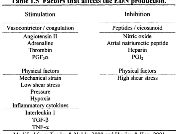
1.2.4.7 Endothelin axis 42
1.2.4.7.1 Endothelin-1 43
1.2.4.7.2 Endothelin receptors 47
1.2.4.7.3 Endothelin system and SSc 48 1.2.5 New insights into the pathogenesis of the disease 50
1.2.5.1 Microchimerism 50
1.2.5.2 Oxidative stress and autoantigens 51
1.2.5.3 Viral infection 52
1.2.6 Animal models 52
1.2.6.1. Naturally occurring animal models 53
1.2.6.1.1 TSKl mouse 53
1.2.6.1.2 UCD 200 line chicken 54
1.2.6.2. Induced animal models 54
1.2.6.2.1 Murine bleomycin induced sclerosis 54 1.2.6.2.2 Murine model of fibrosis with graft vs. host disease 54
1.2.7 Genetic factors 55
1.2.7.1 Analysis of genetic factors 55
1.2.7.2 Genetics of SSc 56
1.2.7.2.1 Immunogenetics 58
12.12.2 Association with other candidate genes 58
1.3. Aims o f the study 61
1.3.1 Specific objectives 61
2. Materials and Methods
63
2.1. Patients and patients’ samples 63
2.1.1 Systemic Sclerosis 63
2.1.2 Primary Raynaud’s phenomenon 65
2.1.3 Autoimmune Raynaud’s phenomenon 65
2.1.4 Control samples 66
2.2. M ethods 66
2.2.1 DNA extraction 66
2.2.1.1 White cell pellet preparation 66
2.2.1.2 Modified saltong out method 66
2.2.2 Identification of polymorphism and primer design 67 2.2.2.1 Selection of the reference sequence 67 2.2.22 Identification of the sequences variation (polymorphisms) 67
2.2.2.3. Primer design 68
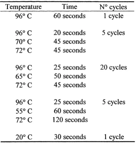
2.2.3 Polymerase chain reaction (PCR) 69 2.2.4 Sequence Specific Primer-PCR (SSP-PCR) 70
2.2.4.1 Criteria for primer design 71
2.2.5 Single Strand Conformational Polymorphism (SSCP) 2.2.5.1 Acrylamide gel electrophoresis
2.2.5.2 Silver staining.
2.3. Statistical analysis
73
73 74 75
3. Results
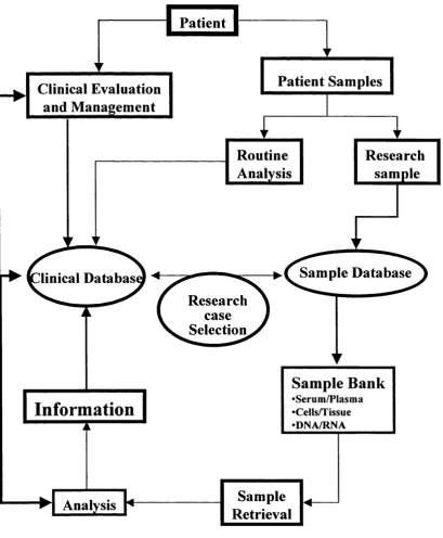
783.1. Clinical Database
3.1.1 Background
3.1.1.1 Historical context
3.1.2 Clinical/serological characteristics of the patients included in the study
3.1.3 Discussion
78 78 79
80 87
3.2 Connective Tissue Growth Factor
3.2.1 Background
3.2.1.1 CTGF gene description and regulation 3.2.1.2 CTGF polymorphism within the gene 3.2.2 Research objectives
3.2.3. Material and Methods
3.2.3.1 CTGF Polymorphism(s) determination 3.2.3.2 Patients and controls
3.2.3.2.1 Control group
3.2.3.2.2 SSc subgroups definition 3.2.3.3 CTGF serum levels determination 3.2.3.4.Statistical analysis
3.2.4.Results
3.2.4.1. CTGF polymorphisms
3.2.4.1.1 CTGF (C-743G) polymorphism 3.2.4.2. Potential transcription factor binding sites 3.2.4.3. CTGF serum levels
3.2.5.Discussion 3.2.6.Conclusions 90 90 90 92 93 93 93 97 97 98 98 98 99 99 101 103 105 105 109
3.3 The endothelin isoforms 1 ,2 and 3 polymorphisms across
the scleroderma disease 110
3.3.1 Background 110
4.3.1.1 Endothelin isoforms, gene structure and regulation 111
4.3.1.2 Endothelin polymorphism 112
3.3.2 Research objective 113
3.3.3 Material and Methods 113
3.3.3.1 Endothelin polymorphism determination 113
3.3.3.2 Patients and controls. 114
3.3.3.3 Statistical analysis 115
3.3.4.1. EDN-1 (T-1370G ) Polymorphism 117 3.3.4.2. EDN-1 (Exon 1 +138 A I/D) polymorphism 117 3.3.4.3 EDN-1 (E106E ) Polymorphism 118 3.3.4.4 EDN-1 (K198N) Polymorphism 119
3.3.5 Discussion 120
3.3.6 Conclusions 122
3.4 The endothelin polymorphisms across the scleroderma disease spectrum: Endothelin Receptor A, Endothelin
Receptor B 124
3.4.1 Background 124
3.4.1.1 Endothelin receptors type A and B, gene structure 124 3.4.1.2 Endothelin receptor polymorphism 125
3.4.2 Research objective 127
3.4.3 Material and Methods 127
3.4.3.1 Endothelin receptors polymorphism determination 127 3.4.3.2 Patient and control groups 128
3.4.3.3 Statistical analysis 128
3.4.4 Results 129
3.4.4.1 Endothelin receptor type A 130 3.4.4.1.1 EDNRA (G-231 A) polymorphism 131 3.4.4.1.2 EDNRA (H323H) polymorphism 132 3.4.4.1.3 EDNRA (H335H) polymorphsim 132 3.4.4.1.4 EDNRA (C228G) polymorphism 132 3.4.4.2 Endothelin receptor type B 133 3.4.4.2.1 EDNRB-3 (C12037A) polymorphism 133 3.4.4.2.2 EDNRB-4 (G11937A) polymorphism 134 3.4.4.2.3 EDNRB-7 (A2841G) polymorphism 135
3.4.5 Discussion 135
3.4.5.1 EDNRA (G-231 A) polymorphism 135 3.4.5.2 EDNRB-3 AND EDNRB-4 polymorphism 137
3.4.6 Conclusions 139
3.5 Polymorphism(s) in the far upstream enhancer element of
the human p roa2(l) Collagen gene 140
3.5.1 Background 140
3.5.1.1 Collagen structure 140
3.5.1.2.Proa(2)l collagen gene regulation 141 3.5.1.3.Collagen degradation: MMP and TIMP 143
3.5.2 Research obj ective 146
3.5.3. Material and Methods 147
3.5.3.1 SSCP of the far upstream enhancer element of the C0L1A2
gene 147
3.5.3.2 Collagen 1 collagenase-1 cleavage site polymorphism 149
3.5.4 Results 149
3.5.6 Discussion 153
4. Discussion and summary
iss
4.1 Construction of a high quality database and its importance in
statistical analysis 156
4.2 Genes and polymorphisms presented in this study 157 4.3 Significance of the CTGF polymorphisms 158 4.4 Significance of the EDN-1 and EDN receptor polymorphisms 159
5.
Future work
i6i
6. Papers and abstracts
u i
7.
Appendix A (Result tables)
i63
8. Appendix B (Primers sequences)
i9o
I ll LIST OF TABLES
Section 1
Table 1.1 Spectrum o f Scleroderma and Scleroderma like disorders Table 1.2 Criteria for the classification o f Systemic Sclerosis
(Scleroderma)
Table 1.3 Systemic Sclerosis subsets classification Table 1.4 Main serological groups in Systemic Sclerosis Table 1.5 Factors that stimulate Endothelin-1 production Table 1.6 Endothelin receptors type A and type B Table 1.7 SSc animal models
Table 1.8 HLA-antibodies associations in SSc patients
Page 17 19 20 28 46 48 53 59 Section 2 Table 2.1 Table 2.2 Table 2.3 Table 2.4
Classification and clinical characteristics o f SSc patient subsets 64
PCR cycling conditions 69
Silver staining procedure 74
The general form o f the contingency tables 76
Section 3.1
Table 3.1 Demographic characteristics o f the patients included into the 82 study
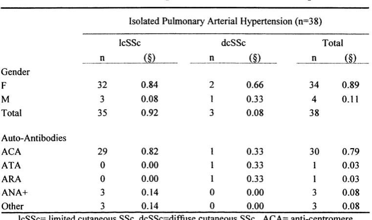
Table 3.2 Clinical and serological characteristics o f IPAH patients 83 Table 3.3 Clinical and serological characteristics o f the FASSc subgroups 84 Table 3.4 Clinical and serological characteristics o f SScRC subgroup 84 Table 3.5 Clinical and serological characteristics o f RC group with no 85
specific biopsy
Table 3.6 Clinical and serological characteristics o f the SSc group 86 Table 3.7 Clinical characteristics o f patients classified by auto antibody 87
profile
Section 3.2
Table 3.8 Characteristics o f the potential CTGF gene polymorphism 97 Table 3.9 Demographic characteristics o f the CTGF study clinical groups 99 Table 3.10 Clinical characteristics o f the SSc patients 100 Table 3.11 M ajor antibody distribution among SSc subgroups 101 Table 3.12 CTGF (C-743G) genotype and allele frequency distribution 102 Table 3.13 C-743G genotype and allele frequency distribution (FASSc and 103
ATA)
Table 3.14 Transcription factor motifs in C-743G polymorphism 104
Table 3.15 Transcription factor motifs m atrix 104
Table 3.16 CTGF serum levels in SSc patients and controls 105
Section 3.3
Table 3.18 EDN-1 polymorphisms association studies
Table 3.19 Initial endothelin axis polymorphisms characteristics Table 3.20 EDN-1 polymorphism studied
Table 3.21 EDN-1 (+138 A I) polymorphism major associations Table 3.22 EDN-1 (K198N) polymorphism m ajor associations
112 113 116 118 119 Section 3.4
Table 3.23 EDN receptors characteristics 125
Table 3.24 EDNRA and EDNRB polymorphism associations 127 Table 3.25 EDNRA and EDNRB polymorphism characteristics 128
Table 3.26 EDNRA and EDNRB final polymorphism 130
T able 3.27 G -231A polymorphism associations with statistical significance 131 Table 3.28 EDNRB-3 polymorphism association with statistical 133
significance
Table 3.29 EDNRB-4 polymorphism association with statistical 134 significance
Section 3.5 Table 3.30
Table 3.31 Table 3.32 Table 3.33
Regions o f high homology between mouse/human for the 143 upstream collagen 1 enhancer.
COL1A2: patients characteristics 150
Collagen 1 cleavage site study: demographic characteristics 152 Collagen 1 cleavage site study: clinical characteristics o f the 152 SSc patients.
Section 7 Table 7.1 Table 7.2 Table 7.3 Table 7.4 Table 7.5 Table 7.6 Table 7.7 Table 7.8 Table 7.9 Table 7.10 Table 7.11 Table 7.12 Table 7.13 Table 7.14
CTGF polymorphisms genotype distribution by clinical groups 163 CTGF (C-743G) genotype and allele frequency distribution by 164 clinical subgroups
CTGF (C-743G) genotype and allele frequency o f SSc 164 serological subgroups
EDN-1 (+138 A 1) genotype distribution by clinical groups. 165 EDN-1 (+ 138 A 1) genotype and allele frequency o f clinical 166 subgroups
EDN-1 (+ 138 A 1) genotype and allele frequency o f serological 166 subgroups
EDN-1 (T-1370G) genotype distribution by clinical groups. 168 ED N -1 (T-1730G) genotype and allele frequency o f SSc clinical 168 subgroups.
EDN-1 (T-1370G) genotype and allele frequency o f SSc 169 serological subgroups
EDN-1 (E 106E) polymorphism genotype distribution by 170 clinical groups
EDN-1 (E 106E) genotype and allele frequency o f SSc clinical 170 subgroups
ED N-1 ( E 106E) genotype and allele frequency o f SSc 171 serological subgroups
Table 7.15 EDN-1 (K198N) genotype and allele frequency o f SSc 173 serological subgroups
Table 7 .16 EDNRA (G -231 A) genotype distribution by clinical groups. 174 Table 7.17 EDNRA (G -231 A) genotype and allele frequency by SSc 175
clinical subgroups
Table 7.18 EDNRA (G -231 A) genotype and allele frequency o f SSc 175 serological subgroups.
Table 7.19 EDNRA (H 323H) genotype distribution by clinical groups 177 Table 7.20 EDNRA (H323H) genotype and allele frequency o f clinical 177
subgroups
Table 7.21 EDNRA (H323H) genotype and allele frequency o f SSc 178 serological subgroups
Table 7.22 EDNRA (E335E) genotype distribution by clinical groups 179 Table 7.23 EDNRA (E335E) genotype and allele frequency o f clinical 179
subgroups
Table 7.24 EDNRA (E335E) genotype and allele frequency o f serological 180 subgroups
Table 7.25 EDNRA (C+228G) genotype distribution by clinical groups 181 Table 7.26 EDNRA (C+228G) genotype and allele frequency o f SSc 181
clinical subtypes
Table 7.27 EDNRA (C+228G) genotype and allele frequency o f SSc 182 serological subgroups.
Table 7.28 EDNRB-3 (C +12037A) genotype distribution by clinical 183 subgroups
Table 7.29 EDNRB-3 (C +12037A) genotype and allele frequency o f SSc 184 clinical subgroups
Table 7.30 EDNRB-3 (C +12037A) genotype and allele frequency o f SSc 184 serological subgroups
Table 7.31 EDNRB-4 (A+11937G) genotype distribution by clinical groups 186 Table 7.32 EDNRB-4 (A+11937G) genotype and allele frequency o f SSc 186
clinical subgroups
Table 7.33 EDNRB-4 (A+11937G) genotype and allele frequency o f SSc 187 serological groups.
Table 7.34 EDNRB-7 (A +284IG ) genotype distribution by clinical groups 188 Table 7.35 EDNRB-7 (A + 284IG ) genotype and allele frequency o f SSc 188
clinical subgroups
Table 7.36 EDNRB-7 (A + 284IG ) genotype allele frequency o f SSc 189 serological groups
Section 8
Table 8.1 CTGF Promoter region primers for the SSCP assay 190 Table 8.2 SSP-PCR specific primer sequence for the CTGF (promoter 191
region)
Table 8.3 SSP-PCR specific primer sequence for the CTGF (coding 192 region)
Table 8.4 SSP-PCR specific prim er sequence for the EDN-3 193 polymorphisms
Table 8.5 SSP-PCR specific primer sequence for the EDN-2 193 polymorphisms
polymorphisms
Table 8.7 SSP-PCR specific prim er sequence for the EDNRA 195 polymorphisms
Table 8.8 SSP-PCR specific primer sequence for the EDNRB 196 polymorphisms
Table 8.9 Col 1A2 primers for SSCP (Human) 197
Table 8.10 Col 1A2 primers for SSCP (Mouse) 197
IV LIST OF FIGURES
Page
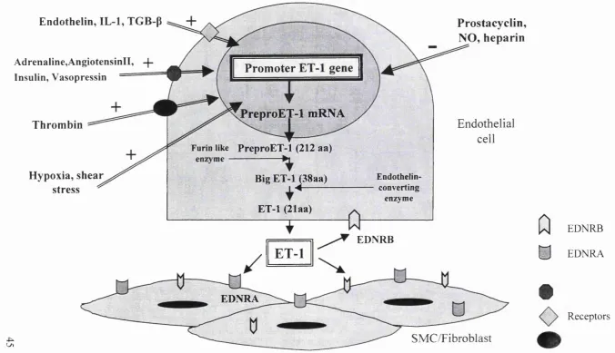
Figure 1 Endothelin-1 biosynthetic pathway 45 Figure 2 Scheme showing principle of Sequence Specific Primer-
PCR (SSP-PCR)
71
Figure 3 Clinical/sample database flow chart 80 Figure 4 Flow diagram of the polymorphisms identification and
selection
95
Figure 5 Single Strand Conformational Polymorphism from the CTGF promoter region
94
Figure 6 Specific sequence polymorphism-PCR. CTGF polymorphism
96
Figure 7 CTGF promoter region, polymorphisms and transcription factor sites.
107
Figure 8 SSP-PCR of the EDN-1, EDNRA and EDNRB polymorphism
115
Figure 9 Auto-antibodies distribution in SSc 123 Figure 10 Auto-antibodies groups distribution in SSc 138 Figure 11 C0L1A2 collagenase-1 cleavage site, from DNA to
protein
145
V. Abbreviations
AA Amino acid
ACA Anti-centromere antibody ACE Angiotensin converting enzyme AECA Anti-endothelial cell antibodies AFA Anti-fibrillarin ( U3-RNP) antibody ANA Antinuclear antibody
APC Antigen presentation cell ARA Anti-RNA polymerase antibody ARP Autoimmune Raynaud's phenomenon ATA Anti-topoisomerase I antibody
BAL Bronchioalveolar lavage cDNA Complementary DNA CENP-1 Centromere protein 1
C0L1A2 Collagen alpha one chain two CTCK C terminal cytokine-knot
CTGF Connective Tissue Growth Factor dcSSc Diffuse cutaneous Systemic Sclerosis EC Endothelial cell
ECE-1 Endothelin converting enzyme 1 ECM Extracellular matrix
EDN-1 Endothelin-1
EDNRA Endothelin receptor A EDNRB Endothelin receptor B
F Female
FASSc Fibrosing alveolitis SSc FV Functional volume FVC Forced vital capacity GvHD Graft versus host disease HLA Human leukocyte antigen
HRCT High resolution computed tomography H-W Hardy-Weinberg
ICAM-1 Intracellular adhesion molecule-1 IFN-gamma Interferon gamma
IL-1 Interleukin-1 IL-4 Interleukin-4
ILD Interstitial lung disease
IcSSc Limited cutaneous Systemic Sclerosis
M Male
MCP-1 Monocyte chemoattractan protein 1 MHC Major histocompability complex mRNA Messenger RNA
MTCD Mixed connective tissue disease NK Natural killer cells
NO Nitric oxide
NSIP Non-specific interstitial pneumonia OR Odds ratio
PAH Pulmonary arterial hypertension PAP Pulmonary artery pressure
PAPE Pulmonary artery pressure in exercise PBC Peripheral blood cells
PCR Polymerase chain reaction PDGF Platelet derived growth factor PF Pulmonary fibrosis
PFR Pulmonary function test PGE Prostaglandin E
RC Renal crisis
RC NSB Renal crisis no specific biopsy RDA Representational difference analysis RNAP RNA polymerase
ROS Reactive oxygen species RP Raynaud's phenomenon RR Relative risk
SMC Smooth muscle cell
SNP Single nucleotide polymorphisms SSc Systemic Sclerosis
SSCP Single Strand Conformational Polymorphism SScRc SSc associated renal crisis
SSP-PCR Sequence Specific Primer-PCR TGF-beta Transforming growth factor beta TCR T cell receptor
TEC Total lung capacity TNF Tumor necrosis factor
UCD University of California at Davis VC Vital capacity
Acknowledgements
This work was performed thanks to a scholarship from CONACyT (Mexico).
Many people have offered me support and help throughout my time at the
Centre for Rheumatology, RFH, and I would like to express my gratitude
towards them, in particular Prof. Carol Black.
Special thanks must also go to Dr. David Abraham for his support and patient
supervision notably concerning the production of this thesis.
I am also grateful to Prof. Ken Welsh for his helpful advice.
I am extremely appreciative of the support and friendship offered to me by
everyone at the Rheumatology research lab. Particular thanks must go to Alan
Holmes for solving everyday problems, Ivan Fisher for his comforting teas,
and Dr. Markella Ponticos for helping me to make sense of a mess of
information.
I am also indebted to Dr. Panos Pantelidis and Dr. Elizabeth Renzoni from the
Royal Brompton Hospital for their advice regarding statistics and PCR know
how.
Also appreciated is the unconditional support of the INER and in particular of
Dr. Moises Selman.
And of course I must thanks to Prof. Alejandro Madrigal for his advice.
Last but not least this thesis could not have been completed without the love
1. INTRODUCTION
1.1 Systemic Sclerosis
1.1.1 Introduction
Systemic sclerosis (SSc) is a clinically heterogeneous rheumatological,
connective tissue disorder (Black, 1995), which is encompassed within the
scleroderma spectrum of conditions (Table 1.1). The disease is of unknown
aetiology but is typically characterised by fibroblast proliferation and the
accumulation of extracellular matrix (ECM) within almost all connective
tissues. This process proceeds vascular injury and immune alteration (Black
and Denton, 1998; Mitchell et al., 1997). The near universal occurrence of
microvascular dysfunction and the associated inflammatory reaction that may
co-exist, preceding fibrogenesis suggest that changes in vascular integrity and
enhanced influx of inflammatory cells are early events in the pathogenesis of
scleroderma (Clements and Furst, 1996; White and Yurovsky, 1995). Virtually
all the organ systems can be affected, most noticeably the skin, but more
clinically important the lungs, kidneys, gastrointestinal tract and heart. The
survival and prognosis are related to the severity and extent of the internal
organ involvement. Currently therapy, although aimed at improving the
vascular complications, suppressing the immune system and preventing the
progressive fibrosis is not generally considered particularly effective in
controlling in a definitive way the disease and its complications (Furst, 2000).
There is therefore an urgent need to develop a staged approach to treatment
based on our increased understanding of disease pathogenesis and careful
/.
L 2 Epidemiology
SSc has a worldwide distribution and affects both males and females. Incidence
rates, from retrospective reviews, vary from 2-19 cases per million population
per year and the prevalence has been reported between 19 and 75 per 100,000
(Maricq et al., 1989).
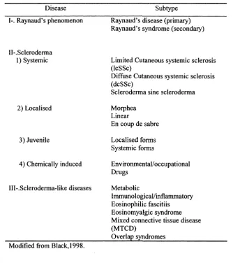
Table 1.1. Spectrum of scleroderma and scleroderma-like diseases.
Disease Subtype
I-. Raynaud’s phenomenon Raynaud’s disease (primary) Raynaud’s syndrome (secondary)
Il-.Scleroderma
1) Systemic Limited Cutaneous systemic sclerosis (IcSSc)
Diffuse Cutaneous systemic sclerosis (dcSSc)
Scleroderma sine scleroderma
2) Localised Morphea
Linear
En coup de sabre
3) Juvenile Localised forms Systemic forms
4) Chemically induced Environmental/occupational Drugs
Ill-.Scleroderma-like diseases Metabolic
Immunological/inflammatory Eosinophilic fascitiis
Eosinomyalgic syndrome Mixed coimective tissue disease (MTCD)
Overlap syndromes Modified from Black, 1998.
In the general population the peak incidence of the disease is during the fifth
and sixth decades, although it can occur much earlier and even in childhood.
more likely to develop SSc. However within this range, variation associated
with the age and environmental factors occur (Silman and Newman, 1996).
The SSc patients overall survival rate is 60-83% at 5 years and 40-75% at 10
years (Bryan et al., 1996) (Bryan et al., 1999), however when the SSc subsets
are analysed, it is apparent that the diffuse form has the worse prognosis with
30% survival at 6 years compared with an 80% figure for the limited form of
the disease over the same period of time (Altman et al., 1991). The current
major cause of mortality within the diffuse disease subset is renal crisis in the
first five years of the disease and pulmonary complications (lung fibrosis and
secondary pulmonary arterial hypertension) after 6 years of evolution (Steen
and Medsger, Jr., 2000). Meanwhile pulmonary arterial hypertension represents
the leading cause of mortality in patients with the limited cutaneous disease
(Medsger and Steen, 1996).
Interestingly, a recent study in the native North American Indian tribe from
Oklahoma showed an increased in prevalence of scleroderma (469 per 100,000
population). The Choctaw Indian adults have a particularly consistent disease
profile with diffuse disease, prominent lung involvement and anti-
topoisomerase I antibodies (Amett et al., 1996).
1.1,3 Classification criteria fo r SSc and SSc subgroups
The preliminary criteria for the classification of Systemic Sclerosis were
proposed by a committee of the American Rheumatism Association (former
name of the American College of Rheumatology) in 1980 (1980) (Table 1.2).
They are based on the presence of proximal scleroderma defined as symmetric
thickening, tightening and induration of the skin of the fingers and the skin
proximal to the metacarpophalangeal or metatarsophalangeal joints. They
propose as minor criteria the presence of sclerodactyly, digital pitting scars and
pulmonary fibrosis. However, the improvement in the detection of patients
with Raynaud’s phenomenon, and more precise autoimmune serology have
preliminary criteria. In this context LeRoy and Medsger (Leroy and Medsger,
Jr., 2001) have proposed a new set of criteria for early SSc, including the new
vascular and serologic observations, in order to include all those patients that
have SSc characteristics but do not fulfil the original criteria.
Table 1.2 Criteria for the classification of Systemic Sclerosis
Criteria Characteristic Definition
A. Major criterion Proximal Scleroderma Symmetric thickening, tightening, and induration o f the skin o f the fingers and the skin proximal to the metacarpophalangeal or metatarsophalangeal joints. The changes may affect the entire extremity, face, neck, and trunk.
B. Minor criterion 1. Sclerodactyly Above-indicated changes limited to the fingers.
2. Digital pitting scars Depressed areas at tips o f fingers or loss o f digital pad tissues as a result o f ischaemia.
3. Bibasilar pulmonary fibrosis.
Bilateral reticular pattern o f linear or lineo-nodular densities most pronounced in basilar portions o f the lungs on standard chest roentgenogram; may assume appearance o f diffuse mottling or honeycomb lung. These changes should not be attributable to primary lung disease.
* For purposes o f classifying patients in clinical trials, population surveys, and other studies, a person shall be said to have SSc if one major or two or more minor criteria are present. Localized forms o f scleroderma, eosinophilic fasciitis, and the various forms o f pseudesclerodema are excluded form these criteria.
§ Preliminary criteria for the classification o f systemic sclerosis (scleroderma). Arthritis Rheum 23:581-590, 1980.
The most commonly adopted classification for SSc subgroups (Leroy et al.,
1988) is based upon the extent of skin involvement and some clinical
laboratory and natural history associations. This classification essentially
divides the systemic disease into limited cutaneous systemic sclerosis (IcSSc)
current classification see Table 1.3. Over 60% of the patients are classified as
IcSSc, where the involvement of internal organs tends to occur late in the
evolution of the disease and it is usually preceded by Raynaud’s phenomenon
(RP), often for many years. On the other hand, the dcSSc type tends to have
much more rapid onset, with organ failure often present within the first 5 years
of the disease (Black and Denton, 1998).
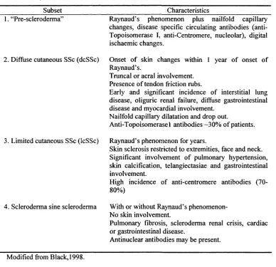
Table 1.3 Systemic Sclerosis subsets classification
Subset Characteristics
1. “Pre-scleroderma’
2. Diffuse cutaneous SSc (dcSSc)
3. Limited cutaneous SSc (IcSSc)
4. Scleroderma sine scleroderma
Raynaud’s phenomenon plus nailfold capillary changes, disease specific circulating antibodies (anti-Topoisomerase I, anti-Centromere, nucleolar), digital ischaemic changes.
Onset o f skin changes within 1 year o f onset o f Raynaud’s.
Truncal or acral involvement. Presence o f tendon friction rubs.
Early and significant incidence o f interstitial lung disease, oliguric renal failure, diffuse gastrointestinal disease and myocardial involvement.
Nailfold capillary dilatation and drop out.
Anti-Topoisomerase 1 antibodies -30% o f patients.
Raynaud’s phenomenon for years.
Skin sclerosis restricted to extremities, face and neck. Significant involvement o f pulmonary hypertension, skin calcification, telangiectasiae and gastrointestinal involvement.
High incidence o f anti-centromere antibodies (70-80%)
With or without Raynaud’s phenomenon-N o skin involvement.
Pulmonary fibrosis, scleroderma renal crisis, cardiac or gastrointestinal disease.
Antinuclear antibodies may be present.
1.1.4 Histopathology
The principal changes of scleroderma are vascular and microvascular
abnormalities, characterized by capillary obliteration, endothelial
injury/activation, intimai proliferation, medial thinning, and perivascular and
interstitial infiltration of mononuclear inflammatory cells with an increased
deposition of normal matrix components in the skin and internal organs (Black,
1995) (O'Angelo et al., 1969) (Harrison et al., 1991).
1.1.5 Clinical features
1.1.5.1 Raynaud s phenomenon (RP)
Raynaud’s phenomenon is frequently (90-95%) the first symptom of the
underlying SSc disease and is considered the most common clinical expression
of vascular derangement (Belch, 1991). The presence of RP may precede skin
changes by several months or years and together with skin thickening is the
most frequent clinical feature of the disease (Mitchell et al., 1997; Wigley,
1996). Raynaud's phenomenon is defined as the episodic vasoconstriction of
small arteries and arterioles of the extremities. The vasospasm events may be
brought on by cold exposure, vibration or emotional stress. Patients experience
pallor and/or cyanosis followed by rubor in re-warming. Pallor and/or cyanosis
are usually associated with coldness and numbness of fingers and rubor with
pain and tingling. The majority of patients with RP (>95%) never develop any
associated connective tissue disease. This group is classified as primary RP.
Peripheral vascular disease and RP has been reported to be more severe within
patients with IcSSc and those with anti-centromere antibodies (ACA) who are
at high risk to develop major peripheral vascular occlusive disease (Herrick et
1.1.5.2. Skin
Involvement of the skin is the hallmark of scleroderma, and there are usually 3
phases; 1) oedematous, 2) indurative and 3) atrophic.
In the early oedematous phase, fingers and hands become puffy and patients
frequently complain of stiffness. Swelling may be also apparent on extremities
(forearms, feet, lower legs) and the face. This phase can last for a few weeks,
months or even years. The skin changes usually begin distally in the
extremities and advance proximally. Subsequently the skin gradually becomes
firm, thickens and eventually becomes tightly bound to underlying
subcutaneous tissue (indurative phase). Flexion contractures develop as a
result of skin thickening and tendon fibrosis. Digital ulcers may develop, with
loss of soft tissue at the finger pads. In patients with the diffuse cutaneous
scleroderma subset, skin changes become generalised, including all the
extremities, face, trunk and abdomen. Rapid progression of these changes over
a relatively short period of time is usually associated with internal organ
affection, especially lungs, kidneys and heart and these features are linked to a
bad prognosis (Clements et al., 2000). The skin changes in this disease subset
usually peak around 3-5 years and then slowly improve. In the localised subset,
the skin events tend to be more gradual, and they are restricted to fingers, distal
extremities and face. After many years the skin may soften and return to the
normal thickness or become thin and atrophic (Black and Denton, 1998;
Clements and Medsger, 1996).
1.1.5.3 Lung disease
Lung disease is a frequent manifestation in SSc with more than 75% of patients
exhibiting some pulmonary involvement. Since the emergence of more
effective treatment for renal involvement, lung disease is now considered as the
primary cause of mortality in patients with SSc (Steen et al., 1994). The course
of lung disease in SSc patients is highly variable, the majority of the patients
stabilize or improve. However in a small subset (-15%) the pulmonary
function test decline is faster in the 3 first years of the disease with a median
survival of 50% at 5 years (Steen and Medsger, Jr., 2000). There are 2 major
types of pulmonary disease affecting SSc patients: fibrosing alveolitis (FASSc)
also known as interstitial lung disease (ILD) and pulmonary arterial
hypertension (PAH) (Bolster and Silver, 1999; Silver, 1996).
1.1.5.3.1 Fibrosing alveolitis in SSc
FASSc usually develops insidiously and tend to remains silent until later stages
of the disease, when it may become a major cause of mortality. The most
common initial symptom is breathlessness on exertion, followed by non
productive cough (Black and Denton, 1998). Bibasal dry crackles are found on
the physical examination. Chest radiography usually shows bilateral
reticulonodular shadowing and honeycombing more marked on basal regions.
This is a relatively insensitive method, and in the past few years high resolution
computed tomography (HRCT) has been more widely adopted technique
approach to identify fibrotic changes, but also to differentiate them from the
early inflammatory phase. This technique is now routinely used in the
evaluation of SSc patients (Diot et al., 1998; Warrick et al., 1991; Wells et al.,
1992). With HRCT the earliest detectable abnormality is a subpleural area of
increased density. When more extensive, the shadowing takes a characteristic
reticulonodular appearance. In more advanced fibrosis this pattern is associated
with honeycomb air spaces and even large cystic air spaces (Wells et al., 1993).
Recent reports have reported a higher prevalence of the recently described non
specific interstitial pneumonia (Katzenstein and Myers, 1998) in patients with
SSc (Veeraraghavan et al., 2001) (Bouros et al., 2002). This is an important
observation because this type of ILD tends to have a better response to the
treatment and improved prognosis (Daniil et al., 1999) (Nicholson et al., 2000).
FASSc tends to be more frequent and more severe in patients with dcSSc and
with those expressing anti-topoisomerase 1 antibodies (ATA) (Diot et al., 1999)
1.1.5.3.2 Pulmonary Arterial Hypertension (PAH)
Pulmonary arterial hypertension (PAH) was previously known as pulmonary
hypertension but it was re-classified following the recent World Health
Organization symposia (Galie and Torbicki, 2001)
Pulmonary arterial hypertension is defined as a median pulmonary artery
pressure higher than 30 mmHg at rest by echocardiography and has a reported
incidence around 10% in patients with SSc. Isolated PAH (IPAH) is more
frequent in patients with IcSSc with an increased association with presence of
anti-centromere antibodies (ACA). The secondary form of PAH occurs in
patients with either dcSSc or IcSSc but always in association with pulmonary
or cardiac disease (Coghlan and Mukerjee, 2001; Yamane et al., 2000). The
presence of PAH represents a bad prognostic factor and is a major cause of
mortality irrespective of whether it occurrs in isolation or in association with
pulmonary fibrosis, with a median survival rate of 12-20 months (Koh et al.,
1996) (MacGregor et al., 2001). The diagnosis of pulmonary hypertension
represents a challenge because its detection on clinical basis is difficult as the
symptoms may appear late in the evolution of the disease. The typical
manifestations are dyspnoea and fatigue, with a loud P2 component of the
second heart sound and a right ventricular S3 gallop. The most common
abnormality in pulmonary function test (PFT) is the decrease of the DLCO
(transfer factor for carbon monoxide), and echocardiography with Doppler
provides a reliable non-invasive measure of pulmonary artery pressure (Denton
et al., 1997a). Recent work performed at Royal Free Hospital has shown that
right heart catheterisation, although is albeit more invasive technique, is a more
reliable method for measurement and evaluation of the pulmonary artery
pressure in SSc (Dr. Coghlan, personal communication)
1.1.5.4. Renal disease
Although histological abnormalities in the kidney are almost always present,
scleroderma renal crisis (SScRC), is reported in 19% of the patients. SScRC is
characterized by accelerated hypertension and/or progressive renal failure, and
80% of the cases tend to occur in the first 4-5 years of disease. Risk factors
include diffuse disease, use of corticosteroids and the presenee of anti-RNA
polymerase III antibody (Steen, 1994) (Steen and Medsger, 2000). The
mortality of this complication has been dramatically modified by the use of
angiotensin-converting enzyme (ACE) inhibitors in the past 10 years, with
survival rates improving from less than 10% at five years before the ACE
treatment to 70% after this treatment was initiated (Steen and Medsger, 2000).
1.1.5.5 Cardiac disease
The clinical manifestations of scleroderma heart disease are variable, usually
subtle in expression and tend to manifest late in the course of the disease.
Symptoms include dyspnoea on exertion and palpitations. Patchy myocardial
fibrosis, areas of contraction band necrosis and vasospasm of distal coronary
vessels are the most frequent alterations in SSc patients (Clements and Furst,
1994; Mitehell et al., 1997).
1.1.5.6 Gastrointestinal disease
Gastrointestinal manifestations are the most common symptoms in SSc present
in up to 90% of the patients. They include oesophageal, small-bowel and colon
alterations. Difficulty in swallowing, dysphagia and heartburn are frequent
complaints, secondary to oesophageal hypo-motility. Small-bowel hypo-
motility can lead to diarrhoea, weigh loss and malabsorption with bacterial
overgrowth as a recurrent problem. Colonic and rectal atony frequently
produces constipation/anal incontinence (Black and Denton, 1998).
1.1.5.7 Macrovascular and microvascular disease
Microvascular disease characterised by endothelial dysfiinetion is a hallmark of
SSc and is noticeable elinically as Raynaud’s phenomenon. More recently
disease. The involvement of macrovascular abnormalities is believed to be
higher than previously thought, and may manifest as intermittent claudication,
ischaemic heart diseases and ischaemic stroke. Whether this macrovascular
disease is secondary to arteriosclerosis or another process associated with the
disease such as vasculitis or antiphospholipid antibody is currently unknown
(Taylor et al., 2002) (Ho et al., 2000) (Wan et al., 2001) (Veale et al., 1995)
L L 6 Serological Features (Autoantibodies)
Antibodies against nuclear antigens (ANA) have been described in up to 95%
of the patients with SSc (Bunn et al., 1998). There are 3 main autoantibodies
association and a number of minor mutually excusive serologic subgroups in
SSc. The major autoantibody specificities are usually associated with
distinctive clinical profiles. Therefore they can be regarded and used as
valuable diagnostic and prognostic factors. (Table 1.4) (Tormey et al., 2001).
1.1.6.1 Anti-topoisomerase I antibody (ATA)
Anti-topoisomerase I antibodies, also termed Scl-70 are found exclusively in
patients with SSc especially with dcSSc subset. These antibodies have been
associated with the presence of heart disease and pulmonary fibrosis (Jacobsen
et al., 2001) (Fanning et al., 1998). Both ATA and AC A titres tend to be stable
over time (Vazquez-Abad et al., 1995) but recently Kuwana et al (2000)
identified a subset of dcSSc patients in which the AT A titre became
undetectable during the course of the disease. In those patients with
undetectable AT A titres the authors reported a more favourable disease
outcome (Kuwana et al., 2000).
1.1.6.2 Anti-centromere antibody (ACA)
ACA is the most common autoantibody occurring in approximately 25% of
patients. These antibodies recognise one or more centromere proteins (CENP-
virtually all patients who express ACA autoantibodies (Bunn and Tormey,
2000). This antibody is found more frequently in patients within the IcSSc
subgroup and has been associated with little internal organ involvement, a
higher risk of peripheral vascular occlusive disease and longest survival
(McCarty et al., 1983) (Weiner et al., 1988) (Wigley et al., 1992).
1.1.6.3 Anti RNA polymerase antibody (ARA)
Anti-RNA polymerase I, II and III (RNAP I, II and III) (ARA) are associated
with higher risk of dcSSc, more extensive and severe cutaneous involvement
and with a greatest risk of renal involvement (Bunn et al., 1998) (Harvey et al.,
1999)
1.1.6.4 Minor autoantibodies specificities
There are also other less frequently occurring autoantibody specificities such as
anti-fibrillarin (U3-Ribonucleoprotein), anti-PM-Scl, anti-Th-RNP, anti-Ku
and anti-Ul-RNP. Anti-fibrillarin antibodies (AFA) or anti-U3-RNP antibodies
are specific for SSc (4%), are more frequently found in Afro-Caribbean
population and identify young dcSSc patients with an increased risk of
developing internal organ involvement, especially isolated pulmonary arterial
hypertension (Tormey et al., 2001) (Sacks et al., 1996). Anti-To antibodies
recognize a 40 kD protein component of the Th RNP particle
(endoribonuclease), which is located in the nucleolus (Yuan et al., 1991).
Antibodies to Th/To ribonucleoprotein (anti-Th/To) are detected in SSc
patients (-4%) and reported to identify those patients who are at greater risk
for reduced survival (Okano and Medsger, Jr., 1990). Anti-RNP antibodies
react with the nuclear RNA splicing particle U 1 snRNP (Reuter and Luhrmann,
1986). This serotype is found in ~ 10% of the SSc patients and has been
associated with a low frequency of renal disease, arthritis and pulmonary
arterial hypertension (Kuwana et al., 1994) (Ihn et al., 1999). The anti-Ku
antibodies are found in a wide spectrum of connective tissue diseases including
involvement are the most frequent clinical features associated with anti-Ku
antibodies (Franceschini et ah, 2002).
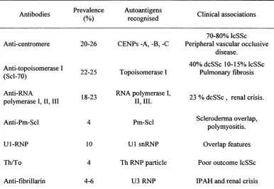
Table 1.4 Main serologic groups in SSc
Antibodies Prevalence (%)
Autoantigens
recognised Clinical associations
70-80% IcSSc Anti-centromere 20-26 CENPs -A, -B, -C Peripheral vascular occlusive
disease.
Anti-topoisomerase I
(Scl-70) 22-25 Topoisomerase I
40% dcSSc 10-15% IcSSc Pulmonary fibrosis
Anti-RNA
polymerase I, II, III 18-23
RNA polymerase I,
II, III. 23 % dcSSc , renal crisis.
Anti-Pm-Scl 4 Pm-Scl Scleroderma overlap,
polymyositis.
Ul-R N P 10 U1 snRNP Overlap features
Th/To 4 Th RNP particle Poor outcome IcSSc
Anti-fibrillarin 4-6 U3 RNP IPAH and renal crisis
IPAH=isolated pulmonary arterial hypertension Modified from Black, 1998 and Harvey, 2000.
1.2 The Pathogenesis of SSc
Fibrosis, the major characteristic of SSc, has many features in common with
the normal physiological reparative response in wound healing, but unlike
wound healing, the scarring phase appears to be dysregulated leading to an
excessive deposition of collagen and ECM. This reparative fibrogenic process
is often preceded by vascular disease and immune activation, which in
association cause the tissue damage leading to organ failure. The initial
stimulus that leads to an abnormal response in SSc is still unknown. However,
damage and activation of the endothelial layer are among the first events
al., 1991). These are apparent as the upregulation of adhesion molecules
platelet aggregation, lymphocyte activation/migration and cytokine production.
These elements can lead to altered vessel permeability with the subsequent
migration of inflammatory cells through the endothelial layer into the
extravascular space where they promote further recruitment of inflammatory
cells. In the normal reparative process, the initial inflammatory response is
self-limited leading to a restricted response. In SSc the inflammation tends to
persist, leading to increased number of activated immune cells (mainly T cells,
but also eosinophils, monocytes and basophils), soluble mediators such as
cytokines, chemokines and growth factors (Black and Denton, 1998).
Interactions between all these factors and resident or recruited cells can lead to
dysregulated responses in the target cells, resulting in a repetitive cycle of
tissue injury and repair.
The changes in fibroblast metabolism that take place include proliferation,
increased production not only of collagen and other ECM elements such as
fibronectin and tenascin, but also cytokines and growth factors (i.e. TGF-p,
PDGF, CTGF, PGE and IL-1) (for a review see Black and Denton, 1996)
which may exert autocrine or paracrine effects, perpetuating the altered fibrotic
response (Trojanowska et al., 1988) (Leroy, 1974) (Cotton et al., 1998).
It is currently unknown whether this altered fibroblast response is wholly
acquired secondary to the environmental stimulus or is partially inherited and
then activated by external factors (Kawakami et al., 1998; Needleman et al.,
1990).
1.2.1 The endothelium and endothelial cells
The universal feature of SSc is the presence of vessel abnormalities. Some of
the first clinical manifestations of these vessel abnormalities are the failure to
re-warm after cold challenge (Raynaud’s phenomenon) and an abnormal
peripheral nailfold capillary pattern, with enlarged loops, mega capillaries and
These observations suggest that changes in vessel function and/or endothelium
integrity are some of the earliest features of Scleroderma pathogenesis. In
general the first noticeable changes that occur in the endothelium include
concentric proliferation of the intimae and perivascular oedema (Campbell and
Leroy, 1975). The described ultrastructural alterations in early SSc comprises
endothelial cell (EC) vacuolisation, granular degeneration of the nucleus,
cellular necrosis, gaps appearing between EC and reduplication of basement
membranes (Fleischmajer and Perlish, 1980). The EC disruption culminates
with altered endothelium permeability (Bollinger et al., 1986) and leaky vessels
that allows increased passage of plasma and mononuclear cells with the
consequent formation of oedema and perivascular infiltrates characteristics of
the early SSc stages.
The precise molecular and cellular changes in blood vessel endothelium that
take place in the early stages of the disease are not known, but these are likely
to have an important impact on the integrity and function of the structural cells
which envelop the blood cells (such as pericytes in capillaries or smooth
muscle cells in small arteries) which in turn are ultimately responsible for
maintaining the vascular tone. The vascular tone is dependent on
vasoconstriction and vasodilatation mechanisms, requiring both an intact
endothelium and vital processes that are under neuronal control (Generini and
Matucci, 1999). In scleroderma, current observations support the presence of
reduced vasodilatation (i.e. relative deficiency of nitric oxide) (Cotton et al.,
1999), increased vasoconstriction (i.e. increased release of endothelin-1)
(Kahaleh, 1991), as well as neurological alterations such as changes in the
levels of substance P (Matucci-Cerinic et al., 1990) (Generini and Matucci,
1999). The net result of the vessel tone alteration in SSc is a sustained
vasoconstriction that impairs the blood flow with consequent changes in
hypoxia, oxidative stress and ischaemia leading to further EC damage and
activation. The EC activation is manifested by augmented production of
cytokines, increased expression of adhesion molecules, and abnormal
(Ames et al., 1997). The expression of adhesion molecules, such as
intracellular adhesion molecule-1 (ICAM-1), vascular cell adhesion-1 (VCAM-
1) and E-selectin on the endothelial cell surface are found not only in the
lesional areas of skin but also in clinically uninvolved regions (Majewski et al.,
1991) (Sollberg et al., 1992) perhaps reflecting an early alteration in EC
activation. Other markers of endothelial activation/damage such as von
Willebrand factor, endothelin-1 and Thrombomodulin (Kahaleh et al., 1981)
(Kahaleh and Leroy, 1999) (Vancheeswaran et al., 1994b) (Ohdama et al.,
1994) are elevated in peripheral circulation of patients. The soluble forms of
some adhesion molecules have also been found increased in SSc patients
serum, these include ICAM-1 and VCAM-1 (Gruschwitz et al., 1995) (Stratton
et al., 1998), although their significance in the disease process remains
unclear.
Endothelial cell adhesion molecules are also known to interact on the specific
integrins on T and B lymphocytes, platelets, neutrophils, monocytes and
natural killer cells (NK). Increased expression of these molecules would
favour their adhesion to the endothelium and subsequent migration and
chemotaxis and recruitment into extra-vascular tissues.
Recent observations linking EC cell apoptosis and anti-endothelial cell
antibodies had been reported from experimental work on an animal model of
SSc, the University of California at Davies Line (UCD) 200 chickens. These
chickens spontaneously develop an inherited scleroderma-like disease, with
immune, vascular and fibrotic characteristics with similarities to the human
disease. Histochemical analysis of UCD 200 chicken skin sections showed that
apoptosis of EC is an early event in the pathogenesis preceding the
mononuclear perivascular infiltration (Sgonc et al., 1996). These observations
have been expanded to include other internal organs (Sgonc et al., 2000). In
fibrotic human skin, apoptotic endothelial cells have only been detected in the
of the temporal relationship between the vascular and immunological
components in SSc (Sgonc et ah, 2000).
Circulating antibodies that bind EC represent an heterogeneous family of
autoantibodies that have been described in several autoimmune diseases with
vascular pathology, including SSc (Renaudineau et ah, 2001). The anti-
endothelial cell antibodies (AECA) have been reported in 40-84% of
scleroderma patients (Salojin et ah, 1997) and they may react with different
structures on the surface of EC (Ihn et ah, 2000). In general the pathogenicity
of these AECA remains uncertain, but there are recent observations that
support a direct damaging role in SSc as some of the AECA can induce
endothelial cell apoptosis (Bordron et ah, 1998), and also increase the
leukocyte adhesion to vascular endothelial cells in vitro (Carvalho et ah, 1996).
It is a matter of speculation if the apoptosis provoked by the AECA has any
direct role in disease pathogenesis.
1,2,2 The immune response and antibodies
There is clear evidence that activation of the immune system is an early event
in the SSc disease process. Mainly T cells but also mast cells, eosinophils and
basophils are found in increased number and in an activated state in the tissues
of SSc patients. These cells are capable of modifying fibroblast and endothelial
cell function through the production of soluble mediators (White, 1996).
There are some observations that support a major role for the T cell in the
development of fibrosis. First there are reports of increased numbers of
activated T cells in the skin of SSc patients mainly as a component of the
perivascular infiltrate that is characteristic of the early stages of the disease.
Another line of evidence that suggests an important role for T cells early in the
pathogenesis of the disease is the tendency of the collagen producing cells, the
fibroblast, to be predominantly located in close proximity to the mononuclear
regard to the T cell subset that predominate in SSc, there seems to be a
relationship to the organ affected and the stage of the disease. For example
CD4+ T cells predominate in the skin (Prescott et al., 1992; Roumm et al.,
1984) whereas in the lungs a mixed pattern had been described with increased
levels of CD8+ T cells in the presence of alveolitis (Yurovsky et al., 1996),
with cells expressing a predominant Th2 cytokine type (IL-4+, IFNy -) in those
patients who were reported to have more aggressive disease (Atamas et al.,
1999; Majumdar et al., 1999). Although in general is there a tendency to
consider the Th2 response as the predominant one in SSc (Mavalia et al., 1997)
(Tsuji-Yamada et al., 2001) there are some reports that Thl (IFNy +, IL-2+)
polarization may occur under specific conditions, but the significance of these
observations still not fiilly understood (Giacomelli et al., 2001) (Valentini et
al., 2001).
The majority of T lymphocytes express T cell receptors (TCR) consisting of
heterodimer of a and p chains (Yoshikai et al., 1984). However a restricted
population (1-5%) of T cells express different TCR components, the y and ô
chains. These subsets display several functional differences with respect to
their counter parts such as major histocompatibility complex (MHC)
unrestricted cytolysis the recognition of intact and unprocessed self-antigens
(Kaufmann, 1996) and in the synthesis of variety of chemokines and cytokines
(Giacomelli et al., 2001). In SSc patients it has been noted that these y/6 T
cells accumulate in skin and lung (Giacomelli et al., 1998) (White and
Yurovsky, 1995) where they enhance interaction and cytotoxicity towards EC
(Kahaleh et al., 1999). It also has been demonstrated that an expansion of
specific y/ô T cells subsets occurs in SSc (Yurovsky et al., 1994). This
observation has led to suggestion that the expansion of these T cells may be
antigen driven.
It is important to mention also that recent reports have linked the presence of
specific HLA molecules, that determine the T cell receptor affinity and
presenting cells (APC), with auto reactivity to specific epitopes on
Topoisomerase-I (Kuwana et al., 1995) (Rands et al., 2000).
The almost universal presence of autoantibodies in scleroderma patients is
another observation that supports the role of the immune system in this disease.
In addition to the autoantibodies already described in SSc there are also reports
identifying anti-endothelial cells and anti-fibroblast autoantibodies although
the target antigens have not been described and their precise role is not known.
In the past years several theories have been proposed to explain the
mechanisms responsible for the loss of self-tolerance and antibody formation
(Kamradt and Mitchison, 2001). They are summarised below:
A) Molecular mimicry with a cross immune reaction of self-epitopes to
foreign antigen(s)
B) The release of anatomically sequestered antigens, that challenges the auto
reactive T cell clones that have not been deleted in the thymus during the
immune system maturation.
C) The cryptic epitope exposition, in which hidden epitopes on cellular
proteins are exposed to the immune system that have not developed self
tolerance for them. This process may be the result of different mechanisms
such as apoptosis (Levine and Koh, 1999) or oxidative stress induced
proteolysis (Casciola-Rosen et al., 1997) (Schachna et al., 2002)
Antinuclear antibodies are present in nearly 95% of the SSc patients, and there
are 3 major specificities that are associated with SSc; AT A, ACA and ARA.
There have been commented upon in section 1.1.6. These antibodies have been
used as an aid to disease classification, as they are usually associated with a
distinctive pattern of clinical features and also as prognostic factors. It is also
important to stress the fact that many of these specific antibodies have been
associated with specific class II alleles of the major histocompatibility complex
Recently serum antibodies reacting with fibroblast plasma membrane antigens
in SSc have been reported to be able to act as an extrinsic stimulus for
fibroblast activation in vitro (Chizzolini et ah, 2002).
Although the notion that these antibodies may have an active pathogenic role in
scleroderma is attractive, up until now there is no experimental evidence to
support a major direct pathogenic role for these autoantibodies in the disease
process.
1,2,3 Fibroblast
Fibroblasts are mesenchymally derived cells that form the major cell type
within soft connective tissue. These cells are responsible for the synthesis of
the ECM, the formation of the connective tissues and for their adequate
maintenance. The function of connective tissue is to provide structural support
and integrity to almost all tissues and organs of the body. In addition, during
tissue repair following injury, the fibroblast coordinates the repair process,
including the degradation of old or damaged connective tissue components and
the synthesis, deposition and assembly of new extracellular matrix.
One of the hallmarks characteristics of SSc is the excessive deposition of ECM
components within connective tissue that leads eventually to fibrosis. Many
studies have shown data to support the notion that the fibroblast is the target
cell responsible for the fibrosis in SSc. These studies have examined the role of
the fibroblast in SSc and have revealed a number of fibroblast characteristics
that have a major impact on the disease process.
These features include;
1.) The fibroblast in scleroderma displays the appearance of highly active cells
(Leroy, 1974) such as skin fibroblast from SSc patients, when cultured in vitro,
produce increased amounts of type I collagen compared with fibroblast from
maintained for many passages in vitro. This observation has been widely
exploited to examine the response of fibroblast in vitro.
2.) The increased biosynthesis of extracellular matrix molecules by SSc
fibroblasts is accompanied by elevation of their mRNA levels in vitro, and
these observations have been confirmed in vivo by in situ hybridisation of SSc
skin biopsies (Herrmann et al, 1991; Jimenez et a l, 1986; Kahari et a l, 1984;
Kahari et a l, 1988). These studies have also demonstrated elevated collagen
mRNA transcripts within dermal skin fibroblasts, especially in cells within the
reticular dermis and in perivascular locations (Peltonen et a l, 1989) close to
the areas of T cell infiltration and inflammation (Scharffetter et a l, 1988).
3.) An increased expression of the cell adhesion molecule ICAM-1 on
scleroderma fibroblast (Abraham et a l, 1991) (Needleman, 1990) that appears
to be responsible for increased binding of T cells to connective tissue
fibroblasts through its interaction with lymphocyte function-associated antigen-
1 (LFA-1) (Shi-wen et al, 1994). This process may be important in the early
inflammatory phase of the disease and favour their recruitment and activation.
4.) The close association of infiltrating immune cells and collagen producing
fibroblasts in SSc tissues suggests that immune cell-fibroblast interactions,
directly or though soluble mediators, may be the responsible for fibroblast
activation in SSc. Thus, T cells, as a rich source of cytokines, are likely to exert
regulatory effects over the fibroblast.
6.) Work by Korn et al (1996) has noted that not all the connective tissue
fibroblasts are activated in SSc to produce more collagen, but that a group or
subpopulation of high collagen producing cells appear to exist. This group has
speculated that these cell populations may be responsible for the increased
extracellular matrix production in SSc (Jelaska et al, 1996) (Jelaska et a l,
7.) Studies by the Kom group have also expanded this idea and proposed that
mechanism(s) of clonal selection are present. This hypothesis suggests that
particular fibroblast subsets are clonally expanded as part of the disease
pathogenesis and that these cell types produce the excess of collagen. The
reasons of this selection remain unknown but altered cell survival, Fas-induced
apoptosis (Santiago et al., 2001) and the action of soluble factors such as
cytokines that stimulate cells leading to the selection of high collagen
producing fibroblast have all been proposed as potential explanations (Jelaska
and Kom, 2000).
There are also other cell types that can differentiate or assume the functions of
fibroblast such as collagen producing cells, for example pericytes and
myofibroblasts. In particular, pericytes that are important cell components of
the microvessels that wrap around endothelial cells. These cells express
platelet-derived growth factor receptors during healing conditions and reports
have shown that a population of pericytes migrate into the extravascular space
and can develop into collagen synthesising fibroblasts (Rajkumar et al., 1999;
Sundberg et al., 1996).
1,2,4 Cytokines and growth factors
The altered production of cytokines, chemokines and growth factors in SSc by
lymphocytes, monocytes and somatic cells, is a well-known event in SSc
pathogenesis. These soluble factors play a major role in the regulation of the
production and metabolism of the ECM at several steps, including collagen
synthesis and deposition, production of ECM degradation enzymes and their
inhibitors and the attraction, activation and proliferation of a wide range of
cells involved in the fibrotic process. They may also have a role in the
initiation and perpetuation of the activated fibroblast phenotype (Denton et al.,