_____________________________________________________________________________________________________ *Corresponding author: E-mail: [email protected];
18(3): 1-5, 2016, Article no.BJMMR.27190 ISSN: 2231-0614, NLM ID: 101570965
SCIENCEDOMAINinternational
www.sciencedomain.org
Maxillary Clear Cell Odontogenic Carcinoma - A
Rare Entity
T. L. Yogesh
1, Hema Keswani
1*, Akshay Shetty
2and Hemavathy S
1 1Department of Oral and Maxillofacial Pathology, Sri Rajiv Gandhi College of Dental Sciences and Hospital, Cholanagar, Hebbal, Bangalore-560032, Karnataka, India. 2Department of Oral and Maxillofacial Surgery, Sri Rajiv Gandhi College of Dental Sciences and Hospital, Bangalore-560032, Karnataka, India.Authors’ contributions
This work was carried out in collaboration between all authors. Authors TLY and HK wrote the draft of the manuscript and managed the literature searches. Author AS provided the case, the figures and contributed to literature search. Author HS managed the correction of draft and supervised the work. All authors read and approved the final manuscript.
Article Information
DOI: 10.9734/BJMMR/2016/27190 Editor(s): (1) James Anthony Giglio, Adjunct Clinical Professor of Oral and Maxillofacial Surgery, School of Dentistry, Virginia Commonwealth University, Virginia, USA. Reviewers: (1) Maya Ramesh, Vinayaka Missions Sankarachariyar Dental Colege, Vinayaka University, India. (2) Shoko Gamoh, Osaka Dental University, Japan. Complete Peer review History: http://www.sciencedomain.org/review-history/16517
Received 23rdMay 2016 Accepted 12thSeptember 2016 Published 12thOctober 2016
ABSTRACT
Clear cell odontogenic carcinoma (CCOC) is a rare odontogenic tumor with predominance in the posterior mandible, usually occurring in the fifth to seventh decades of life, and being found more commonly in females. It is a potentially aggressive tumour, capable of recurring frequently, locally invasive, and with distant metastases. Here we report a case of clear cell odontogenic carcinoma of maxilla in a 70 years old female, with a tumor mass on right side of the palate in the premolar -molar region. Microscopically, biphasic population of cells with characteristic polygonal, clear cells, hyperchromatic and basaloid cells with eosinophillic cytoplasm were seen. There was positive immunoreactivity with markers Ki67 and cytokeratin CK19. The significance of histochemical and immunohistochemical resources in the correct diagnosis of CCOC is presented.
Yogesh et al.; BJMMR, 18(3): 1-5, 2016; Article no.BJMMR.27190
1. INTRODUCTION
Clear cell odontogenic carcinoma (CCOC) is a rare aggressive jaw tumor with female predilection that was first described by Hansen et al in 1985. Its occurrence is most frequent in the posterior mandible [1].
Initially it was called as clear cell odontogenic tumor or clear cell ameloblastoma considering its locally destructive nature. In 1992, World Health Organization (WHO), classified CCOC as benign neoplasm with a capacity for locally invasive growth, and was considered more aggressive than ameloblastoma [1,2].
In 2005 WHO revised the classification of odontogenic tumours, and considered CCOC as a malignant tumor, because CCOC behaves as an infiltrative neoplasm with a marked tendency for local recurrence, regional lymph node metastasis, and distal metastasis, primarily to the lung [2].
Histologically, CCOC was defined as “a benign but locally invasive neoplasm originating from odontogenic epithelium and characterized by sheets, cords and islands of monomorphic, round to oval vacuolated and clear cells” [3]. Under higher magnification, biphasic population of cells characterized by polygonal, clear cells and hyper chromatic, basaloid cells with eosinophillic cytoplasm were seen. Occasionally, these two cell population coexists in a tumor nest, creating a glomeruloid appearance. The islands are separated by zones of a mature, fibrous and partly hyalinised connective tissue stroma [3]. Cellular pleomorphism and mitotic activity are rarely seen.
Tumour cells of CCOC exhibit prominent diastase-digested PAS positive granules. PAS
positive granules show, intracytoplasmic
glycogen deposition [4]. They are
immunoreactive for cytokeratin 8 and cytokeratin 19, Ki-67, EMA, CEA and nonreactive for S-100 protein, glial fibrillary acidic protein, involucrin, vimentin, and smooth muscle actin [5].
2. CASE REPORT
A 70 years old female reported to the Department of Oral and Maxillofacial Pathology, Rajiv Gandhi College of Dental Sciences and Hospital, Bangalore with a history of pain present for one month in the upper right maxilla and palate. Her medical history was unremarkable.
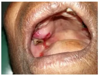
On inspection there was a 3x3 cm, well defined swelling seen over the right posterior maxillary alveolar ridge and palate with an area of central ulceration (Fig. 1).
Fig. 1. Intra-oral view shows swelling on palate with ulcerated surface
On palpation, the findings on inspection were confirmed, the swelling was tender and soft to firm in consistency extending bucally upto the vestibule and palatally just lateral to the midline. Bilaterally, cervical lymph nodes were not palpable. The panaromic radiograph revealed a well delineated, unilocular radiolucent lesion extending from the maxillary right first premolar to first molar [Fig. 2]. With provisional diagnosis of a odontogenic tumor, an incisional biopsy was performed under local anaesthesia.
Fig. 2. Panaromic radiograph showing ill defined radiolucency from maxillary premolar
to molar region
Histological sections stained with H&E [under 4x] revealed sheets and islands of polygonal to round clear cells with eccentric nuclei, separated by fibrous connective tissue septa (Fig. 3).
Under higher magnification, biphasic population of cells with characteristic polygonal, clear cells
and hyperchromatic, basaloid cells with
nuclear pleomorphism was present with rare mitotic figures.
Cytoplasm of the clear cell was Periodic Acid Schiff positive, indicating intracytoplasmic glycogen deposition. Mucicarmine gave a negative result [Fig. 4].
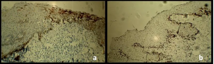
Tumor cells were immunoreactive for CK-19 and Ki-67 [Fig. 5], which showed a strong relation to a homogenous pattern of immunoreactivity.
A final diagnosis of Clear Cell Odontogenic Carcinoma was made.
Fig. 3. H & E stained section (a) under 10x showing numerous polygonal cells and fibrous connective tissue septa (b) under 40x showing clear cells with eccentric nucleus
Fig. 4. (a) Shows PAS positive clear cells containing glycogen (b) mucicarmine test showing negative result
Fig. 5. (a) CK-19 (b) Ki-67 shows positive immunoreactivity towards tumor cells nuclear pleomorphism was present with rare
mitotic figures.
Cytoplasm of the clear cell was Periodic Acid Schiff positive, indicating intracytoplasmic glycogen deposition. Mucicarmine gave a negative result [Fig. 4].
Tumor cells were immunoreactive for CK-19 and Ki-67 [Fig. 5], which showed a strong relation to a homogenous pattern of immunoreactivity.
A final diagnosis of Clear Cell Odontogenic Carcinoma was made.
Fig. 3. H & E stained section (a) under 10x showing numerous polygonal cells and fibrous connective tissue septa (b) under 40x showing clear cells with eccentric nucleus
Fig. 4. (a) Shows PAS positive clear cells containing glycogen (b) mucicarmine test showing negative result
Fig. 5. (a) CK-19 (b) Ki-67 shows positive immunoreactivity towards tumor cells nuclear pleomorphism was present with rare
mitotic figures.
Cytoplasm of the clear cell was Periodic Acid Schiff positive, indicating intracytoplasmic glycogen deposition. Mucicarmine gave a negative result [Fig. 4].
Tumor cells were immunoreactive for CK-19 and Ki-67 [Fig. 5], which showed a strong relation to a homogenous pattern of immunoreactivity.
A final diagnosis of Clear Cell Odontogenic Carcinoma was made.
Fig. 3. H & E stained section (a) under 10x showing numerous polygonal cells and fibrous connective tissue septa (b) under 40x showing clear cells with eccentric nucleus
Fig. 4. (a) Shows PAS positive clear cells containing glycogen (b) mucicarmine test showing negative result
Yogesh et al.; BJMMR, 18(3): 1-5, 2016; Article no.BJMMR.27190
3. DISCUSSION
The term clear cell odontogenic tumor was coined by Hansen et al. [2]. Clear cell odontogenic carcinoma (CCOC) is a rare neoplasm with only few cases reported in the literarure [6]. Clear cell lesions in the head and neck region have a wide range of differential diagnosis that includes odontogenic tumors such
as ameloblastoma, calcifying epithelial
odontogenic tumor, odontogenic carcinoma, and salivary gland tumors like mucoepidermoid carcinoma or hyalinizing clear cell carcinoma. Also included are intraosseous melanocytic tumors, and metastatic tumors from kidney, thyroid, and prostate [2].
In 2005, WHO classified CCOC as malignant tumors characterised by sheets and islands of vacuolated and clear cells. Its local aggressive growth, frequent recurrence, and occasional metastasis are reported in several cases. CCOC primarily affects females. The majority of cases have been diagnosed in patients older than 40 years. The peak incidence is 5th to 7thdecades (mean age 56.5 years: Range 17-89 years) [2]. According to the literature, 73 cases of CCOC have been reported, 57 cases (77.0%) involving the mandible and 17 cases (23%) in the maxilla. The posterior region of jaws is the more frequent site for CCOC in comparison to the anterior region (48% vs 30%). Thus, the mandible is more commonly affected than the maxilla. CCOC of the maxilla as reported here is quite rare [2]. The clinical presentation of CCOC has been reported to present as a painless swelling. Pain and regional tooth mobility are only occasionally present. Radiologically, these tumors exhibit a radiolucent lesion with irregular margins and associated with root resorption [6].
Histologically, 3 patterns have emerged [6] 1) Most common is a biphasic pattern
characterized by nests of clear cells intermixed with smaller islands of
polygonal cells and eosinophillic
cytoplasm;
2) Epithelial islands exclusively composed of clear cells;
3) Least common is clear cell nesst with an ameloblastomatous pattern.
Glycogen storage is common in these tumors and immunoreactivity for cytokeratins (CK 8, 13 & 19) has been reported. Occasionally EMA,
S-100 protein and antiameloblastoma antigen are present. PAS is strongly positive with these tumor cells [7,8]. The present study confirmed immunohistochemical expression of CK-19 and Ki-67.
3.1 Features Suggestive for Differential Diagnosis
- Absence of amyloid deposition and
calcification in stroma to rule out clear cell variant of CEOT
- Lack of intermediate cells, squamous
differentiation and mucin production to exclude mucoepidermoid carcinoma
- Primary intraosseous localization and
absence of salivary gland or mucosal swelling for differentiation with hyalinised clear cell carcinoma of salivary gland
Mucicarmine staining in the present case showed negative results and thus the salivary gland tumors were ruled out. The histopathology showing biphasic pattern of cells and separation of islands by mature fibrous stroma confirmed the diagnosis of CCOC. Several authors have noticed an occurrence of hyalinised or partly hyalinised stroma separating the neoplastic islands. A clear cell component can be expected in odontogenic tumors, as they arise from dental lamina [9].
Treatment of this tumor includes aggressive complete exicision, curettage or enucleation, or surgical resection with or without lymph node dissection. Recurrence rate for this tumor is reported to be about 55% after resection [10]. Adjuvant radiation therapy or chemotherapy may be beneficial in patients with soft tissue or perineural invasion.
4. CONCLUSION
CCOC is a rare odontogenic tumor and very few cases have been reported in the literature. Differentiation from other clear cell lesions is very important. Further studies on clinical, histological and immunohistochemical properties of this tumor need to be considered. Also, due to its aggressive nature long term follow up is mandatory.
CONSENT
ETHICAL APPROVAL
It is not applicable.
COMPETING INTERESTS
Authors have declared that no competing interests exist.
REFERENCES
1. Elbeshir EI, Harris M, Barrett, AW. Clear cell odontogenic carcinoma of the maxilla: Clinical, histological and immunohisto-chemical features of a case Oral Oncology Extra. 2004;40:91–94.
2. Niharika Swain, Richa Dhariwal, Jay Gopal
Ray. Clear cell odontogenic carcinoma of maxilla: A case report and mini review Journal of Oral and Maxillofacial Pathology. 2013;17(1).
3. Elizabeth A. Bilodeau Aaron, Hoschar P,
Leon Barnes E, Jennifer L, Hunt Raja R. Seethala clear cell carcinoma and clear
cell odontogenic carcinoma: A
Comparative Clinicopathologic and
Immunohistochemical Study. Head and Neck Pathol. 2011;5:101–107.
4. Eversole LR, Belton CM, Henson LS.
Clear cell odontogenic tumor:
Histochemical and ultrastructural features.
Journal of Oral Pathology. 1985;14:603-61.
5. Minkyu Kim, Eunae Cho, Jae-Young Kim, Hyun Sil Kim, Woong Nam. Clear cell odontogenic carcinoma mimicking a cystic lesion: A case of misdiagnosis. J Korean Assoc Oral Maxillofac Surg. 2014;40:199-203.
6. Eugenio Maiorano, Mario Altini, Giuseppe Viale, Adriano Piattelli, Gianfranco Favia. Clear Cell odontogenic carcinoma report of two cases and review of the literature. Am J Clin Pathol. 2001;116:107-114.
7. Zahide Mine Yazici, Ozgur Mete, Zubeyde
Elmalı, İbrahim Sayin, Rasim Yilmazer,
Fatma Tulin Kayhan. Clear cell
odontogenic carcinoma of the maxilla ACTA MEDICA (Hradec Králové). 2011; 54(3):122–124.
8. Li TJ, Yu SF, Gao Y, et al. Clear cell odontogenic carcinoma: A clinic-pathologic and immunocytochemical study of 5 cases. Arch Pathol Lab Med. 2001;125:1566–71. 9. Jain G, Hegde P, Shetty P. Clear cell
odontogenic carcinoma: A rare case. Clin Cancer Investig J. 2015;4:408-10.
10. Walia Cheshta, et al. Clinical enigma: A rare case of clear cell odontogenic
carcinoma. Contemporary Clinical
Dentistry 6.4. (2015):559–563. PMC. Web 24 Apr; 2016.
_________________________________________________________________________________
© 2016 Yogesh et al.; This is an Open Access article distributed under the terms of the Creative Commons Attribution License (http://creativecommons.org/licenses/by/4.0), which permits unrestricted use, distribution, and reproduction in any medium, provided the original work is properly cited.
Peer-review history: