PHENETHYL ALCOHOL RESISTANCE I N ESCHERZCHZA COLI.
111. A TEMPERATURE-SENSITIVE MUTATION (dnaP)
AFFECTING DNA REPLICATION
CHIEKO WADA AND TAKASHI YURA Institute for Virus Research, Kyoto Uniuersiiy, Kyoto, Japan
Manuscript received January 4, 1974
ABSTRACT
A temperature-sensitive DNA replication mutant of E. coli K-12 was iso- lated among the mutants selected for phenethyl alcohol resistance a t low tem- peratures. This mutation, designated as dnuPl8, affects sensitivity of the cell to phenethyl alcohol, sodium deoxycholate and rifampicin, presumably due to an alteration in the membrane structure. At high temperatures (e.g., 42'),
synthesis of DNA, but not RNA or protein, is arrested, leading ta the forma- tion of ''filaments'' in which no septum formation is apparent. Nucleoids observed under electron microscope seem to become dispersed and DNA fibrils less condensed, which may explain the loss of viability under these conditions. Genetic analyses, including reversion studies, indicate that a recessive dnaP mutation located between cya and metE on the chromosome is responsible for both alterations of the membrane properties and temperature sensitivity. The dnaPl8 mutation does not affect growth of phage T4 or lambda under condi- tions where host DNA replication is completely inhibited. Kinetic studies of
DNA replication and cell division i n this mutant after the temperature shift from 30 to 42", and during the subsequent recovery a t 30", accumulated evi- dence suggesting that DNA replication comes to a halt at 42" upon completion
of a cycle already initiated before the temperature shift. Since the recovery of DNA synthesis after exposure to 42" does not depend on protein or RNA synthesis or other energy-requiring processes, the product of the mutant dnaP gene appears to be reversibly inactivated at 42". Taken together with the re- cessive nature of the present mutation, it was suggested that one of the mem- brane proteins involved in initiation of DNA replication is affected in this mutant.
A
number of temperature-sensitive mutants primarily affecting DNA repli- cation in Escherichia coli have been isolated and characterized in several laboratories. These mutants were obtained either without any particular selection (KOHIYAMA et al. 1966; WECHSLER and GROSS 1971) or after selection for inability to replicate the chromosomal DNA at high temperatures ( BONHOEFFER and SCHALLER 1965; FANGMAN and NOVICK 1968; KUEMPEL 1969). When cul- tures of these mutants are transferred to high temperatures, DNA synthesis stops either immediately or after a lag which permits completion of the replication cycle already initiated before the temperature shift. At least two genes ( d n d and C ) have been reported to be involved in the initiation of DNA replication in200 C. WADA A N D T. YURA
leads to a n immediate cessation of DNA replication at high temperatures, imply- ing that the chain elongation step is affected in these mutants (see reviews by GROSS 1972; SMITH 1973).
An involvement of the bacterial membrane i n DNA replication has been experimentally substantiated, after it was onginally proposed by
JACOB,
BRENNER and CUZIN (1963) (GANESAN and LEDERBERG 1965; SMITH andH A N A ~ A L T
1967;SUEOKA and QUINN 1968;
FIELDING
and Fox 1970; YAMAGUCHI, MURAKAMI and YOSHIKAWA 1971). Specific alterations of the membrane proteins have also been reported in some temperature-sensitive DNA replication mutants upon shift to high temperatures (INOUYE and GUTHRIE 1969; INOUYE and PARDEE 1970; SHAPIRO et al. 1970; SICCARDI et al. 1971, 1972; LAZDUNSKI and SHAPIRO 1973).Our own approach to this problem has been through analysis of mutants of
E .
coli that are resistant to phenethyl alcohol (PEA) (YURA andWADA
1968; WADA and YURA 1971). In E. coli, PEA is thought to act at the cell membrane (SILVER andWENDT
1967) and to selectively inhibit the initiation of a new cycle of DNA replication without interfering with the completion of a cycle al- ready initiated (TREICK and KONETZKA 1964; LARK and LARK 1966). Thus, we have identified and characterized a chromosomal mutation, pea, responsible for the resistance to PEA in the derivatives of strain C600 (YURA and WADA 1968). We have also studied a mutation at another locus ( m a f ) affecting the maintenance of an autonomous F factor in the same strain. Replication of theF
factor rather than F factor distribution during cell division appeared to be inhibited by PEA when a strain carried the maf mutant allele ( WADA and YURA1971).
In the course of these investigations, a number of temperature-sensitive PEA- resistant mutants were isolated from several PEA-sensitive strains ( WADA and YURA 1968). These mutants were resistant to PEA at 30” and failed to grow at 42’ even in the absence of PEA. DNA synthesis seemed to be specifically affected in some of these mutants, resulting in the formation of “filaments” after pro- longed incubation at high temperatures. The results of detailed analysis of one such mutant (KY2750) are reported in this paper. It was revealed that a single mutation is responsible €or both alterations of the membrane properties (sensi- tivities to PEA, deoxycholate, and to rifampicin) and of temperature sensitivity. The chromosomal replication at the stage of initiation appears to be inhibited when the mutant cells are exposed to high temperatures. In view of these as well as the genetic mapping data presented below, this mutation seems to define a novel gene involved in DNA replication and may tentatively be designated as
dnaPI8 (P stands for phcnethyl alcohol).
MATERIALS A N D METHODS
Bacterial and phage struins: Bacterial strains used are all derivatives of E . coli K12, and their origins and genetic characters are listed in Table 1. Bacteriophages T4D and Plvir were ob- tained from DRS. T. MINAGAWA and J. TOMIZAWA, respectively.
D N A REPLICATION MUTANT O F
E .
ColiTABLE 1
Bacterial strains used and their known genetic characters
201
Strain Sex Genetic characters* Origin+
PA678 KY20rj3 KY2750 KY290O KY290d KY6Oa7 KY9115 CSlOl BC73 AB2277 GP1 W3350 F- F- F- F- F- F- F- Hfr F- F- Hfr F-
thr, leu, thi, lac, gal, mal, xyl, mtl, m a , tonA, an', str his, met, trp; other markers same as i n PA678 dnaPI8; other markers same as in KY2053 mal+; other markers same as in KY2750 t h y A ; other markers same as in KY2750 t h y A ; other markers same as in KY2053
met+ ; other markers same as in KY2750 met, tonA
trp, ilu, phoS, tna, str, lac
pro, f r p , his, ilu, metE, thi, lac, gal, mal,
mtl, ara, str, tsx, tonB ilu, metE, cya, thi gall, gal2, lac
JACOB-WOUMAN PA678 KY2053 KY2750 KY2750 KY2053 KY27501 J. TOMIZAWA
N. OTSUJI
E. ADELBERG
T. YOKOTA
H. OZEKI
~~~~~~~ ~
* Gene symbols are those employed by TAYLOR and TROTTER (1972).
+
Origin as indicated here does not necessarily represent an immediate origin.NaCl and 2 g glucose per liter (pH 7.4). Minimal medium E (VOGEL and BONNER 1956) was usually supplemented with 0.5% glucose, 0.2% Difco Casamino Acids, and 20 pg/ml of L- tryptophan and 2 pg/ml of thiamine (medium E-Casamino acids). Thymine (50 Jug/ml) was further supplemented for experiments with the thymine-requiring strain.
Chemicals: Thymidine-6-3H and uracil-2-14C were obtained from Radiochemical Centre (Amersham), and thymine-6-3H from Dai-ichi Pure Chemicals Co. (Tokyo). L-Arginine [3-3H(N)] and L-arginine-lC(U) were the products of New England Nuclear (Boston), whereas D-glucose-C-d7 (98 atom % as ZH) and 15NH,C1 (95 atom % as 1SN) were purchased from Merck (Canada) and Hikari Kogyo (Tokyo), respectively. Phenethyl alcohol, deoxycholate and rifampicin were supplied by Nakarai Chemicals Co. (Kyoto), E. Merck (Darmstadt), and Lepetit spa. (Milano), respectively. Chloramphenicol was obtained from Sankyo Chemical Co. (Tokyo).
Determination of D N A synthesis: Incorporation of 3H-thymidine or JH-thymine into acid- insoluble fraction was measured to determine DNA synthesis, unless otherwise stated. Thus, either 3H-thymidine (0.4 pc/8 pg/ml) or 3H-thymine (2 pc/50 pg/ml) plus 250 pg/ml of deoxyadenosine was added to PG medium or medium E-Casamino acids. An overnight culture grown in the radioactive medium at 30" was diluted 20-fold in the same medium, incubated further to the log phase and was used for various experiments. Aliquots (0.5 ml) were taken at intervals and were added to 3 m l of cold 5% trichloroacetic acid. After standing for at least 30 min i n ice, cells were centrifuged, washed twice with cold trichloroacetic acid and were sus- pended in 0.5ml2M NH,OH. Contents were then transferred into a vial, 10 m l of Bray mixture added, and radioactivity determined in a liquid scintillation counter.
Isolation of the mutant: PEA-resistant mutants were isolated from a multiply marked strain of E . coli KY2053 that had been derived from strain PAG78. Cells were treated with N-methyl- N-nitro-N-nitrosoguanidine, washed and incubated i n PG medium to allow for segregation, and plated on medium E-Casamino acids containing 0.22% re-distilled PEA and 1.5% agar. After incubation at 30" f o r 3 days, resistant colonies appeared at a frequency of about 10-7. Among
202 C. WADA A N D T. YURA
0
0.
I0.2
0.3PHENETHYL ALCOHOL (%)
FIGURE 1.-Growth of bacteria in the presence of PEA. PG media containing various con- centrations of PEA were inoculated with 106 cells/ml of each strain and were incubated by stand- ing a t 30" for 48 hr. Turbidity was measured in a Klett-Summerson colorimeter with a NO. 54
filter. 0-0, Wild type (KY2053):
@---a,
Mutant (KY2750); A--A, Ts+ revertant# 1 ;
A-A,
Tsf revertant #2.RESULTS
Alteration of the membrane propexties
in
the mutant: As was expected from thc selection employed, the mutant KY2750 was shown to be more resistant toPEA than the parental strain when grown in a liquid medium at 30" (Figure 1 )
.
The mutant was subsequently focnd to be altered in sensitivities to deoxycholate and to rifampicin. As shown in Figure 2, deoxycholate (DOC) at 0.25% strongly inhibited growth of the mutant, but not the wild type, at 30". At 42", the marked loss of colony formers was observed with the mutant in the presence of DOC. The mutant was also shown to be sensitive to low concentrations of rifampicin that had little effect on the wild-type bacteria (Figure 3 ) . These results strongly sug- gest that the structure of some membrane component(s) has been altered in this mutant.Specific cessation of D N A synthesis and cell division at high temperatures: When a mutant culture grown in PG medium at 30" was transferred to 42", DNA
~~ ~
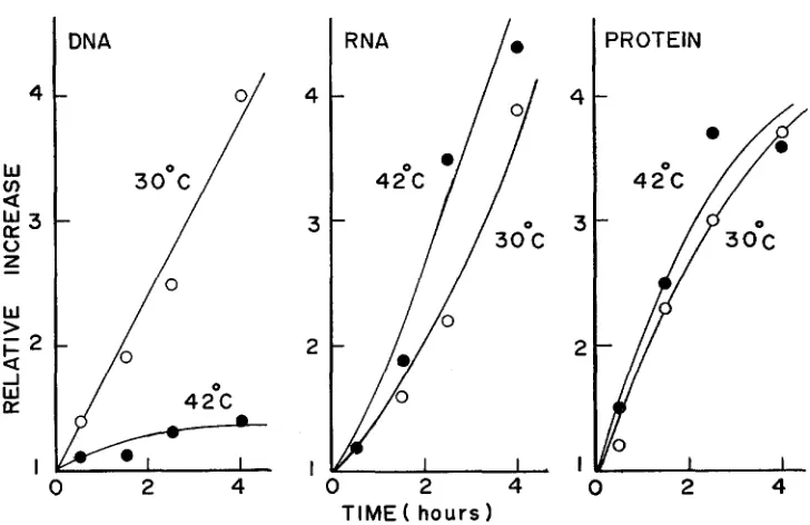
FIGURE 2.-Effect of DOC on the cell viability. Log-phase cultures (PG medium) of wild type (KY2053) and the mutant (KY2750) grown at 30" were diluted several-fold i n prewarmed
PG medium with or without DOC. Cultures were shaken a t 30" (a) or 42" (b), and aliquots taken at intervals were diluted and plated on PG agar to score the number of colony formers at
30". Open symbols are for the wild type, and closed symbols for the mutant. 0, 0 , control;
DNA REPLICATION MUTANT OF
E.
coli 203synthesis stopped after GO to 90 min, resulting in about 50% increase in
DNA
as determined by 3 H - t h p i d i n e incorporation.In
contrast,RNA
synthesis as measured by 14C-uracil incorporation proceeded faster at 42" than at 30°, as didI
10
\
0
I2
3
4
T I M E (
hours
I I I I
0 I 2 3 4
204 C. WADA A N D T. YURA
-
cn
z
w
P
n
0
w
>
F 2
a
4
W
a
I
0 I
2
3 4T I M E ( h o u r s )
FIGURE 3.-Growth of bacteria in the presence of a low concentration of rifampicin. Log-
phase cultures in PG medium of wild type (KY2053) and the mutant (KY2750) were used to inoculate the same medium with or without rifampicin (8 pg/ml) at about 5
x
loT cells/ml. Optical density was followed during shaking at 30".0-0,
wild type; A-A, wild type with rifampicin; -,
mutant;A-A,
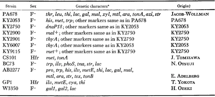
mutant with rifampicin.the parental strain. Similar results were obtained when the temperature was shifted to 40", or when a thymine-requiring derivative (KY2901) of the mutant was examined using 3H-thymine to label the DNA. Figure 4 presents the results of colorimetric determination of DNA, RNA and protein in this mutant grown at 30" or at 42" in medium E-Casamino acids. These results clearly indicate that
DNA synthesis is specifically arrested at high temperatures in the mutant KY2750. I n a separate experiment, cells previously labeled with 3H-thymidine at 30" were washed and incubated in the absence of thymidine at 42O. No differ- ence in acid-soluble radioactivity was found between the mutant and wild-type cultures for at least 1 hr, suggesting that DNA degradation is not accelerated in the mutant.
Cell division also stopped shortly after the cessation of DNA synthesis, leading to about a threefold increase in cell number during 3-hr incubation at
42"
DNA REPLICATION MUTANT OF E . coli 205
DNA
-
- i
/
420c
0 2
4
4
3
2
I
'ROTEIN
0 2 4 0 2 4
TIME ( hours
1
FIGURE 4.-Effect of temperature on DNA, RNA and protein synthesis in the mutant. A log-phase culture of the mutant (KY2901) in medium E-Casamino acids supplemented with tryptophan and thymine was grown to 1.5
x
108/ml, divided into two, and shaken at 30" er 42", respectively. Aliquots were taken at the times indicated for determination of DNA, RNA and protein by colorimetric methods, essentially as described by BERRAH and KONETZKA (1962). Rela- tive increases over the initial values are plotted.tration failed to relieve the effect of high temperatures on DNA synthesis, however.
When mutant cells that had been aerated at 42" for 90 min were infected by phage T4D o r lambda, they could still support the growth of these phages just as well as those grown at 30", in spite of their complete inability to replicate the chromosomal DNA (Table 2). Thus the adverse effect of high temperatures on DNA synthesis in this mutant seems to be restricted to the chromosomal DNA. In this connection, the exposure of the mutant cells harboring an Fgal or ColVBtrp episome to 42" for 2 to 4 hr did not lead to the segregation of clones that had lost the episome at higher frequencies. DNA synthesis in these cells gradually stopped at 42" as in the mutant F- bacteria.
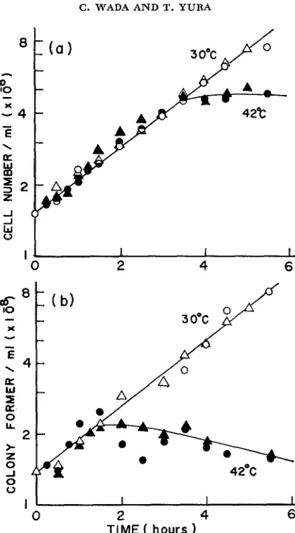
Resumption of DNA synthesis upon return to low temperatures: When a
mutant culture that had Been kept at 42" for 1 to 4 h r was shifted back to 30", DNA synthesis resumed with little lag. Addition of chloramphenicol (100 or
206 C . WADA AND T. YURA
0 2 4 6
I I I
0 2 4 6
TIME ( hours
FIGURE 5.-Effect of temperature on cell division and viability of the mutant. A log-phase culture of the mutant (KY2750) grown in medium E-Casamino acids at 30" was divided into two parts and one aerated at 30" (open symbds) and the other at 42" (closed symbols). Aliquots were taken a t the times indicated for determination of both total cell number (a) and colony formers (b). The cell number was estimated by a Coulter counter (Model B) at the settings of T, = 20, A = and I = i/z. Colony formers were determined by plating appropriate dilutions of each sample on PG agar followed by incubation at 30" for 2 days. Experiment 1 (0, 0 ) ;
Experiment 2 ( A ,
A).
DNA synthesis observed in the presence of chloramphenicol did not vary appreci- ably when +&e length of previous exposure to 42" varied between 1 and 4 hrs. Thus, the initiation capacity does not seem to accumulate during incubation at the restrictive temperature, in contrast to the recent observations with another DNA initiation mutant CT28 ( SCHUBACH, WHITMER and DAVERN 1973). Despite the rapid recoverj- of DNA synthesis observed, cell division hardly occurred when
D N A REPLICATION MUTANT O F E. Coli TABLE 2
Growth of bacteriophages in the mutant bacteria*
20 7
T.CD lambda
Time (min) 30' 42" 30' 42.0
0 2.6
x
103 1.9 x 10³ 6.1x
ID6 4.6,x
10590
-
__ 5.0x
1W 8.7 x 10730 3.6
x
1oj 8.0 x 10¹⁵ __-
120 __
_-
5.5x
108-
* Log-phase cultures of the mutant (KY2750 or KY2900 f o r experiments with T4D or A, respectively) in PG medium were shaken at 30" or 42" f o r 90 min and were infected with T4D
(m.0.i. = 1.6) wr h (m.0.i. = 1.1). After standing f o r 5 (for T4D) or 15 min (for A ) a t each temperature to allow for adsorption, a portion was diluted 1W (for T4D) or 10-2 (for A) into prewarmed PG medium and was further shaken at each temperature. Samples were taken at the times indicated, treated with chloroform, and the phage yields determined with strain W3350 as indicator bacteria.
incubated further for as long as 10 hrs. Apparently, the prolonged incubation at the restrictive temperature irreversibly inactivated the capacity of the mutant cells to carry out further divisions. This may be contrasted to what has been found with other DNA initiation mutants.
In
another experiment, a mutant culture that had been incubated at41"
forw
>
F 3
a
wE
I
0 2 4 6
TIME ( h o u r s )
FIGURE 6.-The recovery of DNA synthesis in the mutant at low temperature. A log-phase culture ( 1 x 10⁸ cells/ml) of the mutant (KY2901) grown at 30" in medium E-Casamino acids containing 3H-thymine was transferred t3 42" at time 0 and was aerated far 2 hr. The culture was then divided into five parts, one kept at 42" (
-
) , whereas the others were returned to 30". 0-0, control at 30". Chloramphenicol (CM, 150 pg/ml) was added 15 min before(A-A)
or at the time of the shift (A-A) t o the low temperature. Rifampicin (50 p g /208 C . WADA A N D T. YURA
I O 0
5 0 0 0
TIME AT 30 C ( minutes)
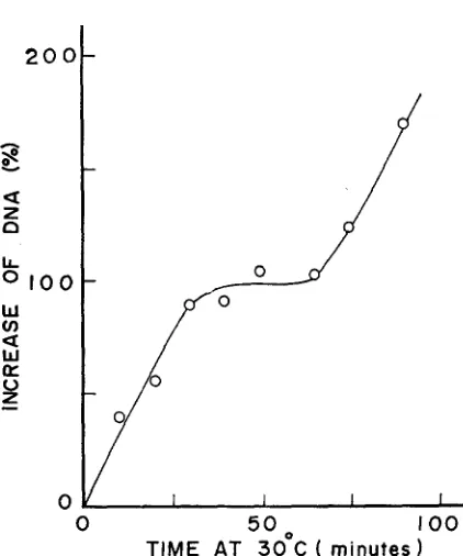
FIGURE 7.-Residual DNA synthesis a t 41" as a function of the length of pulse exposure to low temperature. A culture of the mutant (KY2750) was grown in medium E-Casamino acids containing 3H-thymidine for several generations to about 1.5
x
108 cells/ml at 30". The culture was shifted to 41", shaken for 90 min, and then returned to 30" for further incubation. Aliquots of the culture were taken a t the times indicated on the abscissa, and were shaken a t 41" for an additional 120 min. Radioactivity in acid-insoluble fraction was determined as described i n MATERIALS AND METHODS and is expressed as percent increase over the value (1,050 counts/min) at the time of the shift to the low temperature.90 min to allow cessation of
DNA
synthesis was exposed to a low temperature(30") and then returned to 41" again. Under these conditions, certain fraction of cells underwelit apparently one cycle of
DNA
replication. The amount ofDNA
synthesized, however, varied depending upon the length of the pulse exposure to the low temperature.As
seen in Figure 7, the doubling of DNA in total population took place when the cells were kept at 30" for 30 to 65 min.It
thus appears that about 30-min incubation at 30" is required for the ma- jority of cells to become capable of initiating a new cycle ofDNA
replication. The exposure to the low temperature for 75 min or longer resulted in more than doubling of theDNA,
presumably due to the second round ofDNA
replication. The generation time of this mutant under these conditions was about 100 min at30". It was further shown that chloramphenicol or dinitrophenol added to the medium during the pulse incubation at 30" had little effect on the subsequent resumption of
DNA
replication at 42" (Figure 8).DNA REPLICATION MUTANT OF
E . coli
3 0°C
209
4 2 O C
2.5
>
k5
F
2.00
a
U
a
0
LT
w
+
w
LT
1.5
3
42OC
A%
0 2 4 6
TIM E ( hours)
FIGURE &-Effects of chloramphenicol and dinitrophenol on the recovery of DNA synthesis. A log-phase culture (1
x
108 cells/ml) of the mutant (KY2750) grown a t 30" in medium E-Casamino acids containing 3H-thymidine was transferred to 42" and was shaken further for 3hr. The culture was divided into four parts, with one kept at 42" throughout the experiment
(
e-.),
while the others were transferred to 30" and shaken for 501 min with no addition(0-0)
or with an addition of 150 pg/ml of chloramphenicol(A-A)
or 2x
10-3 M dinitrophenol ( A-A). Each culture was then diluted 20-fold in the same prewarmed medium with no drug and was shaken further a t 42". The radioactivity at time 0, was 4,750 counts/min.during incubation at 30". Moreover, the fact that the amount of
DNA
doubled during recovery in the presence of chloramphenicol (Figure 6 ) , as well as the data presented in Figure 7, suggest that the residualDNA
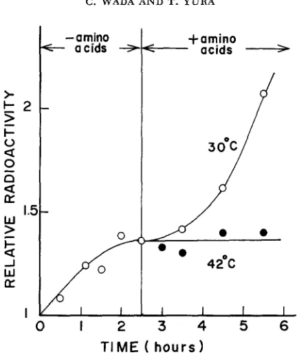
synthesis at high temperatures tends to synchronize the replication cycle, perhaps at the stage of initiation.D N A synthesis following amino acid starvation: To further substantiate the effect of temperature on the initiation of
DNA
replication, the mutant cells were first starved for amino acids at 30', and the recovery ofDNA
synthesis upon re-addition of amino acids was examined at 30" and 42".As
shown in Figure 9,DNA
synthesis resumed at 30" after some lag, but did not resume at 42'. Since amino acid starvation presumably pennits the completion but not the initiation ofDNA
replication in E. coli, these results are also taken t o indicate that the mutant cannot initiate a new round ofDNA
replication cycle at high tem- peratures.21 0 C . WADA AND T. YURA
-
am-ino a a d sI-
o
0
a
w
>
+amino
-
acids+
r'
-
0 .42OC
G
J W EI 1 . 1 1 I I
0
1 2 3 4 5 6TIME ( h o u r s )
FIGURE 9.-Effect of temperature on DNA replication after amino acid starvation in the mutant. A mutant culture (KY2901) in medium E-Casamino acids containing W t h y m i n e was shaken at 30" to about 1.5
x
108 cells/ml. The culture was filtered through a sterile Millipore filter (HA 0.45 a), washed and resuspended in medium E containing 3H-thymine but lacking all amino acids. After shaking for 150 min at 30", the culture was divided into two parts, each shaken at 30" or 42". Amino acids (casamino acids plus tryptophan) were added to both cultures immediately after temperature equilibration. There was no increase in optical density during the amino acid starvation in this experiment. The radioactivity at time 0 was 3,020 counts/min.The following experiments were carried out to see whether the regions of
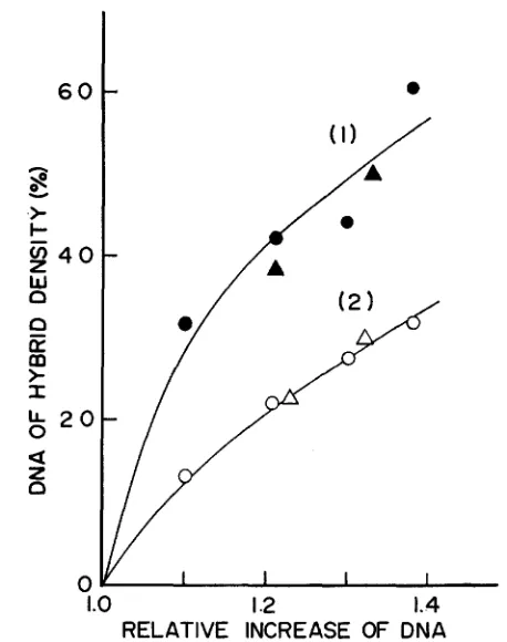
DNA
replicated early during recovery at 30" are fixed or randomly distributed over the entire chromosome i n the mutant cell population. The mutant cells a t the initial phase of the recovcry at 30" after a previous exposure to 41 O were pulse-labeled by 3H-thymidine for 10 min, followed by further incubation for 15 min
DNA REPLICATION MUTANT OF
E.
coli 21 16 0
h
Y
s
>
I-
w
4 0
a
n
E
m
>
I&
2 0
a
a
z
0
1.0 I
.2
1.4RELATIVE INCREASE OF DNA
FIGURE lO.--CsCl density gradient centrifugation of DNA pulse-labeled at the log phase or at the initial phase of the resumed synthesis. (1) A log-phase culture of the mutant (KY2750) grown at 30" i n medium E-Casamino acids was exposed to 41" for 90 min, returned to 30" and was pulse-labeled with 3H-thymidine (20 pc/8 pg/ml) for 10 min (between 5 and 15 min of incubation). Cells were transferred to a non-radioactive medium, shaken for 15 min at 30", ex- posed again t o 41" for 90 min, and finally transferred to the medium containing 15NH4C1 (2
mg/ml), 2H-glucose (2 mg/ml) and an acid hydrolysate of E. coli cells that had been grown on 2H-glucose and 1SNH,C1. During incubation at 30", aliquots were taken a t 30, 60, 80, and 100 min, cells were collected, and DNA's extracted by phenol were centrifuged in CsCl density gradients. (2) As a control, a portion of the same log-phase culture used above was pulse-labeled with 3H-thymidine at 30" for 10 min, transferred to a non-radioactive medium and incubated at 41" for 90 min. Tlio culture was then returned to 30" for 30 min, exposed again to 41" for 90 min, and finally incubated at 30" in the 2H15N-medium. Aliquots were taken, DNA's ex- tracted and centrifuged in the same way as above. The percent of DNA of hybrid density was calculated from the radioactivity profile of each sample (1-2
x
104 counts/min) and was plotted against total DNA as determined by the parallel experiment. Circles and triangles represent the results of two independent experiments.212
0
C. WADA AND T. YURA
I
MOLECULAR WEIGHT(
x
‘
2
LL
0
0
I O
20
30
4 0 50FRACTION NO.
FIGURE 11 . A D S - g e l electrophoresis of envelope proteins. Wild-type (KY2750) cultures were grown at 30” to 3
x
108 celIs/ml in medium E supplemented with 0.1% Casamino acids, 20pg/mI of each amino acid required and 2 pg/ml of thiamine. Both cultures were transferred to
42“ and were labeled with 0.8 @c/ml of 14C-arginine (wild type, 0-0) or 4 pc/ml of 3H-arginine (mutant,
a---@)
for the period that allowed doubling of the optical density. The two cultures were mixed before harvesting cells and the envelope fractions were prepared and analyzed by SDS-gel electrophoresis essentially as described by INOUYE and GUTHRIE (1969). The sample contained 11,400 (3H) and 2,800 (1°C) counts/min of radioactivity. The molecular weight scale indicated has been calculated from the protein profiles of RNA polymerase and 50 Sribosomes of E. coli run simultaneously with other gels.
missing in the mutant preparation. Such a difference was not detected when cells were grown at 30”. This component, having molecular weight o l about 40,000, probably corresponds to
“Y
protein” ( INOUYE and GUTHRIE 1969) o r “MP4O”(LAZDUNSKI and SHAPIRO 1973) that are known to disappear when DNA synthe- sis is inhibited (INOUYE and PARDEE 1970; LAZDUNSKI and SHAPIRO 1973). Thus, no specific alteration of envelope proteins associated with the dnaP mutation could be detected by these experiments.
DNA REPLICATION MUTANT OF E. coli
TABLE 3
Transduction mapping of the dnaP18 mutation by p h g e Pi
21 3
Experiment* Selection
I ilu
+
I1 ilu+
I11 ill;+
Unselecied marker+
__-
tna p h d cya &UP metE
Number of p a n s -
ductants obtained
1 1 0
0 1 1
0 1 0
0 0 1
0 0 0
1 1
1 0
0 1
0 0
1 1 1
1 1 0
1 0 1
1 0 0 0 1 0
0 0 1
0 0 0
2 4 15 18 61
Total 100
2 43 4 43
Total 92 1 19 5 49 1 1 29
Total 105
* Experiment Donor Recipient
I KY2750 (tna+ phoS+ ilu+ dnaP18) BC73 ( t m p h o S i l u d n a P f )
I1 KY9115 (ilu+ dnaPl8 metE+) AB2277 (ilvdnuP+ m t E ) 111 KY2750 (ilu+ cyafdnuP18 metE+) GP1 (ilu cyu dnaP+ m t E ) .I- 1 and 0 represent markers from donor and recipient strains, respectively. The dnaP character was scored by streaking cells from overnight cultures on PG agar followed by incubation at 30"
and 42" to determine temperature sensitivity.
or Mal+ Str-R recombinants tested, some were found to be both temperature- independent (Ts+) and PEA-sensitive (Pea-s), whereas none of the Thr+ Leu+ Str-R recombinants tested inherited these characteristics from the Hfr parent. Further mapping experiments were performed by transduction using phage
P1,
and the results obtained are summarized i n Table 3. It can be seen that the tem- perature sensitivity of the mutant is due to a mutation or mutations occurring between cya and metE. We tentatively designate this gene as
dnaP,
since it seems to represent a novel gene essential for DNA synthesis in E. coli. The probable gene order in this region of the chromosome is: tna-phoS-ilv-cya-dnaP-metE.As was expected from the location of
dnaP,
the episome F14 was found to carry the wild-type allele of this gene,dnaP+.
When F14 was transferred to the mutant KY2750, the resulting merodiploid strain carrying bothdnaP28
anddnaP+
was found to be capable of growing at 42" in PG medium, as is thehaploid wild-type strain. This shows that the
dnaP18
mutant allele is recessiveto
dnaP+.
The F+ derivative of the mutant was still temperature-sensitive, indi-214 C . WADA A N D T. Y U R A
TABLE 4
Properties of temperature-independent revertants*
Phenethyl alcohol Strain ( 0 . 2 % )
Wild type (KY2Q53) S
Mutant (KY2750) R
Revertants S
S S S R R Deoxycholate
( 0 . 5 % )
Rifam idn (8 P A )
R S R S R S S S Number obtained . . 9 12 1 1 1 2 Total 26
* Spontaneous revertants independently obtained from mutant KY2750 were examined for their sensitivity to t h e drugs a t the concentrations indicated. The sensitivities were determined by measuring optical density of the culture after 48 hr (phenethyl alcohol) o r 4 hr (deoxycholate or rifampicin) a t 30" in PG medium.
Temperature-independent reversion: Further evidence that a single mutation at the dnaP locus is responsible for both PEA resistance and teniperature sensi- tivity was obtained by studying temperature-independent revertants spontane- ously obtained from the mutant. As shown in Table 4, many of the revertants underwent simultaneous alterations toward the wild-type phenotype with respect to the sensitivity to PEA, DOC or rifampicin. Effects of PEA concentration on the growth of some of the revertants are included in Figure 1. These results are consistent with the notion that the dnaP18 mutation primarily affects the struc- ture of some membrane component and that this membrane alteration is respon- sible for all the mutant characteristics observed.
When a n F+ factor was introduced into the mutant by conjugation, the result- ing Ff strain underwent reversion to the temperature independence at a fre- quency about one hundred times higher than the original F- strain. Whether this effect of the F+ factor on the reversion frequency is due to the integration of the episome into the chromosome (NISHIMURA et al. 1971) has not been investigated.
DS.\ IWPI.ICATION MUTANT OF E . coli 215
b
'1
I:IGUI~F. 13.--Elcctron niicrographs of thin srrtions of t t i r mutant ( K Y 2 7 5 0 1 g r o \ \ n i t t IY;
mrcliuni a t 30" or 42". Cells were risecl and emhedtlrd by the method of RYTBR i d KFI.I.F.K-
IWI(;ER (1058) with m i n w modifications. a, control (30"). A sequrncr of changrs that apprars t o take placr a t 42" is rrprrscntecl by I), r. cl ant1 e. The portions of c nncl cl indicatecl by arrows
wrrr furthcv mngnifiecl to Rive f and g. respectively. S : nucleoids. a-e, x 35,000; f.g. X 8 0 .
V.
vations may explrin tho irreversible loss of viability of the mctant cells under t h r sr con tl i t ion s.
DISCUSSION
The trml)craturc-sensiti\.c mutation ( d n n P I 8 ) described in this paper may be rcg:irdrd a s a mutation primaril:: affecting the structure and function of some essrntiill componrnt of t!ic bacterial membrane. and DNA replication and cell division become arrcstctl at high trmprraturcs iis the result of this membrane alteration. Firstly. the mutant \vas selcctcd for its resistance to PEA. whose pri- mary site of ;iction appears to be on the membrane (SII.VER and \VENDT 1967); and PEA rcsistancr of certain E. coli strains has been shown to involvc a n alter- ation in the membrane proprrlic*s (Yvn,\ and WADA 1968). Secondly, thr mutant was found to be aIt(w4 in smsitivities to DOC and to rifampicin. The DOC sensi- tivity has been correlatcd with alterations of thc bacterial surfact. strricture ( NAGKI. DE 7 J w . 4 1 ~ and L,I:RI.A 1067; T- TI i im. 4 . Mon~orr and JACOB 1970). and the higher srnsitivity of the mutant to rifampicin presumably reflects the increased j:crmrability to the drug ( h r i . m r 1960). ];inally, all these mutant properties can oftrn be lost simultnnrouslj* by reverse mutations to temperature indcpen- drnce (Table 4). suggrsting that they are due to ii single gcnr mutation.
21 6 C. WADA A N D T. YURA
stopped after some residual synthesis, whereas RNA and protein synthesis con- tinued exponentially for at least two or three generations. Even after the com- plete cessation of DNA synthesis, however, the mutant cells supported the growth of phage lambda as well as T4 at 42" (Table 2 ) , suggesting that the normal functioning of the dnaP+ gene is not required for growth of these phages. The normal burst size of lambda after infection at 42" also suggests that the effect of the mutation does not involve the general damage on DNA synthesis such as depletion o€ the deoxyribonucleotide pools, The F + factor introduced into the mutant cell failed to reverse the temperature-sensitive phenotype of the mutant, indicating that the F+ factor cannot supply the dnaP function. The question of whether replication of the F+ factor itself is arrested at high temperatures like that of the chromosomal DNA has not sc far been investigated.
As to the stage of the chromosomal replication affected by the dnaP18 muta- tion, available data do not permit clear distinction among various possibilities. However, several lines of evidence listed below suggest that replication becomes arrested at or near the initiation of the cycle when the mutant cells are exposed to high temperatures. (1 ) The amounts of residual DNA synthesis at 42" and during amino acid starvation are nearly the same (Figures 6 and 9). (2) After alignment of the DNA at 30" by amino acid starvation, re-addition of amino acids at 42" did not permit further synthesis of DNA (Figure 9 ) . (3) After prior in- hibition of DNA replication at 42", the amount of DNA doubled upon return to 30" in the presence of chloramphenicol or rifampicin (Figure 6). (4) The quan- titative analysis of dependence of the residual DNA synthesis on the length of pulse exposure to low temperature (Figure 7) suggested that the incubation at Lhe high temperature tends to synchronize the replication cycle at the stage of initiation or termination.
(5)
The residual cell division at 42" reached the level (about threefold) that may be expected for an initiation-defective mutant (BEYERSMANN, SCHLICHT and SCHUSTER 1971). In addition, the results of the density-shift experiments presented (Figure IO) and the capacity of the mutant cells to permit replication of independent replicons such as phage lambda and T4 at the restrictive temperature are at least consistent with this interpretation. Many of the characteristics of the mutant described here are similar to what have been reported for other dna mutants defective in the initiation of DNA replica- tion (KOHIYAMA 1968; HIROTA, RYTER and JACOB 1968; HIROTA, MORDOH and JACOB 1970; CARL 1970; ABE and TOMTZAWA 1971;BEYERSMANN,
SCHLICHT and SCHUSTER 1971 ;WECHSLER
and GROSS 1971; WOLF 1972; SCHUBACH, WHITMER and DAVERN 1973).The dnaPI8 mutation has been mapped between ilv and metE on the E. coli
chromosome, the frequency of co-transduction with ilv being about 20%. The DNA initiation mutant CRT46 carries a dnaA mutation closely linked to ilv but on the opposite side of ilv from metE (HIROTA, MORDOH and JACOB 1970). An- other mutation studied by ABE and TOMIZAWA (1971) is also located close to the CRT46 mutation (ABE, personal communication). The relation between dnaP
and other mutations located between cya and metE, including rep (CALENDAR
DNA REPLICATION MUTANT OF E . coli 21 7 and SBAVRONSKAYA 1971), mutU (SIEGEL 1973), and
pdeB
(HORIUCHI
and NAGATA 1973), also remains obscure at the present time, although the normal sensitivity of the present mutant to ultraviolet light tends to preclude a close re- lationship with any of these mutants.The available evidence discussed above leads us to a specific suggestion on the possible nature of the present mutation and its effect on the chromosomal DNA replication in E . coli. Thus, the dnaP18 mutation presumably affects the struc- ture and function of some membrane component of the cell in such a manner that further rounds of DNA replication are prevented upon shift to high tempera- tures. In view of the results presented in Figure 8 on the recovery of DNA repli- cation by a pulse exposure to the low temperature, as well as the recessive nature of the mutation, the dnaP gene product might represent an enzyme that consti- tutes one of the membrane proteins in E. coli. This might be related to a “PEA- sensitive” protein, postulated by LARK and LARK (1966), that is required f o r the initiation of DNA replication.
The present results of electron microscopic observation suggest that the nu- cleoids in the dnaP18 mutant cells become disperse and the DNA fibrils less con- densed as they cease to synthesize DNA at high temperatures. Whether these observations indeed reflect a unique property of the mutant or represent the re- sults of other indirect nature might be worth investigating i n the future. In this connection, the previous observations with the dnaA mutants indicate that the nucleoids are often found in the central region of the elongated cells (KOHIYAMA
et aE. 1966;
HIROTA,
RYTER andJACOB
1968).In addition to the well-known effect of PEA on the initiation of DNA replica- tion in E. coli, strong inhibition of phospholipid synthesis was reported recently that might be related to the membrane alteration brought about by PEA (NUNN
and
TROPP
1972). The formation of active dimers of alkaline phosphatase from inactive monomers-which seems to take place at the membrane-is also in- hibited by PEA (TRIBHUVAN et aZ. 1970). Moreover, a higher concentration of PEA (0.5%) causes the release of DNA from the DNA-membrane complex(MASKER
and EBERLE 1972). Although the precise mechanism of action of PEA on DNA replication is not clear at present, further analyses of PEA-resistant mutants, including the one described here, should offer a profitable approach to the general problems of the role of cellular membrane in DNA replication in bacteria.We are grateful to DR. Y. OZEKI for carrying out electron microscopic observations, and to
DR. A. MATSUMOTO f o r kind advice in operation of the microscope. Helpful discussions and ad- vice by DRS. T. NAGATA and S. HIRGA and assistance by MISS J. SHINOHARA and MRS. A. KOMORI are gratefully acknowledged.
LITERATURE CITED
ABE, M. and J. TOMIZAWA, 1971
BERRAH, G. and W. A. KONETZKA, 1962
Chromosome replication in Escherichia coli K12 mutant af-
Selective and reversible inhibition of the synthesis of
fected in the process of DNA initiation. Genetics 69: 1-15.
21 8
BEYERSMANN, D., M. SCHLICHT and H. SCHUSTER, 1971
BONHOEFFER, F. and H. SCHALLER, 1965
C. WADA A N D T. YURA
Temperature-sensitive initiation of
A method for selective enrichment of mutants based on the high UV sensitivity of DNA containing 5-bromouracil. Biochem. Biophys. Res. Commun. 20: 93-97.
DNA replication in a mutant of Escherichia coli K12. Molec Gen. Genetics 111: 145-158.
CALENDAR, R., B. LINDQVIST, G. SIRONI and A. J. CLARK, 1970 and their interaction with P2 phage. Virology 40: 72-83. CARL, P. L., 1970
Molec. Gen. Genetics 109: 107-122.
FANGMAN, W. L. and A. NOVICK, 1968
FIELDING, P. and C. F. Fox, 1970
GANESAN, A. T. and J. LEDERBERG, 1965
GROSS, J. D., 1972 DNA replication in bacteria. In Curr. Top. Microbiol. Immunol. 57: 39-74.
HIROTA, Y., A. RYTER and F. JACOB, 1968 Thermosensitive mutants of E. coli affected in the processes of DNA synthisis and cellular division. Cold Spring Harbor Symp. Quant. Biol.
33: 677-693.
Characterization of REP- mutants
Escherichia coli mutants with temperature-sensitive synthesis of DNA.
Characterization of two bacterial mutants with temper-
Evidence for stable attachment of DNA to membrane at the ature-sensitive synthesis of DNA. Genetics 60: 1-17.
replication origin oE Escherichia coli. Biochem. Biophys. Res. Commun. 41 : 157-162.
Bhchem. Biophys. Res. Commun. 18: 821-835.
A cell membrane bound fraction of bacterial DNA.
HIROTA, Y., J. MORDOH and F. JACOB, 1970 On the process of cellular division in Escherichia coli. 111. Thermosensitive mutants of E. coli altered in the process of DNA initiation. J. Mol. Biol. 53: 369-387.
Mutations affecting growth of the Escherichia coli cell under a condition of DNA polymerase I deficiency. Molec. Gen. Genetics 123 : 89-1 10.
A mutation which changes a membrane protein of E. coli. Proc. Natl. Acad. Sci. U. S. 64: 957-961.
Changes of membrane proteins and their relation to de- oxyribonucleic acid synthesis and cell division of Escherichia coli. J. Biol. Chem. 245 : 5813-5819.
O n the regulation of DNA replication i n bacteria.
DNA synthesis in temperature-sensitive mutants of Escherichia coli. Cold
Mutants thermosensibles d'Escherichia HORIUCHI, T. and T. NAGATA, 1973
INOUYE, M. and J. P. GUTHRIE, 1969
INOUYE, M. and A. B. PARDEE, 1970
JACOB, F., S. BRENNER and F. CUZIN, 1963
Cold Spring Harbor Symp. Quant. Biol. 28: 329-3443. KOHIYAMA, M., 1968
Spring Harbor Symp. Quant. Biol. 33: 317-324. KOHIYAMA, M., D. COUSIN, A. RYTER and F. JACOB, 1966
coli K12 isolement et charactkrisation rapide. Ann. Inst. Pasteur 110 : 465-486. KUEMPEL, P. L., 1969
of Escherichia coli. J. Bacteriol. 100: 1302-1310. LARK, K. G. and C. LARK, 1966
Temperature-sensitive initiation of chromosome replication in a mutant
Regulation of chromosome replication in Escherichia coli: A comparison of the effects of phenethyl alcohol treatment with those of amino acid starva- tion. J. Mol. Biol. 2 0 : 9-19.
The significance of membrane alterations seen in
Effect of phenethyl alcohol on deoxyribonucleic acid- LAZDUNSKI, A. and B. M. SHAPIRO, 1973
MASKER, W. E. and H. EBERLE, 1972
DNA synthesis mutants of Escherichia coli. Biochim. Biophys. Acta 298: 59-68.
membrane association in Escherichia coli. J. Bacteriol. 109: 11 70-1 174.
DNA REPLICATION MUTANT OF E. coli 219 Chromosome replication in Esch- erichia coli. IV. Control of chromosome replication and cell division by an integrated epi- some. J. Mol. Biol. 55: 441-456.
NUNN, W. D. and B. E. TROPP, 1972 Effects of phenethyl alcohol on phospholipid metabolism in Escherichia coli. J. Bacteriol. 109: 162-168.
OGAWA, H., K. SHIMADA and J. TOMIZAWA, 1968 Studies on radiation-sensitive mutants of Esch- erichia coli. Molec. Gen. Genetics 101 : 227-244.
RYTER, A and E. KELLENBERGER, 1958 Etude au microscope dectronique de plasmas contenant de l'acide dCsoxyribonucMique. Z. Naturforsch. 13b: 597-605.
SCHLEIF, R., 1969 Isolation and characterization of a streptolydigin resistant RNA polymerase. Nature 223: 1068-1069.
SCHUBACH, W. H., J. D. WHITMER and C. I. DAVERN, 1973 Genetic control of DNA initiation in Escherichia coli. J. Mol. Biol. 74: 205-221.
SHAPIRO, B. M., A. G. SICCARDI, Y. HIROTA and F. JACOB, 1970 On the process of cellular division
in Escherichia coli 11. Membrane protein alterations associated with mutations affecting the initiation of DNA synthesis. J. Mol. Biol. 52: 75-89.
SICCARDI, A. G., B. M. SHAPIRO, Y. HIROTA and F. JACOB, 1971 On the process of cellular di-
vision in Escherichia coli IV. Altered protein composition and turnover of the membranes
of thermosensitive mutants defective in chromosomal replication. J. Mol. Biol. 56: 475-490. Interrelationship between membrane protein composition and deoxyribonucleic acid synthesis in Escherichia coli. Biochemistry
11: 1573-1582.
SIEGEL, E. C., 1973 Ultraviolet-sensitive mutator of Escherichia coli K-12. J. Bacteriol. 113:
SILVER, S. and L. WENDT, 1967
SMIRNOV, G. B. and A. G. SKAVRONSKAYA, 1971
SMITH, D. W., 1973
SMITH, D. W. and P. C. HANAWALT, 1967
SUEOKA, N. and W. G. QUINN, 1968
TAYLOR, A. L. and C. D. TROTTER, 1972
TREICK, R. W. and W. A. KONETZKA, 1964
TRIBHUVAN, R. C., A. K. PILGAOKAR, D. S. PRADHAN and A. SREENIVASAN, 1970 NISHIMURA, Y., L. CARO, C. M. BERG and Y. HIROTA, 1971
SICCARDI, A. G., A. LAZDUNSKI and B. M. SHAPIRO, 1972
161-166.
Mechanism of action of phenethyl alcohol: Breakdown of the
Location of uur502 mutation on the chromo-
DNA synthesis in prokaryotes: replication. Progr. Biophys. Mol. Biol. 26:
Properties of the growing point region in the bacterial
Membrane attachment of the chromosome replication
Linkage map of Escherichia coli strain K-12. Bacteriol.
Physiological state of Escherichia coli and the inhi- bition of deoxyribonucleic acid synthesis by phenethyl alcohol. J. Bacteriol. 8 8 : 1580-1584. Effect of phenethyl alcohol on induction of alkaline phosphatase i n Escherichia coli. Biochem. Biophys. Res. Commun. 4 1 : 244-250.
Acetylornithinase of Escherichia coli: Partial purifica- tion and some properties. J. Biol. Chem. 218: 97-106.
Phenethyl alcohol resistance in Escherichia coli: Analysis of a temperature-sensitive mutant. Ann. Report Inst. Virus Res., Kyoto Univ. 11: 47. -, 1971 Phenethyl alcohol resistance in Escherichia coli. 11. Replication of F factor in the re- sistant strain C600. Genetics 69: 275-287.
cellular permeability barrier. J. Bacteriol. 9 3 : 560-566.
some of Escherichia coli K-12. Molec. Gen. Genetics 113: 217-221.
323-393.
chromosome. Biochim. Biophys. Acta 149: 519-521,
origin i n Bacillus subtilis. Cold Spring Harbor Symp. Quant. Biol. 33: 695-705.
Rev. 36 : 504-524.
VOGEL, H. J. and D. M. BONNER, 1956
220 C . WADA A N D T. Y U R A
WECHSLER, J. A. and J. D. GROSS, 1971
WOLF, B., 1972
YAMAGUCHI, K., S. MURAKAMI and H. YOSHIKAWA, 1971
Escherichia coli mutants temperature-sensitive for DNA synthesis. Molec. Gen. Genetics 113: 273-284.
The characteristics and genetic map location of a temperature sensitive DNA
mutant of E. coli K12. Genetics 7 2 : 569-593.
Chromosome-membrane association i n Bacillus subtilis. I. DNA release from membrane fraction. Biochem. Biophys. Res. Commun. 44: 1559-1565.
Phenethyl alcohol resistance in Escherichia coli. I. Resistance of
YURA, T. and C. WADA, 1968
strain C600 and its relation to azide resistance. Genetics 5 9 : 177-190.