International Journal of Emerging Technology and Advanced Engineering
Website: www.ijetae.com (ISSN 2250-2459, ISO 9001:2008 Certified Journal, Volume 7, Issue 12, December 2017)
Advanced Automatic Brain Segmentation Techniques
for MRI using Hybrid Technique
1
M. Bhavana Eswar
Assistant Professor, Dept. of CSEGeethanjali College of Engineering and Technology Hyderabad, Telangana, India
2
Esther Varma
Associate Professor, Dept. of CSEGeethanjali College of Engineering and Technology Hyderabad, Telangana, India
Abstract —This paper displays a study of cutting edge techniques
for dividing the MRI (Magnetic Resonance Imaging) picture of the brain. Division of the brain is a testing errand since it requires more accentuated strategies to separate every one of the districts display in the brain picture. The power contrasts between the diverse districts in the brain MRI picture are less, making it hard to computerize the whole division process. Henceforth, a careful comprehension of the current division calculation is basic for exact division. The division calculations reviewed in this work are Neural Network Model, Self Organizing Maps, Radial Basis Function, Back Propagation, Fuzzy C-Means, Deformable Models, Level Set Models, Genetic
Algorithm, Differential Evolutionary Algorithm, Hybrid
Clustering and Artificial Intelligence. Such an overview would be useful for specialists working in the field of brain picture division. The paper talks about the complexities in the division calculation and furthermore the difficulties in dividing the brain MRI pictures. The division yields and examination of the current writing has additionally been talked about. The real criteria and their points of interest in the division of every calculation have been accounted for in like manner in the perceptions.
Keywords —Magnetic Resonance Imaging (MRI); brain picture;
division; neural systems; deformable models; fluffy c-implies
1. INTRODUCTION
A brain tumor is a gathering (or mass) of unusual cells in the brain. A tumor may prompt malignancy, which is a noteworthy driving reason for death and in charge of around 13% of all passing around the world. Disease rate is developing at a disturbing rate on the planet. So discovery of the tumor is imperative in prior stages. Incredible learning and experience on radiology are required for exact tumor identification in restorative imaging. X-ray is the most adaptable of our symptomatic imaging modalities, having the capacity to describe an extensive variety of parameters in the living subject and give choice spatial determination. Brain tumor distinguishing proof frame attractive reverberation imaging (MRI) comprises of a few phases. Division is known to be a fundamental however troublesome advance in medicinal imaging characterization and investigation. Consequently, it is profoundly important that division of the MRI pictures must be done precisely before requesting that the PC do the correct determination. This audit displays a diagram of attractive reverberation imaging (MRI) - based restorative picture investigation for brain tumor considers.
Brain
Together, the brain and spinal line (the focal sensory system (CNS)) control the physiological and mental elements of our body. For the most part our brain incorporates three noteworthy parts: 1. Brain. It controls considering, learning, critical thinking, feelings, discourse, perusing, composing, and intentional development. 2. Cerebellum. It controls development, adjust, and act. 3. Brain stem. It associates the brain to the spinal rope, and controls essential capacities in human body, for example, engine, tactile pathways, heart, vault and reflexes [1].
The brain is made out of two tissue writes, to be specific dark issue (GM) and white issue (WM). Dark issue is made of neuronal and glial cells, otherwise called neuroglia or glia that controls brain movement and the basal cores which are the dim issue cores found profound inside the white issue. The basal cores include: caudate core, putamen, pallidum and claustrum. White issue filaments comprise of numerous elinated axons which associate the cerebral cortex with other brain locales. The left and the correct sides of the equator of the brain are associated by corpus callosum which is a thick band of white issue filaments. Both, cerebellum and brain have a thin external cortex of dim issue, inside white issue and little however profoundly arranged masses of the dark issue. The spinal rope is situated toward the base of the brain. It has three structures: the midbrain, pons and medulla oblongata [2]. The brain additionally contains cerebrospinal liquid (CSF) which comprises of glucose, salts, compounds, and white platelets. This liquid circles through channels (ventricles) around the brain and the spinal rope to shield them from damage. There is likewise another tissue called meninges which are the layer covering the brain and spinal rope [2].
International Journal of Emerging Technology and Advanced Engineering
[image:2.595.43.290.142.277.2]Website: www.ijetae.com (ISSN 2250-2459, ISO 9001:2008 Certified Journal, Volume 7, Issue 12, December 2017)
Fig. 1 Structure of Brain and its Functions.
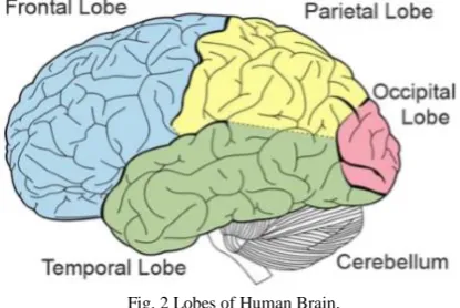
Biggest piece of the brain. It is associated with the cognizant musings, development and sensations. It additionally comprises of two parts, the privilege and the left halves of the globe. Every control the contrary side of the body. In addition, every half of the globe is separated into four projections: the frontal, fleeting, parietal and occipital flaps as appeared in Fig. 2 [3].
Fig. 2 Lobes of Human Brain.
Elements of projections are: 1. Frontal Lobe. It is a segment of the cerebral cortex lying simply behind the brow. It is engaged with talking and muscle developments and in making arrangements and judgments. 2. Occipital Lobe. It is a bit of the cerebral cortex lying at the back of the head. It incorporates regions that get data from the visual fields. 3. Parietal Lobe. It is a segment of the cerebral cortex lying at the highest point of the make a beeline for the back. It gets tangible contribution for touch and body position. 4. Worldly Lobe. It is a part of the cerebral cortex lying generally over the ears. It incorporates the sound-related regions, each accepting data essentially from the contrary ear [3].
Brain Tumor
Brain tumor is an unusual development of tissue in the cerebrum or focal spine that can upset appropriate brain work and makes an expanding weight in the cerebrum. Because of expanded weight on the cerebrum, some brain tissues are moved, pushed against the skull or are in charge of the harm of the nerves of the other sound cerebrum tissues [4].
Brain and spinal rope tumors are diverse for everybody. They frame in various zones, create
A. From various cell writes, and may have distinctive treatment choices. Researchers have arranged cerebrum tumor as indicated by: 1. The type and grade (how aggressive it is),
B. Whether it is a primary or a secondary tumor, C. If it is cancerous (malignant) or not (benign), and D. Where in the brain the tumor is located [5].
The slightest forceful kind of cerebrum tumor is frequently called an amiable brain tumor. They begin from cells inside or encompassing the brain, don't contain disease cells, develop gradually, and regularly have clear outskirts that don't spread into other tissue. They may turn out to be very vast before creating any side effects. In the event that these tumors can be expelled, they tend not to return. All things considered, they can cause noteworthy neurological manifestations relying upon their size, and area close different structures in the cerebrum. Some favorable tumors can advance to end up threatening. Harmful brain tumors contain disease cells and frequently don't have clear fringes. They are thought to be hazardous on the grounds that they develop quickly and attack encompassing brain tissue. Albeit threatening cerebrum tumors infrequently spread to different zones of the body, they can spread all through the brain or to the spine. These tumors can be treated with surgery, chemotherapy and radiation, however they may repeat after treatment. Regardless of whether carcinogenic or benevolent, tumors that begin in cells of the brain are called essential cerebrum tumors. Essential brain tumors may spread to different parts of the cerebrum or to the spine, yet infrequently to different organs. Metastatic or auxiliary cerebrum tumors start in another piece of the body and after that spread to the brain. These tumors are more typical than essential brain tumors and are named by the area in which they start. They are dealt with in view of where they begin, for example, the lung, bosom, colon or skin. Every one of these tumors has extraordinary clinical, radiographic and natural attributes [4].
2. RELATED WORKS
[image:2.595.57.265.395.534.2]
International Journal of Emerging Technology and Advanced Engineering
Website: www.ijetae.com (ISSN 2250-2459, ISO 9001:2008 Certified Journal, Volume 7, Issue 12, December 2017)
In recent years, those techniques have been combined with artificial neural networks (ANNs)[5], genetic algorithm (GA)[6], fuzzy logic[7], and Texture features are used for classification and segmentation in Ref.[8,9,10,11]. Recent works Zhang Nan 2011, have shown that classification of human brain in MRI images is possible via multi-kernel support vector machine (SVM) and adaptive training is designed to follow-up the changes of tumors during several MRI examinations. In Ref. [3], the author perform unsupervised brain tumor segmentation, and region detection using hybrid intelligent fuzzy Hopfield neural network. The authors in Ref. [2] develop a segmentation technique initially performs classification process by utilizing Fuzzy Inference System (FIS) and FFBNN. In several of previous works [8, 9, 10, 11] demonstrated the effectiveness of texture features in characterizing brain tumor tissue and analyzed the irregular texture variations of tumors in MRI. M. Jafari and S. Kasaei suggest a novel neural network-based classifier to distinguish normal and abnormal (benign or malignant) brain MRIs[12]. V. Sheejakumari and B. S. Gomathi presents optimal features for classifying tissues from the testing image dataset using Hybrid Genetic Algorithm-Neural Network (HGANN). The contribution of this paper is the integration of an efficient feature extraction tool and a robust classifier to perform a more robust and accurate automated MRI normal/abnormal brain images classification. The proposed hybrid technique initially performs classification process by utilizing Fuzzy Inference System (FIS) and FFNN. Both classifiers are utilizing the extracted image features as an input for the classification process.
Image segmentation [16] is the process of partitioning a digital image into multiple segments (sets of pixels, also known as super pixels). The goal of segmentation is to simplify and/or change the representation of an image into something that is more meaningful and easier to analyze. Image segmentation is typically used to locate objects and boundaries (lines, curves, etc.) in images. More precisely, image segmentation is the process of assigning a label to every pixel in an image such that pixels with the same label share certain visual characteristics.
In case of medical image segmentation the aim is to [16]: Study anatomical structure,
Identify Region of Interest i.e. locate tumor, lesion and other abnormalities,
Measure tissue volume to measure growth of tumor (also decrease in size of tumor with treatment), and
Help in treatment planning prior to radiation therapy; in radiation dose calculation.
Using segmentation in medical images is a very important task for detecting the abnormalities, study and tracking progress of diseases and surgery planning. Segmentation must not allow regions of the image to overlap.
Objective of this review section is to present literature survey of image segmentation methods. The main goal is to highlight advantages and limitations of these methods. Key image processing techniques for brain MRI image segmentation are classified as thresholding, region-growing, clustering, edge detection, atlasbased, other methods etc. All these techniques are explained further in the following sections and many notable methods have been reviewed.
3. IMPLEMENTATION OF PROPOSED METHODOLOGY
MR Image Data
All cuts in this work were accumulated on 1.5T attractive field, by PHILIPS MR Scanner produced by Philips Medical System/Netherlands with a serial number of 20415 having the accompanying highlights: a DCM coding plan designator having a pixel data transfer capacity of 235.3 Hz and a pixel introduction of Monochrome and a 2D securing type. Thickness of the cut is 5mm with 2 mm hole amongst cuts and aggregate number of cuts of 22 cut. The view areas are the sagital, coronal and hub with all the T1W-FFE, T2W-TSE, T2W-Fair and MR review.
Preparing and Testing Data
International Journal of Emerging Technology and Advanced Engineering
[image:4.595.54.281.144.335.2]Website: www.ijetae.com (ISSN 2250-2459, ISO 9001:2008 Certified Journal, Volume 7, Issue 12, December 2017)
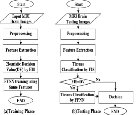
Figure 3: Proposed Classification Method (a)Training Phase (b)Testing Phase
According to the classification results, the proposed fuzzy system is not enough for some cases when the output between (0.4-0.6) Decision Value. Therefore, neural classifier are added for these cases utilizing same features as input to the additional classified stage which perform by feed forward neural network as shown in below figure .
Figure 4: Flow Chart of the Proposed Hybrid Technique
A three layer neural system was made with five hubs in the main (input) layer, one to five hubs in the concealed layer, and one hub as the yield layer. The preparation required 3368 ages to choose genuinely about the presence of tumors having a mean square blunder around 4.14e-7 and aggregate time around 44 sec utilizing Levenberg-Marquardt calculation of preparing as appeared in figure 7. The quantity of hubs in the concealed layer are fluctuated from (1to 5) in a recreation to decide the ideal number of shrouded hubs. Because of equipment impediments, three hubs in the concealed layer were chosen to run the last reenactment.
4. SCAN AND IMAGING TECHNIQUES A scan is the first step to identify whether a brain tumor is present or not, and if present, locate exactly where it is growing. A scan creates computerized images of the brain and spinal cord by examining it from different angles. Some scans use a contrast agent (or a dye) to allow the doctor to see the difference between normal and abnormal tissue. A patient may need more than one type of scan to diagnose a tumor, depending on its type and location.
Commonly used scanning and imaging techniques [1]: Computed Axial Tomography (CAT or CT Scan) is a
computerized x-ray that can show a combination of soft tissue, bone, and blood vessels. This is often the first test a person will receive in an emergency room (i.e. after a seizure).
Magnetic Resonance Imaging (MRI) can create clear and detailed three dimensional images of a brain tumor. An MRI is not often used with people who have a pace maker or other metal device.
Magnetic Resonance Spectroscopy (MRI Spect or MRS), measures the levels of metabolites in the body. An MRS can detect irregular patterns of activity to help diagnose the type of tumor, evaluate its response to therapies, or determine aggressiveness of a tumor.
Perfusion MRI examines the flow of blood into the tissues to help assess the grade/aggressiveness of tumors and differentiate a recurrent tumor from dead tumor tissue.
Functional MRI (fMRI) tracks the use of oxygen and blood flow in the brain as patients perform tasks. An fMRI can identify the motor, sensory, visual and language centres of the brain which helps your doctor carefully plan for surgery.
Positron Emission Tomography (PET) scan uses a radioactive substance to visualize hypermetabolic activity such as with malignant cells, or abnormalities from a tumor or scar tissue. PET is also used during brain mapping procedures.
International Journal of Emerging Technology and Advanced Engineering
Website: www.ijetae.com (ISSN 2250-2459, ISO 9001:2008 Certified Journal, Volume 7, Issue 12, December 2017)
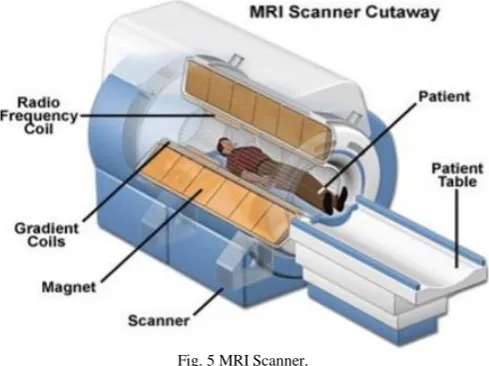
[image:5.595.312.553.177.309.2]4.1 Magnetic Resonance Imaging and Brain Tumors Raymond V. Damadian developed MRI in 1969 and was the primary individual to utilize MRI to research the human body [6]. As appeared in Fig. 3 [8], Magnetic Resonance Imaging (MRI) is an effective representation system that enables pictures of interior life systems to be obtained in a safe and non-obtrusive way. It depends on the standards of Nuclear Magnetic Resonance (NMR). Amid MR imaging, the patient is put in a solid attractive field which causes the protons in the water particle of the body to adjust in either a parallel (low vitality) or hostile to parallel (high vitality) introduction with the attractive field. At that point a radiofrequency beat is presented which powers the turning protons to move out of harmony state. At the point when a radio recurrence beat is ceased, the protons come back to harmony state and create a sinusoidal flag at a recurrence subject to the neighborhood attractive field. At long last, a radio recurrence loops or resonators inside the scanner distinguishes the flag and makes the picture [7]. This imaging medium has been of specific significance for delivering pictures of the cerebrum, because of the capacity of MRI to record flags that can recognize distinctive „soft‟ tissues, (for example, dark issue and white issue). Cerebrum pictures in MRI sweep can be typical or unusual. The typical mind is portrayed by having dark issue (GM), white issue (WM) and cerebrospinal liquid (CSF) tissues. The unusual cerebrum for the most part contains dynamic tumor, rot and edema notwithstanding typical mind tissues. Corruption is a dead cell situated inside a dynamic tumor, while edema is situated close dynamic tumor outskirts. Edemas, which comes about because of nearby disturbance of blood cerebrum hindrance, frequently cover with typical tissues and it is constantly hard to recognize from alternate tissues [9].
Fig. 5 MRI Scanner.
An axial MRI looks at the brain from below in a series of images starting at the chin and moving to the top of the head. A sagittal MRI looks at the brain from the side in a series of images starting at one ear and moving to the other.
[image:5.595.313.553.376.509.2]A coronal MRI looks at the brain from behind in a series of images starting at the back of the head and moving to the face as shown in Fig. 4 [10].
Fig. 6 Brain MR Images from a) Axial Plane, b) Sagittal Plane, and c) Coronal Plane.
The signal processing has three different images that can be achieved from the same body: T1weighted, T2- weighted and PD-weighted (proton density) as shown in Fig. 5 [11].
Fig. 7 Brain MR Images from Axial Plane a) T1-w, b) T2-w, and c) PD-w image.
[image:5.595.41.286.527.710.2]International Journal of Emerging Technology and Advanced Engineering
Website: www.ijetae.com (ISSN 2250-2459, ISO 9001:2008 Certified Journal, Volume 7, Issue 12, December 2017)
The first or shorter echo (TE < 30msec) is proton density (PD) weighted or a mixture of T1 and T2. This image is very helpful for evaluating periventricular pathology, such as multiple sclerosis, because the hyperintense plaques are contrasted against the lower signal CSF. More recently, the FLAIR (Fluid Attenuated Inversion Recovery) sequence has replaced the PD image. FLAIR images are T2weighted with the CSF signal suppressed [12]. When reviewing an MR image, the easiest way to determine which pulse sequence was used, or the "weighting" of the image, is to look at the cerebrospinal fluid (CSF). If the CSF is bright (high signal), then it must be a T2-weighted imaged. If the CSF is dark, it is a T1-weighted image. Thereafter, look at the signal intensity of the brain structures. On MR images of the brain, the primary determinants of signal intensity and contrast are the T1 and T2 relaxation times. The contrast is distinctly different on T1 and T2-weighted images. Also, brain pathology has some common signal characteristics [12].
4.2. Difficulties in segmentation of brain MRI
Cortical segmentation has not been made fully automated and operated at high speed because of the reliability of the MRI with regards to the homogeneity of its magnetic field. The problems of MRI include:
Noise: There is random noise connected with MR imaging system. This is known to have a Rician distribution [13].
Intensity non-homogeneity also called bias field or shading artifact: The non-uniformity in the radio frequency (RF) field during data collection results in shading effect [14].
Partial volume effect: In this type of effect more than one type of class or tissue occupies one pixel or voxel of an image. The pixels or voxels are called mixels [15].
5. CONCLUSION
Picture division is broadly utilized as a part of various biomedical-imaging applications, e.g., the measurement of tissue volumes, investigation of anatomical structure, finding, restriction of pathology, treatment arranging and PC incorporated surgery. Presently e-days, speed of calculation is not any more an issue for specialists. In this way, the concentration is coordinated toward change of data from pictures acquired through the cut introduction and idealizing the procedure of division to get a precise photo of the cerebrum tumor. As diagnosing tumor is a convoluted and touchy errand; in this manner, precision and unwavering quality are constantly allocated much significance. Thus, an explained system that features new vistas for growing more strong picture division method is much looked for.
REFERENCES
[1]. Cancer Support Community, “Frankly Speaking About Cancer: Brain Tumors”, National Brain Tumor Society, New York, Final Rep. June 2013.
[2]. Charles R. Noback, Norman L. Strominger, Robert J. Demarest and David A. Ruggiero, the Human Nervous System: Structure and Function, 6th ed., Humana Press, 2005.
[3]. The Nervous System, [Online]. Available: http://humananatomy2013.weebly.com/nervous-system.html, Accessed on: April 20, 2015.
[4]. Louis D.N., Ohgaki H., Wiestler O.D, Cavenee W.K. (Eds.), WHO Classification of Tumors of the Central Nervous System, International Agency for Research on Cancer (IARC), Lyon, France, 2007. [5]. Jan C. Buckner, et al., ―Central Nervous System Tumors, Mayo Clinic
Proceedings, Vol. 82, No. 10, pp. 1271-1286. 2007.
[6]. Damadian, R., Goldsmith, M. & Minkoff, L. NMR in Cancer: XVI. FONAR Image of the Live Human Body. Physiological Chemistry and Physics, Vol.9, No.1, pp.97- 100, ISSN: 0031-9325. 1977.
[7]. A. O Rodriguez, Principles of Magnetic Resonance Imaging, Revista Mexicana de Fisica, Vol. 50, No. 3, pp. 272-286. 2004.
[8]. Rotator Cuff MRI Shoulder Scan & Arthrogram (MRA), [Online]. Available: http://www.colinmcnulty.com/blog/2011/08/04/myrotator-cuff-mri-shoulder-scan-and-arthrogram-mra/, Accessed on: April 20, 2015.
[9]. [Online]. Available: http://www.radiologyassistant.nl/, Accessed on: April 20, 2015.
[10].[Online]. Available:
http://www2.massgeneral.org/livingwithtsc/affects/brain-popup_anatomy.htm, Accessed on: April 20, 2015.
[11].Different types of MRI, [Online]. Available: https://www.ole.bris.ac.uk/bbcswebdav/institution/Faculty%20of%20M edicine%20and%20Dentistry/MB%20ChB/Radiology/MRI %20e-tutorial/page_04.htm, Accessed on: April 20, 2015.
[12].Magnetic Resonance Imaging of the Brain, [Online]. Available: http://spinwarp.ucsd.edu/neuroweb/rad401/syllabus.htm Accessed on: April 20, 2015.
[13].Prima, S., Ayache, N., Barrick, T. & Roberts, N. Maximum Likelihood Estimation of the Bias Field in MR Brain Images: Investigating Different Modelings of the Imaging Process, Processings of Medical Image Computing and Computer-Assisted Intervention (MICCAI' 2001), Vol.2208, pp.811-819, DOI: 10.1007/3-540-45468-3_97. 2001. [14].Li, X., Li, L., Lu, H., Chen, D. & Liang, Z. Inhomogeneity Correction
for Magnetic Resonance Images with Fuzzy CMean Algorithm, Processings of SPIE, Vol.5032, 2003.
[15].Ruan, S., Jaggi, C., Xue, J., Fadili, J. & Bloyet, D. Brain Tissue Classification of Magnetic Resonance Images Using Partial Volume Modeling. IEEE Transactions on Medical Imaging, Vol.19, No.12, pp.1179- 1187, ISSN: 0278-0062. 2000.
[16].Image Segmentation, [Online]. Available: http://en.wikipedia.org/wiki/Image_segmentation, Accessed on: April 20, 2015.
[17].Sezgin M. and Sankur B., Survey over image thresholding techniques and quantitative performance evaluation, Journal of Electronic Imaging, Vol. 13, No. 1, pp. 146– 165. 2004.
[18].Rastgarpour M., and Shanbehzadeh J., Application of AI Techniques in Medical Image Segmentation and Novel Categorization of Available Methods and Tools, Proceedings of the International Multi Conference of Engineers and Computer Scientists 2011 Vol. I, IMECS 2011, March 16-18, 2011, Hong Kong.
[19].Y. Zhang, H. Qu, Y. Wang, “Adaptive Image Segmentation Based on Fast Thresholding and Image Merging”, Artificial reality and Telexistence-Workshops, pp. 308-311, 1994.
[20].T. Lindeberg and M.X. Li "Segmentation and classification of edges using minimum description length approximation and complementary junction cues", Computer Vision and Image Understanding, Vol. 67, No. 1, pp. 88-98, 1997.
International Journal of Emerging Technology and Advanced Engineering
Website: www.ijetae.com (ISSN 2250-2459, ISO 9001:2008 Certified Journal, Volume 7, Issue 12, December 2017)
[22].W. X. Kang, Q. Q. Yang, R. R. Liang, “The Comparative Research on Image Segmentation Algorithms”, IEEE Conference on ETCS, pp. 703-707, 2009.
[23].H. Zhang, J. E. Fritts, S. A. Goldman, “Image Segmentation Evaluation: A Survey of unsupervised methods”, computer vision and image understanding, pp. 260-280, 2008.
[24].H. G. Kaganami, Z. Beij, “Region Based Detection versus Edge Detection”, IEEE Transactions on Intelligent information hiding and multimedia signal processing, pp. 1217- 1221, 2009.
[25].T.U Paul and S.K. Bandyopadhyay, “Segmentation of Brain Tumor from Brain MRI Images Reintroducing K – Means with advanced Dual Localization Method”, International Journal of Engineering Research and Applications, June 2012.
[26].K Keshamoni, M Tripathi, “DELIBERATION AND EXERTION OF WIRELESS BODY AREA NETWORKS FOR EXCLUSIVE HEALTH CARE SUPERVISION OVER IOT”, 2017.
[27].K Keshamoni, S Hemanth.”Smart Gas Level Monitoring, Booking & Gas Leakage Detector over IoT” - Advance Computing Conference (IACC), 2017 IEEE , 2017.
[28].G. Heo and C. G. Small. Form representions and means for landmarks: A survey and comparative study. Computer Vision and Image Understanding, 102(2):188–203, 2006.
[29].J. C. Dunn, A Fuzzy Relative of the ISODATA Process and Its Use in Detecting Compact Well-Separated Clusters, Journal of Cybernetics, Vol. 3, No.3, pp. 32-57. 1973.
[30].Megha. P. Arakeri · G. Ram Mohana Reddy “Computeraided diagnosis system for tissue characterization of brain tumor on magnetic resonance images” SpringerVerlag London. 2013.
[31].A. Rajendran and R. Dhanasekaran, A hybrid Method Based on Fuzzy Clustering and Active Contour Using GGVF for Brain Tumor Segmentation on MRI Images, European Journal of Scientific Research, Vol. 61, No. 2, pp. 305-313. 2011.
[32].Ze-Xuan Ji,Quan-SenSun ,De-ShenXia “A framework with modified fast FCM for brain MR images segmentation” Pattern Recognition 44, pp. 999–1013. 2011.
[33].Ze-Xuan Ji, Quan-Sen Sun, De-Shen Xia “A modified possibilistic fuzzy c-means clustering algorithm for bias field estimation and segmentation of brain MR image” Computerized Medical Imaging and Graphics 35, 383–397. 2011.
[34].Pal N.R, Pal K, Keller J.M. and Bezdek J.C, “A Possibilistic Fuzzy c-Means Clustering Algorithm”, IEEE Transactions on Fuzzy Systems, Vol. 13, No. 4, pp. 517–530, 2005.
[35].Talairach J, Tournoux P. Co-Planar Stereotaxic Atlas of the Human Brain, 3-Dimensional Proportional System: An Approach to Cerebral Imaging. Stuttgart, Ger. Thieme, p. 122. 1988.
[36].Rajarethinam NC, Andreasen R, Cizadlo T, Arndt S, Swayze VW, et al. Automatic atlas-based volume estimation of human brain regions from MR images. J. Comput. Assist. Tomogr. 20 pp. 98–106. 1996. [37].Gibbs P, Buckley D, Blackb S, Horsman A. Tumour determination
from MR images by morphological segmentation. Phys Med Biol, 41, pp. 2437–46. 1999.
[38].Salman Y, Badawi A, Assal M, Alian S. New automatic technique for tracking brain tumor response.Int Conf Biol Med Phys, pp. 1–4. 2005. [39].Sato M, Lakare S, Wan M, Kaufman A. A gradient magnitude based