1 Chapter 1:
Introduction
1.1 Thesis Introduction
A prosthesis is an artificial device that replaces a part of the human body which is absent due to illness, injury or deformity. The design and construction of any prosthesis depends both on the complexity of the body part being replaced and the rehabilitative requirements of the prosthesis user.
Upper limb prostheses are prescribed for those individuals who have either partial or complete upper limb absence, which may either be acquired (through amputation) or congenital (absent at birth). They are fitted by healthcare professionals called Prosthetists, who work as part of a multi-professional rehabilitation team, usually within dedicated disablement service centres within the United Kingdom. Upper limb prostheses should primarily be comfortable to wear, functionally useful and aesthetically compatible with the requisite body parts. Comfort is achieved through an intimately matched prosthetic interface, called a socket, which directly encloses the remains of the most distal arm segment, or residual limb. The socket attaches to other components within the prosthesis, which normally include a hand or other terminal device or active prehensor (grasping device) (1).
There are three types of upper limb prostheses that are commonly available for potential prosthesis users; myoelectric prostheses, body-powered prostheses and cosmetic prostheses (2). Myoelectric prostheses are controlled by electro-myo-graphic (EMG, or myoelectric) signals generated from skeletal muscles within the remaining distal segment of the arm, known as the residual limb. The myoelectric signals are acquired by surface electrodes that are housed within the walls of the prosthetic interface, (known as the socket), which fits over the residual limb. Matching the socket shape and size with the residual limb shape and volume is essential for effective prosthesis fitting (3).
2 Body-powered prostheses are purely functional, cosmetically poor and have changed little since their inception over a century ago (4, 5). They require an external harness, which uses specific body movements to actively control joint segments and prehensile (grasp) function. They are still widely regarded as the most functional upper limb prosthesis type (6). By contrast, cosmetic prostheses are primarily aesthetic, but can also be used for simple daily tasks, such as holding or steadying objects, even though they have no active prehensile capability (6, 7).
Myoelectric prostheses combine aspects of both cosmetic and body-powered prostheses, incorporating electrically-powered active prehension and a relatively cosmetic appearance. In contrast to body-powered prostheses, myoelectric prostheses have the significant advantage of requiring no external harness for prosthesis control, and have also undergone significant improvements to their technical specifications during the last 30 years. The development of next-generation multifunctional hands with numerous degrees of freedom (8) combined with control system improvements such as myoelectric signal pattern recognition (9, 10) has greatly increased the potential capability of myoelectric prostheses. However, evidence suggests that these improvements are not being matched by a proportional increase in prosthesis user satisfaction (5, 11, 12) and usage rates (13-21); suggesting that other elements may be restricting prosthesis functionality.
One prosthetic element that has received relatively little attention over recent years within upper limb prostheses is the prosthetic socket, despite the fact that it is generally regarded as crucial to prosthesis usage (22–25). The upper limb prosthesis socket is a rigid, usually laminated, plastic shell (figure 1.1a), which normally completely encloses the residual limb. The role of the Prosthetist is primarily concerned with providing a well-fitting socket for the prosthesis user (1).
3 25), and still rely on principles developed and implemented before the requirements of clinical myoelectric control systems were fully recognised and understood (26-29).
Figure 1.1: Electrodes fixed into rigid socket (A); Impression on skin from electrode contact (B) (30)
One particular problem that can affect myoelectric control is relative movement between the electrode and the surface of the skin. Where surface electrodes are used in other applications, for example in the measurement of heart rhythm, they are adhered to the skin’s surface (31). However, in myoelectric sockets, attachment of the electrodes to the skin in addition to the socket is not possible because the user will need to don and doff their prosthesis. For this reason, the electrodes are located within fixed housings which are integrated within the prosthetic socket wall (figure 1.1). Consequently, this means that any movements, which occur between the socket and the skin, may also occur between the electrode and the skin.
4 with respect to prosthesis assessment and functionality, and are described in more detail within chapter 2.
The loads imparted at the interface between the socket and the skin in myoelectric upper limb prostheses and the subsequent relative movement may vary, but little is known regarding how much movement occurs and whether this is large enough to affect and alter electrode-to-skin contact.
Systems such as roll-on sockets have been devised to minimise the effects of these movements (33-35), but they are not frequently used in upper limb prostheses and have other disadvantages, which will be examined later in the thesis. The normal socket types used in myoelectric prostheses incorporate methods of socket suspension, fitting and attachment that are inherently similar to most types of prostheses currently available, and can in theory allow such movements to occur (28, 36, 37).
In body-powered and cosmetic prostheses, socket movement with respect to the skin will have little effect on prosthesis capability as they play no part in the prosthesis control processes. However, in myoelectric prostheses, the electrodes used to acquire the myoelectric signals require secure contact with the skin at all times for optimum function (38-40). Movement between the electrode and the surface of the residual limb could lead to the production of motion artifacts, or ‘false’ myoelectric signals, which may interfere with prosthesis control (38-40). Although the socket is rigid, the residual limb is fleshy and inherently mobile. This means that movement between the skin’s surface and the socket wall, and hence the electrode, is distinctly possible (41, 42). Although the resultant effects on signal acquisition are widely acknowledged (9, 10, 39, 40), and clinical anecdotal evidence regarding the problems with myoelectric prosthesis control are widespread, little documented evidence exists regarding the extent of the effect of these movements on prosthesis functionality.
5 The skills base of the Prosthetist has changed significantly in recent decades; the reasons behind these changes, and their potential consequences with regard to the area of the thesis, are highlighted and discussed within Appendix E- The changing education of Prosthetists.
Anecdotal evidence suggests that some Prosthetists will use elastic bands, secured around the external surface of the electrode, to try to restrict electrode movement or electrode detachment from the skin’s surface. Anecdotal evidence also suggests that felt washers or plastic inserts may also be used between the inner and outer socket (if an outer socket is used) to increase contact security between the electrode and the skin. However, both of these aforementioned methods are relatively crude and inaccurate.
The need for more evidence as to what effect immobile electrodes (which are fixed and contained within upper limb prosthetic sockets in simple housings) have on myoelectric prosthesis functionality prompted the development of this thesis. The evidenced high rates of rejection of myoelectric prostheses by upper limb amputees, linked to lack of prosthesis functionality, suggested that changes in design to electrode housing mechanisms and development of new novel designs could significantly reduce prehensile disruption, thereby improving prosthesis functionality and user uptake.
More research is also needed to confirm the extent to which surface electrodes fixed within myoelectric prosthetic sockets allow motion artefacts to occur during typical ADLs, thereby disrupting prehensile prosthesis control and resulting in restricted prosthesis functionality. In addition, the mechanisms and solutions to this phenomenon needed to be investigated in order to provide enhanced functionality for upper limb myoelectric prosthesis users.
1.2 Thesis overview
To meet the requirements outlined previously, the following investigations were undertaken:
6 2. An investigation contrasting current best practice in the field of myoelectric signal
acquisition with current clinical practice employed within myoelectric prostheses. 3. The development of potential improvements to signal acquisition within current types
of prosthetic socket by means of a novel electrode housing device.
4. An analysis comparing common upper limb movements and activities with their potential effect on signal acquisition.
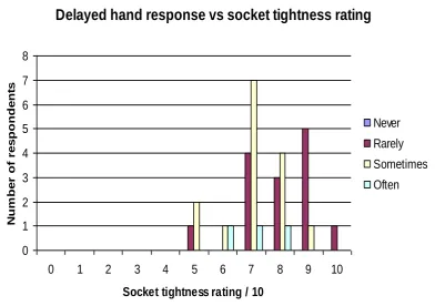
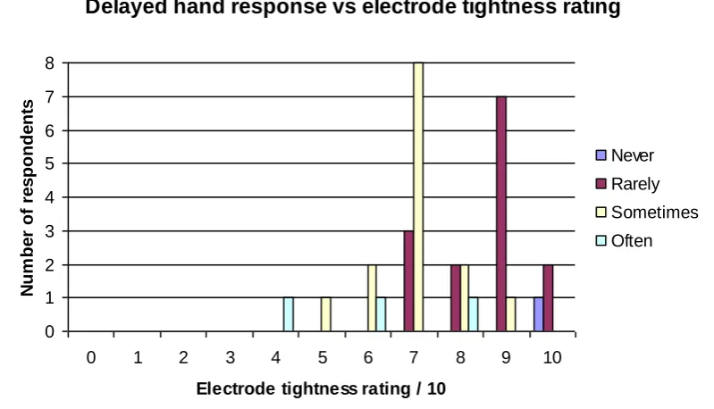
Firstly, evidence needed to be gathered from current myoelectric prosthesis users highlighting any difficulties with prosthesis control due to variations in their perceived electrode to skin contact security and socket tightness. These would need to be linked to their ability to perform ADLs, since these represent the movements, and the subsequent control interruptions, most likely to be employed and encountered by amputees. Variations in prosthesis loading, the effects of carrying, lifting and pulling, and the effect of different movements on specific electrode contacts, were also investigated to provide more specific data regarding individual movement effects and their consequences on functionality.
Additionally, it was thought prudent to consider what changes could be made to electrode housing arrangements; given the lack of adjustment and adaptability currently available with myoelectric sockets. The effects of using socket-housed electrodes with no adjustment within the socket was therefore compared to alternative electrode attachment methods in order to provide more evidence as to their efficacy in providing functionality.
7 From this background evidence and analysis, the following investigations were subsequently developed and performed, which form the basis of this thesis:
1) A prosthesis-user analysis study.
This involved the distribution of a questionnaire to current and past myoelectric prosthesis users. This provided data on the levels and significance of poor myoelectric control and its relationship with socket and electrode tightness and contact security, plus
This investigation is presented in chapter 3. It was recognised that the potential effects of socket movement and prosthesis control are acknowledged in the literature. However, it was thought important to obtain the views and experiences of prosthesis users to gain evidence as to how they perceived these effects impacted on their ability to perform ADLs. The way of achieving this was by obtaining prosthesis users’ views relating socket and electrode contact, and how this influenced prosthesis control and their ability to perform these ADLs. This chapter therefore describes the processes involved in the collection and collation of data using a specifically-designed questionnaire, and contrasts the results obtained with those from other studies relevant to this subject area.
2) An electrode contact functionality assessment.
Chapter 4 describes a pilot study which examined three different test conditions, linked to electrode housing and contact security. These conditions involved the use of myoelectric electrodes which were either, intentionally disconnected from the prosthetic socket, arranged with enhanced perceived contact security to the skin of the residual limb, or housed within the standard electrode housing arrangement. This comparative assessment was undertaken to demonstrate which test condition provided greater levels of myoelectric prosthesis control and resultant functionality. The method of functionality assessment was specifically-chosen as it was designed to include the assessment of prostheses as well as the natural hand.
8 acquired from analysing the efficacy of facilitating different electrode orientations within the prosthetic socket.
3) Development of a novel bespoke electrode housing unit.
Chapter 5 describes a follow-on study which investigated various electrode-to-skin contact security and alignment test conditions via the use of a novel bespoke electrode housing unit, which allowed finite adjustments to be administered to both electrode contact security and alignment. These adjustments were assessed with respect to prosthesis control via the use of a validated, reliable functionality assessment.
This bespoke novel electrode housing unit was devised and developed to offer the possibility of altering electrode/skin contact security by facilitating positional adjustment to either end of the electrode (and therefore the electrode contacts) whilst also offering varying alignment positions. Its effect on prosthesis functionality was compared to that afforded by the current typical clinical standard using a validated, reliable functionality assessment.
4) A motion analysis study.
This study examined the effects of specific movements which were designed to represent those used during normal daily upper limb activities, and the respective changes in prosthesis load on the production of motion artefacts.
This investigation is presented in chapter 6. It was developed to provide an enhanced understanding of the frequency of unwanted activation of terminal device events caused by production of motion artifacts whilst performing ADLs when wearing a myoelectric prosthesis. Three reproducible upper limb movements were selected, which had previously been validated as being representative of a range of common, daily upper limb activities. Prosthesis users were asked to perform these movements whilst their myoelectric prosthesis was connected to a clinical assessment system which recorded the motion artifacts generated during each motion. A bespoke pulley apparatus was also used; with the loading mechanism attached to the prosthetic hand facilitate the use of varying loads, which were representative of those associated with normal daily activities.
9 are contrasted with the changing nature of prosthesis provision and the delivery of clinical myoelectric prostheses, with particular regard for Prosthetist experience and specialist expertise within this area.
1.3 Chapter Summary
Prosthesis control has been highlighted at recent conferences as being the key factor in increasing myoelectric prosthesis usage and functionality (43). Much of the work that has been undertaken within this field has been completed by therapists, seeking to improve functionality assessment techniques, and engineers, improving technical specifications relating to signal processing and prehensor capability (43). A need to quantify the effects of socket fit and different electrode orientations with respect to the skin, both in intimacy of contact and orientation, on prosthesis functionality, prompted the need for this thesis. There was also a need to develop a novel electrode housing to potentially improve functionality.
The research presented in this thesis is therefore primarily related to the role of the prosthetic socket, and the design of its integral parts, and the way in which each individual bespoke socket may be designed and fabricated; which is intrinsically linked to the knowledge and skill of the Prosthetist. The thesis employs specific investigations to contrast the effects of socket tightness and various parameters linked to electrode contact and orientation, with resultant myoelectric prosthesis control and functionality. Whilst there are various hypotheses cited within the text, the overall hypothesis is that myoelectric prosthesis control is adversely affected by relative movements between currently employed socket designs plus their electrode housings, and the residual limb within the socket. The null hypothesis is that the socket and the electrode housings do not significantly affect prosthesis functionality within standard socket designs.
10 Chapter 2:
The natural upper limb, prosthetic replacement and myoelectric control
2.1 Introduction
The natural human upper limb provides a multitude of uses and functions, making its complete or partial absence a potentially debilitating factor for any affected individual, regardless of their age, gender or social status (44-46). There are two main factors to consider with regard to upper limb absence:
1. The actual physical loss of all or part of the upper limb, and its subsequent effect on body image and symmetry, and
2. The reduction in the available levels of upper limb functionality and functional usefulness.
All human cultures exhibit a ‘normalised’ standard body type based on appearance, and unfortunately also a stigma of disability which may affect social interaction for many affected individuals making the cosmesis of prostheses a priority (47). Replacing the physical elements of the natural upper limb with an effective prosthesis is reasonably successful for most levels of upper limb loss; a fact borne out by the large numbers of prosthesis users who regularly wear cosmetic type prostheses (6, 7, 13-18, 20, 21, 44, 46, 48-51). However, replacement of upper limb functionality is much more challenging, with many prosthesis users abandoning functional prostheses completely (6, 48, 52, 53).
11 Additionally, the comparative capabilities of upper limb prostheses with regard to the natural arm, particularly in replacing optimum levels of functionality, are also clearly important to this study. Consequently, the next part of this chapter examines functionality assessment methodology, modelling techniques and specific functionality assessments designed for prosthesis users that are comparable to the capabilities of the natural upper limb.
Finally, this chapter identifies and examines prosthesis designs and prosthetic components, which are currently used to replace and replicate the natural upper limb. The causes of limb absence and amputation techniques are examined, along with currently-available prosthesis types; with myoelectric prostheses and myoelectric control being the primary focus. In particular, socket design and function is contrasted with myoelectric signal acquisition, as these two areas are fundamental to the hypothesis.
2.2 The Natural upper limb: overall structure and function
The natural upper limb includes 32 bones and 3 major joints; the shoulder, the elbow and the wrist, along with numerous smaller joints located within the hand (54-56). The upper limb segments and major joints within the arm work in tandem with a complex control system to correctly locate and position the hand for object manipulation and activity completion (57).
The following sub-sections describe the anatomical structures, ranges of motion and functional control processes that are normally present in the natural upper limb. In addition to hand functionality, particular attention is also paid to the following:
Joint movements and functional ranges of motion for the major upper limb joints These play a significant part in the complex movement capabilities which are available within the natural arm, and are particularly relevant to the activities included within chapters 4, 5 and 6;
The anatomical structure of the elbow and other upper limb joints
12 important because specific anatomical landmarks within its structure are also used suspend standard transradial prostheses;
The muscles of the forearm.
These are used to effect movements within the natural hand, and remnants of these muscles are also used to operate commonly prescribed myoelectric transradial prostheses.
2.2.1 The hand
The hand is the ‘end effecter’ of the natural upper limb and is its most complex and functionally important structure (55). The hand consists of 27 relatively small, distinct bones which are grouped into three main categories; the carpals (located most proximally), the metacarpals (within the upper palmar region) and the phalanges (the digits) (54-56). These are manoeuvred and articulated by numerous muscles and their associated tendons, which are categorised as extrinsic (located within the forearm) and intrinsic (located within the hand) (54, 56). Movements of the natural hand have been classified as either prehensile (object held) or non-prehensile (object manipulated) (58).
13
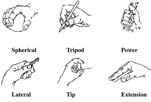
Spherical Tripod Power
[image:13.595.85.383.72.273.2]Lateral Tip Extension
Figure 2.1: The six defined grip patterns for the natural human hand (60)
A fully-functioning natural hand is able to interchange these grip types quickly and effectively, enabling the subject to employ the hand in a variety of ADLs (section 2.18.1), with each activity requiring a different combination of grip types (57). Consequently, the effective functionality or functional capability of the hand may be determined by its speed and ability to conform to each grip type accurately and to apply this form to a requisite activity (57).
However, the hand is not merely employed as a functional tool. It is also used for social contact and physical expression and as such has a clear aesthetic value as well as a functional value (55). As a purely functional tool, the hand is actually restricted by its evolutionary and aforementioned social requirements (58). Since effective prosthesis replacement will be determined by all the above factors, these social and aesthetic considerations will have significance in terms of prosthesis design and user acceptance. Furthermore, to operate effectively, the hand must be positioned optimally during any activity; requiring coordinated movements of the wrist, elbow and shoulder joints (57).
2.2.2 The wrist joint
14 contrast, the ulna is the largest forearm bone intersecting at the elbow (54). The radius and ulna intersect with the carpal bones at the base of the hand, forming the wrist joint, and enabling the hand to flex, extend, abduct and adduct with respect to the forearm. Pronation and supination effectively occurs at the distal radio-ulnar joint, not at the intersection with the carpals, but despite this technical anomaly pronation and supination are normally considered wrist movements (54, 56).
Figure 2.2(a): Functional wrist ranges of motion (61, 62) and Figure 2.2(b): movements (63)
2.2.3 The elbow joint
The elbow is a single axis hinge-type joint with a normal flexion range of between 0 degrees and 142 degrees and (within normal elbow joints) no available hyperextension (64) (see figure 2.3, below). The elbow and shoulder joints enable the hand and the wrist to be placed in the correct position for optimum functionality. The range of motion for individuals with a congenital limb absence will usually be slightly greater, as malformation of the elbow joint ligaments often leads to joint laxity and pronounced hyperextension of the elbow joint (66).
The elbow joint structures are particularly relevant for transradial socket fitting. The Prosthetist must contour the socket around the residual limb in order to maintain suspension (where applicable) and comfort during prosthesis usage. These aspects of socket fitting are inherently essential to the basis of this thesis and will be dealt with in more detail later in this chapter.
Movement Functional range of
motion°
Flexion 54°
Extension 60°
Adduction (Radial
deviation)
17°
Abduction (Ulnar
deviation)
40°
Pronation 20°
15 Figure 2.3: Anatomical structures of the elbow joint (65)
2.2.4 The shoulder joint
The relatively restricted motion of the elbow is compensated for by the extensive range of motion available from the shoulder, which is a shallow ball-and–socket joint offering abduction / adduction, flexion / extension and rotation in a traversing arc as shown in figure 2.4:
Figure 2.4(a): Functional shoulder motion (67); Figure 2.4 (b): Shoulder arc of motion (68)
Movement Functional range of
motion(°)
Flexion 159°
Extension 50°
Adduction 40°
Abduction 164°
16 This extended range of movement also allows the subject to compensate if either wrist or elbow movements are reduced due to injury, deformity or prosthesis movement restrictions.
2.3 Muscle groups within the natural upper limb
Provision of such an intricate movement capability, and to control each of the many joints within the wrist and hand effectively, requires numerous muscular insertions and an efficient control system. As a consequence, the natural forearm is distinguished by its complex muscular patterns formed from each of the many muscles required to accomplish the finite movements of the hand and wrist. This is worth noting even at this stage, since muscles which are effectively independent could provide contrasting and potentially conflicting myoelectric signals, which in turn may interfere with the potential clarity of the myoelectric activation signal.
17
Muscle Action Origin Insertion
Flexor carpi Radialis
Wrist Flexion
Med. humeral epicondyle Bases of 2nd and 3rd metacarpals Flexor Carpi
Ulnaris
Med. humeral epicondyle Pisiform
Extensor Carpi
Radialis longus Wrist Extension
Lateral supracondylar ridge of the humerus
Base of 2nd Metacarpal Extensor Carpi
Radialis Brevis
Lateral epicondyle of the humerus
Base of 3rd Metacarpal Extensor Carpi
Ulnaris
Lateral epicondyle of the humerus
5th Metacarpal
Pronator Quadratus
Pronation of the wrist
Distal quarter of the anterior ulna
Distal quarter of anterior radius Pronator
Teres
Pronation of the wrist
Medial epicondyle of the humerus (humeral head); Coronoid process of the Ulna (Ulnaral head)
Middle and lateral surface of the radial body
Brachioradialis Flexion/
supination of the wrist
Lateral supracondylar ridge of the humerus
Styloid process of the Radius
Supinator Brevis
Supination of the wrist
Lateral epicondyle of the humerus; supinator crest of ulna; radial collateral & annular ligament
Upper/outer surface of Radius
18
Muscle Action Origin Insertion
Flexor digitorum superficialis
Flexion
of the proximal 2nd – 5th finger joints
Medial epicondyle of the humerus (humeral head); Coronoid process of the Ulna (Ulnaral head); Oblique line of the radius
Bodies of the middle phalanges of the fingers
Flexor digitorum profundus
Flexion
of the distal 2nd – 5th finger joints
Proximal/anterior surface of the ulna
/ interosseous membrane
Bases of distal 2nd-5th phalanges
Flexor pollicis longus
Flexion of the thumb
Anterior surface of radius /interosseous membrane
Base of distal 1st phalange
Extensor digitorum
Extension of the 2nd-4th fingers
Lateral epicondyle of the humerus
Extensor expansions of the 2nd-4th phalanges
Extensor digiti minimi
Extension of the little (5th) finger
Lateral epicondyle of the humerus
Extensor expansions of
the 5th phalange Abductor pollicis
longus
Extension
and abduction of the thumb
Rear aspect of the proximal ulna, radius and interosseous membrane
Base of 1st
metacarpal
Extensor pollicis longus
Extension of the thumb
Rear aspect of the ulna and interosseous membrane
Base of distal phalanx of 5th phalange
Extensor indicis Extension of the 2nd (index) finger and wrist
Rear aspect of distal ulna and interosseous membrane
Extensor expansion of 2nd Phalange
Table 2.2:Muscles affecting Hand grip function (adapted from 54, 56)
19 identify from a basic examination of the forearm muscle mass (54, 56). The numerous forearm muscles present a complex layout, and are much less easily determined than the relatively large biceps and triceps upper arm muscles which flex and extend the elbow.
Following amputation or due to congenital deformity, the remnants of the forearm muscles and upper arm muscles are used to control transradial and transhumeral myoelectric prostheses respectively, and are therefore of interest to this study (69, 70, 71). However, myoelectric prostheses are not commonly used at more proximal levels, when complete upper limb absence at the shoulder has occurred, and transhumeral prosthesis usage is generally less common than transradial prosthesis usage due to the loss of the natural elbow in addition to the wrist and hand (6, 72, 73).
The mechanical change in shape of a contracting muscle is important to recognise, since this will naturally change the overall external contour, or topography, of a body part, or more pertinently, a residual limb. The prosthetic socket shape will be effectively fixed in a rigid socket, and therefore will not change to accommodate the changing shape of the residual limb. Therefore, if the residual limb changes in shape whilst encapsulated within a prosthetic socket (for instance during muscle contraction), then this will affect the stiffness of the interface, and potentially produce movement between the residual limb and the electrode which is fixed in the housing in the socket wall (42).
2.4 The control mechanisms of the natural upper limb
The large numbers of muscle groups that are available to move the upper limb structures and form the grip types of the hand, require an accurate and coordinated control system. This control is regulated by the central nervous system, primarily the cerebral cortex, which is the part of the brain and is able to make conscious and sub-conscious decisions initiating upper limb movements (54, 74, 75). The primary motor cortex, situated on the frontal lobe of the cerebral cortex, includes distinct regions on its surface that correlate point for point with specific areas of the body (56). The hand and arm in lie in close proximity to the trunk on one side, and to the face on the other (56).
20 finite activities available from the hand are more numerous and complex than lower limb and trunk activities, and hence requires more ‘brain power’ for effective control.
The upper limb also receives inputs from the secondary motor cortices, located around the primary motor cortex. These are used to gauge the position of an object in space, thereby allowing the hand to reach its target effectively and quickly (54, 75). In addition, these areas of the brain determine the grip strength required to complete a task, depending on the nature of the object being targeted.
Although the initial activities using the hand are consciously controlled, the remaining factors would be performed at a subconscious level. This enables the subject to focus on other aspects of their environment without having to make time-consuming decisions about every aspect of the primary task being completed (54, 75).
These functions take place almost instantaneously, and without the subject requiring conscious control of them. For example, a subject may wish to grasp an object, but they may achieve this almost subconsciously. To control the natural hand, the central motor network also employs a ‘feed-forward’ control mode that activates separate muscles within a coordinated pattern to provide a fast and effective grip management system (74, 75). The motor control network plans the approach of the hand towards its target at a subconscious level, enabling the person to focus on one or more elements of the task, or indeed on other tasks that may be completely separate from the predetermined movement. This natural control system also employs the muscles required for task completion in groups, whereby these muscles are activated in a preordained sequence, removing the need for time consuming signals and messages to be sent to each individual muscle used (75, 76).
The following section analyses the concept of upper limb functionality and its measurement, which provides the benchmark for the provision of successful prosthesis replacement.
2.5 Upper limb Functionality and its measurement
21 very adaptable and versatile; making them adept at completing a wide variety of tasks in a whole host of various circumstances. It is this versatility and adaptability that makes the assessment of upper limb functionality very challenging. Indeed, even defining it can be seen as problematic; given the variances in each individual’s requirements and applications. Light et al (1999), defined functionality as “suitability to the task”, before proceeding to examine a number of prosthesis function and functionality assessments (57). The relationship between the level of functionality and any give task is not always clearly defined however, making even this correlation a potentially subjective assessment in its own right.
To assess the effectiveness of a prosthesis, there is a requirement for a ‘gold standard’ or benchmark against which the functional capabilities of the prosthesis may be compared. For the purposes of this investigation, the fully-functioning natural upper limb provides that standard as achieving this level of functionality is the ultimate goal of prosthetic rehabilitation. It is important to accurately determine and categorise the capabilities of the natural upper limb even though, as upper limb functionality as a concept is not easy to define. An accurate benchmark must be achieved if modern prostheses options are to be categorised and prescribed effectively, and prosthesis users are to acquire optimal functionality.
2.5.1 Activities of daily living
Most modern functionality assessments quantify the subject’s ability to undertake specific activities of daily living (ADLs) (77). These activities are chosen because they realistically encompass the range of upper limb and natural hand activities, which would normally be employed on a regular daily basis. ADLs were first developed as a concept in 1935, but it was not until 1968 that they became a term of reference within journal articles (78).
There are a number of reasons for evaluating functionality in terms of ADLs (79): They provide an overview of functional status;
They determine activity limitations; They establish a baseline for treatment;
They provide a guide for intervention planning;
22 They measure outcomes of rehabilitation;
They provide data for evidence-based practice.
Functionality assessments based on ADLs are usually preferred by therapists and clinicians, since they offer a more realistic view of the capabilities of the prosthesis user than simple, timed tests (57, 77). The disadvantages of ADL-based assessments are that they are often more difficult to measure accurately than basic tests, and may be subjective with regard to the assessor (57, 77). However, in overall terms they can offer a more accurate functionality assessment than those afforded by a singular comparative approach (57, 77). However, there may still be restrictions based on the numbers of prosthesis users that are included, due to the one-to-one nature of the assessment methodology.
2.5.2 Alternative functionality assessments
Other methods of assessment may involve the use of simple, timed tests, or may also involve interviews or questionnaire-based surveys. Timed tests are particularly common with regard to prosthesis assessments, but are restrictive in terms of the outcomes measures that may be achieved (57, 77). They do however provide an accurate set of data, and the testing regime can easily be repeated and is not subjective with respect to the clinical assessor (57, 77). They are useful in providing data, which specifically contrasts and compares a distinct function or functions between a select number of terminal devices or prosthetic components. However, the particular function chosen may not accurately represent upper limb functionality as a whole and therefore may be misleading (57, 77).
23 (81). However, questionnaires are an extremely useful tool for widespread population samples, and the development of a questionnaire for use as part of this study is shown and detailed in chapter 3.
2.5.3 Modelling functionality assessments
Modelling the success or otherwise of functionality assessments is clearly not straightforward, given the potentially subjective nature of functionality. Reliability and validity are key features of any potential functionality assessment tool, but are by no means the only factors requiring consideration. Fess (1986) listed the following as requisite standardised criteria which should be used in these types of assessments (82):
a) A statement defining the purpose of the test;
b) A detailed description of the equipment to be used for the assessment procedures; c) The dissemination of normative data into appropriate categories;
d) Specific administrative / scoring instructions;
e) For validity studies, the suitability of the assessment to the task;
f) For reliability studies, the repeatability of the data from varying assessors.
Many assessments which have sought to measure functionality have not always met these criteria. Validation of assessments has been particularly problematic, given the huge variation in activities and functions employed by the natural hand within the sphere of functionality. One model used to help achieve validity of assessment is the ‘Rasch’ model, so-named after its originator, Danish mathematician Georg Rasch (83, 84). The Rasch model provides a benchmark for successful assessment by rating the response from each user against the ability of the user and the latent difficulty of that particular task (83, 84). An assessment that meets the criteria within the Rasch model will exhibit a proportional and rational correlation within the measurement data (83, 84). In essence, the model determines that the data must meet the model’s criteria if the assessment technique is to be deemed as valid and therefore useful.
24 there may have been some discrepancies with this assessment and its relationship with the Rasch model which were associated with language difficulties and the use of certain key words and interpretations (82).
The commonly accepted benchmark for functionality assessment verification tools is the International Classification of Functioning, Disability and Health (ICF), developed by the World Health Organisation to classify the levels of health and disability amongst subjects’ worldwide (85). Ensuring that best practice standards are set is essential if functionality assessments are to achieve accurate outcomes.
These outcomes are also equally important in prosthesis user functionality assessment; having benchmarks that apply to both prosthetic and natural upper limb usage is essential if parity is eventually to be achieved. The following section examines the assessments that have been implemented for prosthesis user functionality assessment, but are still based around standard concepts that are used for the natural upper limb.
2.5.4 Functionality assessments for prosthesis users
Lindner et al (2010) compared functional outcome measures for 68 upper limb prosthesis user surveys with the ICF, by analysing the following (86):
1. Data extraction;
a. Test / retest reliability; b. Inter-rater reliability; c. Intra-rater reliability; 2. Internal consistency;
3. Content validity; 4. Construct validity; 5. Responsiveness.
25 capability of each subject are noted whilst the subject completes each task. Hermansson et al (2005) showed that the ACMC assessment method did, generally at least, also correlate well with the Rasch model (84). However, there were some variations, which were potentially linked to the different interpretations made by clinical assessors regarding the capabilities of each subject undertaking the respective individual tasks. The subjectivity of this assessment again highlights the difficulties associated with functionality assessment and the prospective outcomes of any assessment method.
Hermansson et al (2006) found that intra-rater reliability of the ACMC, or the reliability of scoring by the same clinical assessor for the same subject at different times, was excellent (87). However, inter-rating reliability of the ACMC, or the reliability of scoring between different assessors, did exhibit some discrepancies (87). This was afforded to the different levels of experience that each assessor had with the ACMC assessment prior to the subjects’ evaluation (87).
The unique characteristics of upper limb prosthesis usage have brought into question the validity of ‘standard’ natural upper limb assessments when applied to their usage. Assessments, such as the ACMC, have attempted to redress this situation but the tendency historically has been for prosthesis assessments to focus on simple, timed tests, not ADLs (57, 77, 84). Often, newer prosthesis variants, such as myoelectric prehensors, have been compared with the split-hook, the latter being used as the benchmark for prosthesis functional capability (57). Meredith (1994) performed such a comparative assessment, investigating the functional capabilities of three prehensors, including a myoelectric hand and a split hook with a harness (88). The main drawback with this approach was that the split hook was not validated as the most functional device, and was not comparable to the natural hand, the latter being a more appropriate benchmark for any new prosthesis or design.
26 index to calculate the hand or prosthesis functionality. This is, by definition, producing an assessment that will exhibit a proportional and rational correlation within the measurement data, as required within a Rasch model.
The SHAP requires the subject to complete 26 tasks, and is completely reliant on the subject themselves to achieve this, thereby eliminating any influence that the assessor may inadvertently impart on the assessment outcome. The respective functionality of the hand is determined by a summative score, calculated from timed contributions from the individual tasks that collectively constitute the overall assessment. A fully functioning natural hand score is rated as 100 (although it is technically possible to achieve a score higher than this) (89). The lowest score possible is zero. Therefore, any functionality assessment performed using the SHAP will be scored between 0 and 100; a higher score indicates a greater level of hand functionality exhibited by the subject. The SHAP also allows the assessor to distinguish the contributions of each of the grip types within the assessment, enabling them to determine whether specific hand movements are restricted. This is useful in deciding on the course of rehabilitation that may be employed to improve the subject’s hand functionality (89-92).
Each task relates to a specific grip pattern, and the SHAP assesses the contribution of each grip by recording the time taken for each task. The following formulae (equations (1) and (2), below) are then employed to calculate an overall functionality index score based on a Euclidean squared distance calculation, which is a measure of the prosthesis user functionality and combines the varying contributions of the six prehensile patterns (see figure 2.1), represented by ‘i’ (where ‘i’=1-6) . The Euclidean distance ‘d’ is given by equation (2), where the value of ‘z’ is given for each of the 6 prehensile variants; see equation (1), and is a multivariate (six varying pattern contributions, with varying times) metric in each case (89):
zi = x - x
si
(2)
d=
6 ∑ (zi) 2 i = 1
27 The SHAP specifically attempts to negate the effects of the other elements of the upper limb in order to achieve a functionality assessment score that is comparable to the fully-functioning natural hand (89). It is stated by the originators of the SHAP that these other upper limb elements may distort the perceived functionality of the hand, and not enable an accurate score to be summated (89). There is logic to this argument, as engineers and designers seeking to improve hand functionality require an accurate benchmark against which any ‘new’ design may be compared and measured.
Despite the task not being completed by the hand, it may be argued that the manner of completion is not necessarily the most important aspect of the task for many individuals. Many individuals with upper limb absence, who choose not to wear a prosthesis, acquire excellent functional skills from using and adapting other body parts, including their residual limbs, feet and even their mouth (93).
The natural upper limb, and particularly the hand, clearly provides the subject with a potential plethora of functional movements, activities and applications, coordinated and implemented via an intricate and highly responsive control system. Despite this, an amputation should not always be considered as a medical failure; particularly if the pre-amputation condition was functionally restrictive (94). Indeed, pre-amputation should be considered as the first step on the rehabilitation pathway for many individuals with seriously debilitating conditions or injuries (94, 95).
Retaining the maximum amount of potential functionality is a key factor in determining the type and level of amputation that occurs (71, 94, 96). The following section identifies the causes of limb absence and amputation, and their ramifications.
2.6 Limb absence and amputation 2.6.1 Causes of limb absence
28 upper limb absence will normally be less than one year old and with healthy young males the most likely to be affected (71, 94, 95, 97-100).
2.6.2 Congenital absence
Approximately 4 in 10,000 live births in the United States of America are affected by congenital limb absence (103). Approximately 40% result in a transradial level of limb absence, with an approximate 2: 1 majority affecting the upper rather than the lower limb (97, 103, 104). Causative factors are not clearly established, but poor diet, genetic predisposition and exposure to toxins or specific environmental conditions may predispose a congenital absence (71, 105).
Congenital absence is the commonest cause of limb absence in children under ten years of age (71, 101, 105), and generally occurs between weeks 3-8 following gestation (104). Congenital absence often requires no further surgery, and is normally transverse not longitudinal, with finger remnants or ‘buds’ often remaining at the most common transradial level (105).
Parents should be counselled on the effects of limb absence (99) as limb loss correlates with a lack of self-esteem in young adulthood (97), and treating congenital cases can be challenging as parents often have issues with guilt (106). However, subjects who have congenital limb absence are generally well adjusted to their body image, and will often develop individual techniques to accomplish basic daily activities without the use of prostheses (71, 93).
The congenital distribution of remaining muscle tissue will affect the ability of the affected individual to produce usable myoelectric control signals. Similarly, the surgical processes involved in amputation will determine the distribution, availability and effectiveness of the remaining muscles in much the same way (105).
2.6.3 Levels of limb amputation and absence
29 for yearly referrals at the transradial level, and shows that the main level requiring prosthetic treatment is the transhumeral level at 25% (107).
This study focuses primarily on users of transradial prostheses following amputation, as these are the most common users of myoelectric prostheses (6, 72) but also includes information regarding transhumeral limb absence as this too is clearly an important and relatively prevalent level (6, 72).
2.6.4 Amputation principles and surgery
Amputation surgery will affect the rehabilitation outcome for a number of reasons. Physically, lesions, burns or other such impairments will affect the potential myoelectric signal acquisition from key musculature within the residual limb, with skin grafting also a distinct possibility (108). The decision to amputate will be made with reference to the severity of the injury, bone and tissue damage and nerve damage (71).
The indication for any amputation is to promote rehabilitation via the removal of painful, damaged or diseased tissue with a functioning, painless residual limb (94-96, 98-100, 103, 108). Saving as much of the limb as possible (particularly the elbow joint), is a priority (94-96, 98-100, 103, 108). A longer residual limb will also provide a more effective mechanical lever arm and at the transradial level, may also retain pronation and supination (71, 94-96, 103). However, the provision of a prosthetic replacement will also depend on the room that is available to fit the necessary componentry; if a residual limb is left too long, there may be insufficient space for an effective wrist unit, elbow unit or roll-on socket or liner; thus compromising the effectiveness of the prosthesis (1, 24).
2.6.5 Amputation techniques
30 Antagonistic muscles are sutured over bone, in a technique known as myoplasty, with the muscle groups anchored to each other (98, 100). This allows the muscles to contract under resistance, thereby generating the myoelectric signal that is essential for prosthesis control. Skin coverage should be complete and undamaged, but unlike muscle, skin should not be adhered to the bone (94-96). If this occurs, then any subsequent tension on the skin (from the prosthetic socket for example) may pull on the adherent area, and potentially separate the amputation scar or generally lead to discomfort for the prosthesis user (1).
Myoplasty techniques have been developed to provide an appropriate limb shape, to fixate the bone and give good kinaesthetic feedback (103). Alternative techniques such as the fillet flap may be used to preserve tissue length over the residual limb. The fillet flap technique uses tissue from the amputated forearm to provide distal cover over the bone ends (109).
Equal skin flaps are generally recommended with a residual limb that is hemispheric and non-bulbous (except for wrist and elbow disarticulations, where the bone ends may be employed for prosthesis suspension) (94-96, 110). In some instances, muscle tissue is insufficient to cover the bone ends; in these cases muscle tendons may be used, although the lack of soft tissue bone end coverage that results may require relief at the distal end of the prosthetic socket prior to fitting (94).
31 2.7 Prosthetic rehabilitation
The quality of the amputation, its underlying causes and the resulting residual limb that presents will have a key bearing on the success of the fitted prosthesis and the potential for successful rehabilitation. Psychologically, the nature of the injury, delays in healing and the unrealistic expectations noted above, for both children and adult potential users, with respect to the prosthesis replacement will all impact on the overall rehabilitation outcome to varying degrees depending on the circumstances and the individual concerned (97, 100).
A number of professional staff will be involved with the rehabilitation of potential prosthesis users, along with the Prosthetist. Within the United Kingdom, a consultant in Rehabilitation medicine will normally oversee the rehabilitation process for each potential prosthesis user, and will usually be joined by the respective Prosthetist, an Occupational Therapist, a Nurse and a Physiotherapist. Other professionals, such as counselling staff, may also be involved within the rehabilitation process. For upper limb prosthesis users, the role of the Occupational Therapist is particularly pertinent, along with the Prosthetist, especially with regard to rehabilitation planning and prosthetic training. The relatively large number of journal articles and papers written on the subject of upper limb prosthesis assessment, by Occupational therapists from around the world, add credence to their involvement within these processes (19, 45, 46, 48, 49, 77, 84, 86, 87, 90). The role of the Rehabilitation Consultant should not be underestimated either, and these individuals have contributed greatly to the groundswell of literature and prosthetic analysis that is currently available (21, 71, 110).
Anecdotal evidence suggests that introducing potential myoelectric prosthesis users to current myoelectric prosthesis users, within similar age groups and limb absence levels, assists the multi-professional team with respect to prosthesis rehabilitation. Multi-professional team working has been shown to greatly enhance the quality of the rehabilitation available for the potential prosthesis user (4, 71). It is essential that the Prosthetist contributes effectively to research processes within upper limb prosthetics, in tandem with other health care professionals, to ensure that appropriate developments are provided to upper limb prosthesis users (43).
32 their original functions. Prosthetic training is therefore used to enable many prosthesis users to become adept at controlling these remaining muscles, thus theoretically enabling them to be able to operate a myoelectric prosthesis.
The provision of a suitable prosthesis relies upon the effective decision making of the rehabilitation multi-professional team with regard to the specific requirements of the potential prosthesis user. The actual production of a well-fitting socket however relies solely on the skill of the Prosthetist to carry out a suitable clinical casting and assessment process, and a requisite cast rectification. The socket must also be constructed and manufactured to suit the other appropriate prosthetic components that have been decided upon by the rehabilitation team (4). The first stage in this process is to produce a three-dimensional model of the residual limb. This process is briefly outlined below.
2.8 The Upper limb Prosthesis socket 2.8.1 The casting and rectification process
The Prosthetist will need to accurately reproduce the residual limb in the form of a three-dimensional model, in order to manufacture a socket which closely matches its shape and volume. This is essential for correct prosthetic fitting (1, 3, 24, 36). Normally, a plaster of Paris cast of the residual limb, called the ‘negative plaster cast,’ is taken and the residual limb’s anatomical structures are marked on a casting sock prior to the application of the plaster bandages (111). Depending upon the type of socket being prescribed, the residual limb will be positioned in the correct casting angle, and the Prosthetist will apply suitable amounts of pressure over the areas that are to tolerate more of the prosthesis load. When the cast will lead to the provision of a myoelectric socket, the Prosthetist will also assess the remaining musculature for suitable signal strengths, and will contour the cast appropriately over what will be the electrode sites within the finished socket. It is essential for optimum prosthetic fit that the Prosthetist has suitable levels of experience for this task (4, 112).
33 The prosthetic socket will then be manufactured over this positive cast, thereby creating an intimate interface with the residual limb.
2.8.2 Upper limb socket types
There are a number of socket types that have been developed, but most have similar basic characteristics and requirements. Before examining the specific requirements for myoelectric prosthesis sockets, it is worth noting the generic factors that are also required for all sockets (23, 24, 25), which will now be detailed.
2.8.2.1. Enclosure and protection of the residual limb
Many residual limbs have sensitive areas or vulnerable tissue that may be prone to further damage or pain if left unprotected. A correct socket fit is therefore imperative to reduce levels of discomfort (113, 114). Some sockets include either soft liners, which are usually separated from the hard outer socket, or soft linings that are secured within the inner surface of the socket, or soft fabric socks (115). All of these options may increase sweating. However, they are particularly useful if the residual limb has scarring, which is more common among those prosthesis users whose limb absence was caused by traumatic injury (71, 98, 108).
2.8.2.2 The capability of connecting other elements within the prosthesis
The socket will often provide the attachment point for other components, such as a forearm unit in transradial prostheses, or an anchor point for a harness and / or a control cable for body-powered functional prostheses. The prosthesis user may place the cosmetic hand, active terminal device or other prehensor into the desired position via the residual limb‘s interface with the socket (23).
2.8.2.3 Provision of proprioceptive feedback
34 2.8.2.4 Distribution of the prosthesis load
The prosthesis will place a load on the residual limb, which will vary depending on the weight of the prosthesis and its relative applications. Usually, a cosmetic prosthesis will deliver fewer loads to the residual limb than a functional prosthesis, because it is lighter, and is not employed for active prehensile tasks. However, when a harness is employed, this will absorb the majority of the load that is generated from body-powered prosthesis usage (45).
2.8.2.5 Prosthesis suspension
Upper limb sockets which provide prosthesis suspension may remove the need for a harness when used within cosmetic and myoelectric prostheses at the transradial level (28, 37, 116). It is worth noting that there is a substantial difference between the weight of a typical cosmetic prosthesis and a typical myoelectric prosthesis; the latter being substantially heavier (117-119). This means that providing effective suspension is inherently more difficult within a myoelectric socket; particularly if the residual limb is relatively short with the incumbent lever effect of the prosthesis acting on the socket. The weight of the prosthesis will be concentrated within the myoelectric hand, which will be at the distal end of the prosthesis, and therefore the load on the residual limb will also be increased.
2.8.2.6 Functional prosthesis operation
Sockets within myoelectric prostheses will normally house the electrodes which are used to control the prosthesis, or in some cases electrical touch switches ( although these are far less frequently employed, and are normally only used in prostheses for more proximal levels of limb absence) (40, 70, 72, 120, 121). The prosthesis user will require conscious control of at least one muscle group within the residual limb in order to be able to operate a myoelectrically-controlled prehensor or other terminal device using these socket-housed electrodes.
2.9 Socket design and construction
35 user; albeit through a soft lining/liner if this is applicable (113, 115). For the socket to keep its shape over the residual limb and to provide a secure fastening for other elements of the prosthesis there is a requirement for it to have rigidity within its construction (42). The distribution of prosthesis load will also demand a rigid interface in all but the lightest of usage.
2.9.1 Socket manufacture
The majority of upper limb prosthesis sockets consist primarily of composite thermosetting plastics, and are manufactured via a process known as lamination (111, 122). In this process, layers of nylon stockinet are applied over the relevant positive plaster cast, which should be an accurate model of the prosthesis user’s residual limb. These layers of stockinet provide the tensile strength within the socket, and are covered with a polymer resin that becomes rigid during the lamination process via the addition of a chemical catalyst (111, 122). The resin provides the socket with increased compressive strength and the nylon / resin mix is vacuum formed over the plaster model for accurate replication (111, 122).
The composition of the socket may be altered to suit the requirements of the prosthesis users’ applications and aesthetic considerations; for example, thin walled sockets are provided for paediatric individuals, allowing more flexibility for residual limb growth (123).
When a myoelectric socket is being manufactured, the lamination process will also include the addition of ‘dummy’ electrodes which are secured to the positive plaster cast prior to socket manufacture. These will create secure electrode housings, which enable the electrodes to fit intimately within the socket walls. More detailed information regarding these electrode housings are provided in chapters 5 & 6. It is worth noting at this point that these housings are inherently fixed within the socket wall, and will be subject to the same movements which occur between the socket and the residual limb during usage.
2.9.2 Protective elements of the socket
36 sockets (113, 114). Wearing socks or using soft linings will again protect against chafing and discomfort (115), and will be compatible with cosmetic prosthesis usage and body-powered prosthesis usage (1).
2.9.3 Socket contouring and suspension
The socket must be contoured around the remaining elements of the residual limb and, within standard sockets as previously described, pressure must be applied only to those areas that can tolerate loading, such as soft tissues, particularly if no socks are being worn (25, 124). The hard nature of the plastic socket will clearly be uncomfortable for the prosthesis user if direct loads are applied over the bony prominences or other sensitive areas (124). However, the Prosthetist must still endeavour to produce a plaster model that distributes the pressure imparted by the prosthesis in the most effective manner even if socks are worn over the residual limb (36).
If the socket is contoured effectively, suspension may be afforded for some levels of limb absence if the correct shape and volume match is produced (28, 37, 116, 124). Specific socket types for the most common levels of limb absence are detailed later within this chapter, but sockets which have been specifically designed to improve suspension across most levels of limb absence are now available and are clearly of relevance to upper limb prosthetic replacement.
2.9.4 Transradial socket variations
Most current transradial prostheses incorporate anatomically self-suspending sockets primarily because these remove the need for an external harness (28, 37, 116). Prior to 1960, transradial prostheses were almost always used in conjunction with a harness and incorporated a type of socket called a ‘cup socket’ that did not extend beyond the humeral epicondyles and the olecranon (4).
37 Germany (28). The ‘Munster’ socket employed higher proximal trim lines than the standard cup sockets that had been previously used for transradial prostheses. The anterior trim line enclosed the biceps tendon, and the socket was indented either side of this. A posterior counterforce was provided with an equally high posterior trim line that enclosed the olecranon and indented above this bony prominence (28, 116). The socket was cast at an angle of 90 degrees which, in tandem with the socket impressions previously described, provided suspension for the attached prosthesis; even though the range of motion at the elbow was considerably restricted (28, 116).
The ‘Munster’ socket was the first self-suspending transradial socket, and thus was the first to be incorporated within cosmetic upper limb prostheses (116). This was extremely significant, since many users had not adapted to functional prostheses, and would perform many tasks without employing their prosthesis at all (6, 93). In addition, subsequent studies demonstrated that the cosmetic prosthesis, despite its name, could be used to provide a range of passive functions that would meet the functional requirements of many prosthesis users (6, 7).
Problems with the limited flexion range available at the elbow when wearing the Munster type socket were subsequently improved by the introduction of the ‘North-Western supracondylar’ socket in 1972 (37). This socket did not enclose the biceps tendon at all, but had a relatively low anterior trim line that provided the user with a full range of elbow flexion. Instead, the suspension for this socket came to a large extent from the higher medial and lateral trim lines, that extended up, and indented over, the medial and lateral humeral epicondyles either side of the elbow joint. The posterior trim line also enclosed the Olecranon, but was significantly shallower, indenting and finishing a mere finger’s width above the superior border of this bony prominence. The socket was again cast at 90 degrees. Although the lower posterior trim line did provide slightly less restriction to elbow extension, in reality elbow extension was still significantly restricted.
38 (37). By contrast, the ‘Munster’ socket is recommended for shorter limbs, primarily due to difficulties with donning and doffing (116).
It was this lack of motion available at the elbow that prompted Prosthetists in the United Kingdom and elsewhere to modify the ‘North Western supracondylar’ design, and create a ‘hybrid’ socket. The principle was simple; reduce the casting angle to between 60 and 70 degrees, not 90 degrees, and increase the height of the posterior wall and the anterior trim line to compensate for the reduction in suspension that would occur by reducing the casting angle. In this way, shorter residual limbs could potentially be accommodated. Suspension is reduced slightly by these changes, but unlike the previous socket variants described, the ‘hybrid’ socket is successfully employed on a range of residual limb lengths, although evidence for this particular socket is very much anecdotal. Other hybrid designs, using similar principles, have been postulated by Radocy and Brown (1986) (125), Lake and Dodson (2006) (126), and Sauter (1986) (127).
Many prosthesis users have readily accepted prostheses with a slightly looser fit, as this also provides an increase in the range of motion, and a more natural appearance. This is particularly important for those wearing light cosmetic prostheses, where aesthetics is the most important factor. It should be noted however that for all these designs, the Prosthetist must be skilled in producing sockets that match the specific user requirements. Prior to the 1990’s, in the UK a plaster technician would normally complete the rectification process, but this is now undertaken entirely by the Prosthetist (128).
2.9.5 Transhumeral socket variations