0095-1137/05/$08.00⫹0 doi:10.1128/JCM.43.12.5940–5944.2005
Copyright © 2005, American Society for Microbiology. All Rights Reserved.
Comparison of Two Target Genes for Detection and Genotyping of
Giardia lamblia
in Human Feces by PCR and PCR-Restriction
Fragment Length Polymorphism
Isabelle Bertrand,* Laetitia Albertini, and Janine Schwartzbrod
LCPME-UMR 7564 CNRS-UHP, Equipe Microbiologie et Physique, Faculte´ de Pharmacie, 5 rue Albert Lebrun, BP 80 403, 54001 Nancy Cedex, France
Received 16 June 2005/Returned for modification 2 August 2005/Accepted 28 September 2005
A PCR assay targeting thetpigene was developed to detect and to genotypeGiardia lambliain human feces.
Our assay was specific and discriminated betweenG. lambliaassemblages A and B.G. lambliacysts isolated
from human feces were also analyzed with two previously described PCR-restriction fragment length
poly-morphism (RFLP) assays, which are based on the detection of tpi or gdh genes. These RFLP analyses
distinguished groups I and II within assemblage A or groups III and IV within assemblage B. Among 26 fecal
samples from patients with sporadic giardiasis diagnosed by hospital laboratories, thetpigene was amplified
from 25 (96%) with our PCR assay, whereas only 21 (81%) samples were positive when the gdhgene was
targeted. Of the 25 positive samples, nine (36%) contained assemblage A and 16 (64%) contained assemblage B. Thus, RFLP analysis classified eight samples (32%) in assemblage A group II, eight (32%) in assemblage B group III, and five (20%) in assemblage B group IV. The group could not be specified for four samples. The
tpiandgdhgenes ofG. lambliaassemblage B were amplified from 14 (93%) of 15 samples collected only from
French soldiers coming back from the Ivory Coast. All of these contained assemblage B group III. The PCR
method developed is sensitive, simple, and specific and shows that thetpigene is well adapted forG. lamblia
genotyping.
The intestinal protozoanGiardia lamblia(synonyms,G.
in-testinalis and G. duodenalis [1]) is a cosmopolitan parasite frequently involved in human parasitic gastroenteritis
through-out the world. Transmission of theG. lambliacyst to humans
occurs mainly following ingestion of contaminated water. Clin-ical manifestations of symptomatic giardiasis include greasy stools, flatulence, diarrhea, and abdominal cramps (9). How-ever, the majority of cases are asymptomatic or minimally symptomatic in immunocompetent individuals.
Among the six species identified in theGiardiagenus, only
G. lambliainfects humans and numerous other mammals as
well (1, 25). Moreover, isolates ofG. lambliaare classified into
seven assemblages, based on the characterization of the
gluta-mate dehydrogenase (gdh), small-subunit (SSU) rRNA, and
triosephosphate isomerase (tpi) genes (12, 18, 20, 21).
Assem-blages A and B infect humans and a broad range of other hosts, including livestock, cats, dogs, and wild mammals. The assem-blage A isolates have been further grouped into subgroups I and II. The assemblage B isolates have been separated into subgroups III and IV (17, 24). Genetic assemblages C, D, E, F, and G appear to be host restricted to domestic animals, live-stock, and wild animals (19, 21).
At present, antigen detection immunoassays forGiardiaare
used as the routine diagnostic procedure of choice in many hospitals and public health laboratories (8, 13, 27). However,
these methods are unable to differentiate between the genetic
asemblages ofGiardia lamblia. Molecular detection methods
based on PCR have been developed to detectG. lambliacysts
in feces. These techniques have numerous advantages in terms of sensitivity, speed, and specificity in comparison to conven-tional methodologies (3, 16). Moreover, these molecular
tech-niques may allow the genotyping ofGiardia lambliacysts (3,
4, 20).
We previously described a method for the successful extrac-tion and detecextrac-tion of giardial DNA from naturally contami-nated wastewater (5). In the present study, we evaluated
prim-ers for rapid and sensitive classification of G. lamblia cysts
from human feces into assemblages A and B. These primers were previously designed for the detection and quantification ofG. lambliaassemblages A and B in environmental samples by real-time PCR (data not shown). In this study, we evaluated the distribution of these major assemblages in sporadic human giardiasis in France and also in samples from 15 French sol-diers coming back from the Ivory Coast. Moreover, the simul-taneous use of our assay and two previously described PCR-restriction fragment length polymorphism (RFLP) assays
allowed comparison of thetpiandgdhgenes for detection and
genotyping ofG. lamblia.
MATERIALS AND METHODS
Purified suspensions.Giardia lambliaassemblage B cysts, produced by pas-sage of human strain H3 (10) ofG. lambliathrough Mongolian gerbils, were obtained from Waterborne Inc. (New Orleans, La.). Prior to delivery, cysts were 95 to 99% purified by sucrose and Percoll density gradient centrifugation and washing with water, and then stored in phosphate-buffered saline (PBS, pH 7.4) with antibiotics.Giardia lamblia(assemblage A) cysts were purified from one human feces sample by the ethyl acetate procedure (see below) followed by
* Corresponding author. Mailing address: LCPME-UMR 7564 CNRS-UHP, Equipe Microbiologie et Physique, Faculte´ de Pharmacie, 5 rue Albert Lebrun, BP 80 403, 54001 Nancy Cedex, France. Phone: 33-(0)3-83-682-292. Fax: 33-(0)3-83-682-301. E-mail: Isabelle.Bertrand@pharma .uhp-nancy.fr.
5940
on May 15, 2020 by guest
http://jcm.asm.org/
Percoll/sucrose flotation at a 1:10 dilution and a sucrose density gradient (7). PurifiedGiardia muriscysts of the Roberts-Thompson isolate were obtained from Waterborne Inc.
DNA samples.Three DNA samples ofGiardia lambliafrom cattle, sheep, and pigs were included in this study. A sequencing analysis had showed 100% matches between the amplified products (165 bp) obtained with these samples and the sequence with GenBank accession number AF069559 corresponding to
G. lambliaassemblage E strain P-15 (data not shown). DNA recovered from
Cryptosporidium parvum(Institut National de la Recherche Agronomique, Nou-zilly, France),Entamoeba histolytica,Entamoeba dispar(Institut de Parasitologie, Strasbourg, France), Campylobacter jejuni (ATCC 29428; Central Hospital, Nancy, France),Salmonella entericaSerovar Typhimurium WG49, and Esche-richia coliK12 Hfr was also included in this study.
Fecal samples.Forty-one fecal samples from patients in whichGiardiacysts had been detected by conventional techniques were collected during this study; 26 fecal samples originated from patients with sporadic cases of giardiasis diag-nosed between October 2000 and September 2004. Of these, 18 samples were provided by three civilian hospitals (Centre Hospitalier Universitaire Brabois, Nancy; Pitie´-Salpeˆtrie`re Hospital, Paris; and Bichat Hospital, Paris, France) and eight samples from adults were sent by a military hospital (Legouest Army Hospital, Metz, France). Of the 18 patients in civilian hospitals, 61% were adults aged between 21 to 61 years and 78% were males (Table 1). No information about the immune status of these patients was provided by the laboratories. Fifteen samples from French soldiers (males aged between 23 and 50 years) were also collected from Legouest Army Hospital. These 15 cases of giardiasis were diagnosed in April 2004 after a 4-month stay in the same city in Ivory Coast (West Africa).
Cyst purification from fecal samples.Cysts were purified either by gel filtra-tion chromatography as previously described (23) or by the ethyl acetate proce-dure adapted from the formalin ethyl acetate method (26). The ethyl acetate procedure was performed with a maximum of 5 g feces suspended in 20 ml deionized water and 6 ml ethyl acetate.
Microscopy.The staining procedure was carried out as previously described (5).
DNA extraction.All buffers and reagents used in this step are provided in the QIAamp DNA stool kit (QIAGEN, Courtaboeuf, France), except buffer AVL and RNA carrier (Qiagen). The DNA extraction was carried out on a 200-l purified sample. Three modifications of this protocol were employed as previ-ously described (5). (i) Cyst wall lysis and adsorption of impurities were improved by increasing the time of incubation at 95°C to 10 min and the time of incubation with InhibitEx to 3 min; (ii) 5l RNA carrier (1g/l in buffer AVL) was added during the protein digestion step; and (iii) protein digestion and column purifi-cation were applied twice per sample. After the first elution of DNA extract from the column, the protocol was started again at the protein digestion step. The DNA was then stored at⫺80°C.
[image:2.585.44.545.89.410.2]PCR amplification and restriction fragment length polymorphism analysis. (i) Oligonucleotide primers.Five sets of oligonucleotide primers obtained from Proligo France were used for the analysis of fecal samples. Two sets of primers for detection ofGiardia lambliaassemblages A and B were designed against the coding region of thetpigene using the Primer Express Oligo Design software (v. 1.5; Applied Biosystems). ClustalX (v. 1.8) was used for the determination of the variations of sequence between the followingGiardiaspecies and genotypes (GenBank accession number):G. lamblia assemblage A: WB (L02120), JH (U57897), Ad-1 (AF069556), Ad-2 (AF069557), isolate 2907 (AY228647), iso-late from wild deer (AY302562); assemblage B: GS/M (L02116), BAH-12
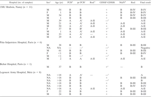
TABLE 1. Results of PCR and PCR-RFLP analyses of theGiardia lamblia tpiandgdhgenes amplified from 26 samples corresponding to sporadic cases
Hospital (no. of samples) Sexa Age (yr) PCRd tpiPCRe RsaIb GDHiF-GDHiR NlaIVb RsaI Final result
CHU Brabois, Nancy (n⫽11)
M 54 B B ⫹ B B-IV B-IV
F 1 B B ⫹ B B-IV B-IV
M 34 B B ⫹ B B-IV B-IV
M 1 B B ⫹ B B-III B-III
M 39 A Ac A-II —c A-II
M 21 A Ac A-II ⫹ A-II A-II
M 50 A Ac A-II ⫹ A-II A-II
F 37 B Bc ⫹c B B-III B-III
M 1 A Ac A-II ⫹ A-II A-II
M 33 A Ac — ⫹ A-II A-II
M 29 A A A-II ⫹ A-II A-II
Pitie-Salpetriere Hospital, Paris (n⫽6)
M 38 B B ⫹ B B-III B-III
NA NA — —c —c Negative
M 61 B B ⫹ B B-III B-III
F ⬍1 B B ⫹ B B-III B-IV
M ⬍1 B —c —c B
M 2 A A A-II ⫹ A-II A-II
Bichat Hospital, Paris (n⫽1)
M 37 B B ⫹c — B
Legouest Army Hospital, Metz (n⫽8)
NA ⬎18 A Ac — —c A
NA ⬎18 B B ⫹ B B-III B-III
NA ⬎18 B B —c B
NA ⬎18 B B ⫹ B B-III B-III
NA ⬎18 B B ⫹ B B-IV B-IV
NA ⬎18 A A A-II ⫹ A-II A-II
F 22 B B ⫹ B B-III B-III
M 24 B B ⫹ B B-III B-III
aM, male; F, female; NA, not available.
b—, insufficient quantity of PCR product for RFLP analysis. cDNA amplification by double PCR.
dA-PCR and B-PCR. —, negative results. eTPIA-PCR and TPIB-PCR.
on May 15, 2020 by guest
http://jcm.asm.org/
(AF069561), Ad-19 (AF069560), isolate 2924 (AY228628), isolate 2582 (AY228629), isolate 2506 (AY228630), isolate 2887 (AY228631), isolate 2902 (AY228632), isolate 2877 (AY228633), isolate 2900 (AY228634), isolate 2901 (AY228635), isolate 3470 (AY228636), isolate 3565 (AY228637), isolate 3577 (AY228638), isolate 1758 (AY228639); assemblage C: Ad-136 (AF069563), late 2643 (AY228641), isolate 2669 (AY228642), isolate 2674 (AY228643), iso-late 2665 (AY228644); assemblage E: P-15 (AF069559), isoiso-late 109 (AY228645), isolate 15 (AY228646); assemblage F: Ad-23 (AF069558); and assemblage G: Ad-157 (AF069562), isolate 2135 (AY228640);G. ardeae(AF069562);G. mi-croti: isolate 3463 (AY228648), isolate 3460 (AY228649);G. muris(AF069565). The primers used for assemblage A amplification were forward (A-for) 5⬘ -GGAGACCGACGAGCAAAGC-3⬘(positions 839 to 857 on the WB sequence, GenBank no. L02120), and reverse (A-rev), 5⬘-CTTGCCAAGCGCCTCAA-3⬘ (positions 970 to 986 on the WB sequence). A 148-bp fragment of the assemblage A gene was amplified with primers A-for and A-rev (A-PCR). The primers used for assemblage B amplification were forward (B-for), 5⬘-AATAGCAGCACA RAACGTGTATCTG-3⬘ (positions 126 to 150 on the BAH-12 sequence, GenBank no. AF069561), and reverse (B-rev), 5⬘-CCCATGTCCAGCAGCATC T-3⬘(positions 188 to 206 on the BAH-12 sequence). An 81-bp fragment of assemblage B gene was obtained with primers B-for and B-rev (B-PCR). Primers sets TPIAF-TPIAR (TPIA-PCR) and TPIBF-TPIBR (TPIB-PCR) were used as previously described (2) for amplification of theG. lamblia tpigene from assem-blages A and B, respectively. Finally, primer set GDHiF-GDHiR was used for amplification of theG. lamblia gdhgene, as previously described (20).
(ii) PCR amplification and RFLP analysis.Amplification of thetpiandgdh
genes was performed as a single PCR. Amplification reactions (50l) contained 5l of DNA, 1⫻PCR buffer corresponding to a final concentration of 1.5 mM MgCl2(Qiagen), each deoxynucleotide triphosphate at a concentration of 200
M (Applied Biosystems), each primer at a concentration of 0.5M, and 2.5 U of HotStarTaq DNA polymerase (Qiagen). Cycling parameters were 15 min at 95°C (initial heat activation step), followed by 50 cycles of 30 s at 94°C, 30 s at 62°C, and 30 s at 72°C, with a final extension of 7 min at 72°C. Both positive and negative controls were included in each PCR to validate results. Quantities of DNA equivalent to 600G. lambliaassemblage A (purified from feces) cysts and 4,500G. lambliaassemblage B (strain H-3) cysts were used as the templates for the positive controls, and distilled water was used as the template for negative controls throughout.
RFLP analysis was performed by digesting 2.5l of PCR product with 2 U of RsaI (Promega) or 2 U of NlaIV (New England Biolabs) in 1⫻enzyme buffer in a final volume of 20l for 3 h at 37°C. The RsaI digestion allowed the distinction between assemblage A group I and group II after amplification with the TPIAF and TPIAR primers (2). The NlaIV digestion was used for the distinction between assemblage A group I, assemblage A group II, and assemblage B after amplification with the GDHiF and GDHiR primers. RsaI digestion distinguished between assemblage B group III and assemblage B group IV after use of the GDHiF and GDHiR primers (20).
(iii) PCR product and restriction fragment detection. PCR products and restriction fragments were separated by horizontal electrophoresis in 2 and 3.2% agarose gels, respectively, with ethidium bromide (0.6g/ml) staining. A 100-bp DNA ladder (Promega) was included as a size marker. PCR products and restriction fragments were recorded by UV transillumination.
RESULTS
First, the genotype ofGiardia lambliacysts from one fecal
sample classified in assemblage A with TPIA-PCR and TPIB-PCR (2) was reconfirmed by sequencing analysis. This analysis showed 100% matches between the amplified product obtained with TPIA-PCR (577 bp) and the sequence with GenBank
ac-cession number U57897 corresponding toG. lamblia
assem-blage A group II. The sensitivity of A-PCR and the specificity of both A-PCR and B-PCR could be evaluated with this puri-fied suspension of cysts.
The specificity of our PCR assay was first evaluated by sub-jecting the primers to a BLAST test (http://www.ncbi.nlm.nih .gov/BLAST/). For the A-for and A-rev primers, the BLAST
tests returned sequences other thanG. lambliaassemblage A,
and only one sequence showed a 100% match with the A-rev
primer. However, the sequences other thanG. lamblia
assem-blage A returned by these two BLAST tests were different. For the B-for and B-rev primers, the BLAST tests returned
se-quences other thanG. lambliaassemblage B with one to eight
mismatches. Furthermore, the mismatched sequences re-turned by these two BLAST tests were different.
The specificity was then examined by performing PCR
as-says. By using a quantity of DNA equivalent to 600Giardia
lambliaassemblage A cysts per reaction, the predicted 148-bp product was obtained by A-PCR, but no product was amplified by B-PCR. Conversely, by using a quantity of DNA equivalent
to 4,500Giardia lamblia assemblage B (strain H3) cysts per
reaction, the predicted 81-bp product was observed with B-PCR, but no product was obtained with A-PCR. No product was amplified by performing either A-PCR or B-PCR with
DNA extracted from purified Giardia lamblia assemblage E
from cattle, sheep, and pigs, Giardia muris, Cryptosporidium
parvum, Entamoeba histolytica, Entamoeba dispar, Campy-lobacter jejuni,Salmonella enterica, orEscherichia coli.
To estimate the sensitivities of our PCR assays, 10-fold di-lutions of purified cysts were performed in PBS (pH 7.2) prior to DNA extraction. The amplification step was then performed
in triplicate on 10-fold serial dilutions of cysts. ForG. lamblia
assemblage A, the predicted 148-bp product was obtained for each well of the triplicates for concentrations ranging from 600
to 6 cysts per reaction mixture. ForG. lambliaassemblage B,
the 81-bp product was observed for each well of the triplicates for concentrations ranging from 4,500 to 4.5 cysts per reaction mixture.
Among the 26 fecal samples corresponding to sporadic gi-ardiasis diagnosed in hospital laboratories, the presence of
Giardia cysts was reconfirmed by immunofluorescence (IF) microscopy for all of these during our study. On the other hand, the number of positive samples by PCR varied according to the PCR assay employed and the target gene amplified.
Indeed, thetpigene was amplified from 25 samples (96.1%)
with A-PCR and B-PCR developed in our laboratory. The same target gene was amplified from 24 samples (92.3%) with TPIA-PCR and TPIB-PCR. However, only 18 positive samples (69.2%) were obtained with a single PCR. A double PCR allowed the achievement of six additional positive samples. For
the gdh gene, 21 samples (80.7%) were positive with the
GDHiF and GDHiR primers. Moreover, a single PCR resulted in 19 positive samples (73.1%) and a double PCR was needed for two other samples.
Regarding classification into assemblages A and B (Table 1), the results of the various PCR and PCR- RFLP methods were in agreement for the sporadic cases. Assemblage A was de-tected in nine (34.6%) and seven (26.9%) samples by using the
tpiand gdhgenes as the target, respectively. When targeting
thetpigene, assemblage B was identified in 16 (61.5%) and 15
(57.6%) samples with B-PCR and TPIB-PCR, respectively.
However, when using thegdhgene as the target, assemblage B
was detected in only 13 (50%) fecal samples.
A more specific analysis of these sporadic cases was then conducted with the distinction between assemblage A groups I and II and assemblage B groups III and IV on the basis of their fragment patterns obtained by RFLP analysis (Table 1). The results obtained with the two published methods were in agree-ment for sporadic cases. Whatever the gene used, subgroup I was never observed in these fecal samples. On the other hand,
on May 15, 2020 by guest
http://jcm.asm.org/
six samples were genotyped as assemblage A group II with both RFLP techniques and two samples were classified in this group with only one of these methods. For one sample
classi-fied in assemblage A by PCR assays targeting thetpigene, the
group could not be specified. Indeed, the quantity of PCR product obtained with A-PCR was insufficient for RFLP anal-ysis, and no product was amplified with the GDHiF and GDHiR primers.
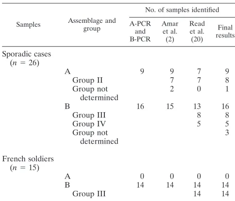
Only one method used in our study allowed distinction be-tween assemblage B groups III and IV (20). The RFLP anal-ysis resulted in eight and five samples classified in assemblage B group III and group IV, respectively. For three samples identified as assemblage B with our PCR assay, the group could not be specified with the method described by Read and colleagues (20). Of these, two samples were negative after double PCR with the GDHiF and GDHiR primers. For the third sample, the quantity of PCR product obtained with the GDHiF and GDHiR primers was insufficient for RFLP anal-ysis. The genotyping results are summarized in Table 2.
Finally, 31% of the samples were classified in assemblage A group II and in assemblage B group III as well. Assemblage B
group IV was detected in 19% of the samples. TheG. lamblia
cysts used for validation of our A-PCR assay were classified in assemblage A group II by the RFLP analysis, as they were by the sequencing analysis carried out at the beginning of our study.
Among the 15 cases of giardiasis diagnosed in samples from French soldiers coming back from the Ivory Coast, the
pres-ence of Giardia cysts was reconfirmed by IF microscopy for
100% of the samples. TheGiardia lambliaassemblage B
ge-notype was isolated in 93.3% of the samples with the three
methods, and tpi orgdh gene fragments were not amplified
from only one sample. Moreover, the PCR-RFLP analysis de-veloped by Read and colleagues (20) allowed the detection of assemblage B group III in all of these samples (Table 2).
DISCUSSION
In the present study, we developed a conventional PCR
assay allowing the rapid detection and distinction ofG. lamblia
assemblages A and B in human feces. When applied to 10-fold
serial dilutions of purified G. lamblia cysts, this PCR assay
showed a high degree of sensitivity. Thus, a threshold of 6 and 4.5 purified cysts per reaction was reached by single PCR for assemblages A and B, respectively. Moreover, our primers were validated by the comparison between the genotyping re-sults obtained with our PCR assay and previously described PCR-RFLP methods (2, 20). For sporadic cases of giardiasis
confirmed by IF analysis (n⫽26), our PCR resulted in 96%
positive samples with a single amplification step. The negative results observed for 4% of the samples could be explained by the presence of parasites at a very low level or degradation of parasite DNA during transport of the samples to our labora-tory. The amplification of these 26 samples with the primers described by Amar and colleagues (2) and Read and col-leagues (20) allowed us to obtain 92% and 81% positive sam-ples, respectively. However, double amplification was then needed for six and two samples, respectively, with these pre-viously described methods.
In the study conducted by Amar and colleagues (2),
ampli-fication of the tpi gene was performed as a two-step PCR
(seminested PCR) and resulted in 94% positive samples (n⫽
35). The ethyl acetate procedure, which is adapted from the formalin ethyl acetate method, was tested for fecal samples.
This method allowed rapid and efficient purification ofGiardia
cysts prior to DNA extraction. Indeed, at least 15 fecal samples were simultaneously purified with this method, whereas the gel filtration chromatography method was time-consuming and limited to four samples per purification step. The ethyl acetate
procedure is well adapted for the purification ofGiardiacysts
in wastewater samples prior to quantification by a real-time PCR method (data not shown). Moreover, this purification
method could be used for the purification ofGiardiacysts in
sludge samples.
The present study provides, for the first time, information on
the distribution of the genotypes ofG. lamblia from humans
with sporadic giardiasis in France. This work was based on a relatively small group of patients, but we are still pursuing analysis of sporadic giardiasis. However, our observation that the majority of sporadic giardiasis case isolates were assem-blage B genotype (61.5%) corresponds to the findings of
sev-eral studies conducted in India (100%,n⫽10 [21]), Peru (76%,
n ⫽ 25 [21]), United States (80%, n ⫽ 15 [11]), and United
Kingdom (64%,n⫽35 [2]). However, an Italian study reported
80% assemblage A in 30 stool samples examined by sequencing
or PCR-RFLP analysis of the-giardin gene (6). The
predom-inance of assemblage B in samples collected in sewage treat-ment facilities was shown in one study (10). On the other hand, two other studies concluded that these was a majority of as-semblage A in wastewater samples (6, 22). The use of RFLP analysis resulted in the same percentage for assemblage A group II and assemblage B group III, whereas assemblage B group IV was less common. The absence of assemblage A group I in these 26 fecal samples corresponds to the findings of the study conducted in the United Kingdom (2).
Among the 15 cases of giardiasis diagnosed in French
sol-TABLE 2. Summary of genotyping results obtained with the three methods
Samples Assemblage and group
No. of samples identified
A-PCR and B-PCR
Amar et al. (2)
Read et al. (20)
Final results
Sporadic cases (n⫽26)
A 9 9 7 9
Group II 7 7 8
Group not determined
2 0 1
B 16 15 13 16
Group III 8 8
Group IV 5 5
Group not determined
3
French soldiers (n⫽15)
A 0 0 0 0
B 14 14 14 14
Group III 14 14
on May 15, 2020 by guest
http://jcm.asm.org/
[image:4.585.41.283.87.294.2]diers coming back from the Ivory Coast, the detection of as-semblage B group III in 14 confirmed the hypothesis of a common source of contamination. At present, only two studies have reported the genotyping results from outbreak-associated giardiasis. Among 24 samples from a nursery outbreak, Amar
and colleagues (2) detected G. duodenalis assemblage B in
88% of the samples. Sulaiman and colleagues (21) detected assemblage B in two isolates from a food-borne outbreak by
sequencing analysis of thetpigene.
In our study, G. lamblia assemblage A and assemblage B
were never detected together, whereas a mixture of these as-semblages has been reported previously in a few studies (2, 11, 12, 14, 15). Thus, Amar and colleagues (2) observed a mixture of assemblage A group II and assemblage B in 9% of 35 samples, whereas Lu and colleagues (15) simultaneously de-tected assemblage A group I and assemblage B in 33% of only three samples.
In summary, the PCR assay developed in this study, com-bined with the ethyl acetate procedure, allowed rapid detection
and genotyping ofG. lambliacysts from clinical samples. Our
results show that thetpigene is better adapted than the gdh
gene for efficient discrimination between the two major assem-blages. Thus, detection methods targeting loci with a high
degree of polymorphism such as tpican be extremely useful
when a common source of contamination is certainly involved. This work provides the first information about the
distribu-tion of the two major assemblages of G. lambliain sporadic
human giardiasis in France. However, further studies with a larger series of fecal or environmental samples could lead to better knowledge of the distribution of these assemblages in humans as well as the role of domestic animals and livestock as a potential source of infection for humans.
ACKNOWLEDGMENTS
We thank B. Fortier and J. Collomb (CHU Brabois, Nancy), J. Puyhardy (Hopital Militaire Legouest, Metz). R. Haus-Cheymol (De ´-partement d’Epide´miologie et de Sante´ Publique Nord, Paris), N. Kapel (Hopital de la Pitie´-Salpeˆtrie`re, Paris), and S. Houze´ (Hopital Bichat, Paris) for providing stool samples. For other DNA samples, we thank B. Fortier and J. Collomb, M. Naciri (INRA, Institut National de la Recherche Agronomique, Nouzilly), O. Villard (Institut de Para-sitologie, Strasbourg), A. Lozniewski, F. Mory (Hopital Central, Nancy). and S. Caccio (Istituto Superiore di Sanita, Rome, Italy).
This work was supported by European Community contract number TOFPSW EVK1.2000.22080.
REFERENCES
1.Adam, R. D.2001. Biology ofGiardia lamblia. Clin. Microbiol. Rev.14:447– 475.
2.Amar, C. F. L., P. H. Dear, S. Pedraza-Diaz, N. Looker, E. Linnane, and J. McLauchlin.2002. Sensitive PCR-restriction fragment length polymorphism assay for the detection and genotyping ofGiardia duodenalisin human feces. J. Clin. Microbiol.40:446–452.
3.Amar, C. F. L., C. East, E. Maclure, J. McLauchlin, C. Jenkins, P. Duncan-son, and D. R. A. Wareing.2004. Blinded application of microscopy, bacte-riological culture, immunoassays and PCR to detect gastrointestinal patho-gens from faecal samples of patients with community-acquired diarrhoea. Eur. J. Clin. Microbiol. Infect. Dis.23:529–534.
4.Aydin, A. F., B. A. Besirbellioglu, I. Y. Avci, M. Tanyuksel, E. Araz, and A. Pahsa.2004. Classification ofGiardia duodenalisparasites in Turkey into groups A and B using restriction fragment length polymorphism. Diagn. Microbiol. Infect. Dis.50:147–151.
5.Bertrand, I., C. Gantzer, T. Chesnot, and J. Schwartzbrod.2004. Improved specificity forGiardia lambliacyst quantification in wastewater by develop-ment of a real-time PCR method. J. Microbiol. Methods57:41–53. 6.Caccio, S. M., M. De Giacomo, and E. Pozio.2002. Sequence analysis of the
-giardin gene and development of a polymerase chain reaction-restriction fragment length polymorphism assay to genotypeGiardia duodenaliscysts from human faecal samples. Int. J. Parasitol.32:1023–1030.
7.Chesnot, T., and J. Schwartzbrod.2004. Quantitative and qualitative com-parison of density-based purification methods for detection of Cryptospo-ridiumoocysts in turbid environmental matrices. J. Microbiol. Methods58:
375–386.
8.Garcia, L. S., and R. Y. Shimizu.1997. Evaluation of nine immunoassay kits (enzyme immunoassay and direct fluorescence) for detection ofGiardia lambliaandCryptosporidium parvum in human fecal specimens. J. Clin. Microbiol.35:1526–1529.
9.Gardner, T. B., and D. R. Hill.2001. Treatment of giardiasis. Clin. Microbiol. Rev.14:114–128.
10.Guy, R. A., P. Payment, U. J. Krull, and P. A. Horgen.2003. Real-time PCR for quantification ofGiardiaandCryptosporidiumin environmental water samples and sewage. Appl. Environ. Microbiol.69:5178–5185.
11.Guy, R. A., C. Xiao, and P. A. Horgen.2004. Real-time PCR assay for detection and genotype differentiation ofGiardia lambliain stool specimens. J. Clin. Microbiol.42:3317–3320.
12.Hopkins, R. M., B. P. Meloni, D. M. Groth, J. D. Wetherall, J. A. Reynold-son, and R. C. A. Thompson.1997. Ribosomal RNA sequencing reveals differences between genotypes ofGiardiaisolates recovered from humans and dogs living in the same locality. J. Parasitol.83:44–51.
13.Johnston, S. P., M. M. Ballard, M. J. Beach, L. Causer, and P. P. Wilkins.
2003. Evaluation of threee commercial assays for detection ofGiardiaand
Cryptosporidiumorganisms in fecal specimens. J. Clin. Microbiol.41:623– 626.
14.Lalle, M., E. Pozio, G. Capelli, F. Bruschi, D. Crotti, and S. M. Caccio.2005. Genetic heterogeneity at the -giardin locus among human and animal isolates ofGiardia duodenalisand identification of potentially zoonotic sub-genotypes. Int. J. Parasitol.35:207–213.
15.Lu, S. Q., A. C. Baruch, and R. D. Adam.1998. Molecular comparison of
Giardia lambliaisolates. Int. J. Parasitol.28:1341–1345.
16.McGlade, T. R., I. D. Robertson, A. D. Elliott, and R. C. A. Thomspon.2003. High prevalence ofGiardiadetected in cats by PCR. Vet. Parasitol.110:
197–205.
17.Monis, P. T., G. Mayrhofer, R. H. Andrews, W. L. Homan, L. Limper, and P. L. Ey.1996. Molecular genetic analysis ofGiardia intestinalisisolates at the glutamate dehydrogenase locus. Parasitology112:1–12.
18.Monis, P. T., R. H. Andrews, G. Mayrhofer, and P. L. Ey.1999. Molecular systematics of the parasitic protozoanGiardia intestinalis. Mol. Biol. Evol.
16:1135–1144.
19.Monis, P. T., R. H. Andrews, G. Mayrhofer, and P. L. Ey.2003. Genetic diversity within the morphological speciesGiardia intestinalisand its rela-tionship to host origin. Infect. Genet. Evol.3:29–38.
20.Read, C. M., P. T. Monis, and R. C. A. Thompson.2004. Discrimination of all genotypes ofGiardia duodenalisat the glutamate dehydrogenase locus using PCR-RFLP. Infect. Genet. Evol.4:125–130.
21.Sulaiman, I. M., R. Fayer, C. Bern, R. H. Gilman, J. M. Trout, P. M. Schantz, P. Das, A. A. Lal, and L. Xiao.2003. Triosephosphate isomerase gene characterization and potential zoonotic transmission ofGiardia duode-nalis. Emerg. Infect. Dis.9:1444–1452.
22.Sulaiman, I. M., J. Jiang, A. Singh, and L. Xiao. 2004. Distribution of
Giardia duodenalisgenotypes and subgenotypes in raw urban wastewater in Milwaukee, Wisconsin. Appl. Environ. Microbiol.70:3776–3780. 23.Thiriat, L., F. Sidaner, and J. Schwartzbrod.1998. Determination ofGiardia
cysts viability in environmental and fecal samples by immunofluorescence, fluorogenic dye staining and differential interference contrast microscopy. Lett. Appl. Microbiol.26:237–242.
24.Thompson, R. C. A.2000. Giardiasis as a re-emerging infectious disease and its zoonotic potential. Int. J. Parasitol.30:1259–1267.
25.Thompson, R. C. A., R. M. Hopkins, and W. L. Homan.2000. Nomenclature and genetic groupings ofGiardiainfecting mammals. Parasitol. Today16:
210–213.
26.Weber, R., R. T. Bryan, and D. D. Juranek.1992. Improved stool concen-tration procedure for detection ofCryptosporidiumoocysts in fecal speci-mens. J. Clin. Microbiol.30:2869–2873.
27.Zimmerman, S. K., and C. A. Needham.1995. Comparison of conventional stool concentration and preserved-smear methods with Merifluor Cryptospo-ridium/Giardiadirect immunofluorescence assay and ProSpecTGiardiaEZ microplate assay for detection ofGiardia lamblia. J. Clin. Microbiol.33:
1942–1943.