http://dx.doi.org/10.4236/jbnb.2016.72011
How to cite this paper: Sadat, S.M.A., Jahan, S.T. and Haddadi, A. (2016) Effects of Size and Surface Charge of Polymeric Nanoparticles on in Vitro and in Vivo Applications. Journal of Biomaterials and Nanobiotechnology, 7, 91-108.
http://dx.doi.org/10.4236/jbnb.2016.72011
Effects of Size and Surface Charge of
Polymeric Nanoparticles on
in Vitro
and
in
Vivo
Applications
Sams M. A. Sadat, Sheikh Tasnim Jahan, Azita Haddadi
*Division of Pharmacy, College of Pharmacy and Nutrition, University of Saskatchewan,
Saskatoon, Canada
Received 15 October 2015; accepted 19 April 2016; published 22 April 2016
Copyright © 2016 by authors and Scientific Research Publishing Inc.
This work is licensed under the Creative Commons Attribution International License (CC BY).
http://creativecommons.org/licenses/by/4.0/
Abstract
Biodegradable polymeric materials are the most common carriers for use in drug delivery
sys-tems. With this trend, newer drug delivery systems using targeted and controlled release
poly-meric nanoparticles (NPs) are being developed to manipulate their navigation in complex
in vivo
environment. However, a clear understanding of the interactions between biological systems and
these nanoparticulates is still unexplored. Different studies have been performed to correlate the
physicochemical properties of polymeric NPs with the biological responses. Size and surface
charge are the two fundamental physicochemical properties that provide a key direction to design
an effective NP formulation. In this critical review, our goal is to provide a brief overview on the
influences of size and surface charge of different polymeric NPs in vitro and to highlight the
chal-lenges involved with in vivo trials.
Keywords
Nanoparticle, Size, Surface Charge, In Vitro, In Vivo
1. Introduction
Manufacturing effective drug delivery system is a critical challenge in nanomedicine since nanocarriers are
ex-pected to reach and accumulate in the site of interest. As a consequence, numerous drug delivery systems have
been investigated in vitro and in vivo to deliver a wide range of drugs and molecules. To conquer the challenge,
with the aim of avoiding uncontrolled biodistribution, rapid clearance and systemic toxicities in healthy tissues
polymeric nanoparticles (NPs) have gained higher interest among all the novel formulations. Research during
the past few decades proves their beneficial features in formulation design, characterization, behavior and
ap-plication [1]. The in vitro and in vivo fate of NPs is particularly depended on uniformity of particle size and zeta
potential. Change in these properties has significant biological implications on cellular internalization,
pharma-cokinetics, and bio-distribution [2]. These characteristics of NPs facilitate the opportunities for therapeutic
ap-plication, which can be confirmed by in vitro and in vivo studies [3]. The aim of most nano-devices is to prevent
the degradation of drug followed by higher bioavailability in cellular level and to regulate its pharmacodynamics
profile. Thus, the nanomedicine platforms could serve as a drug delivery system that is able to transport a high
dose of therapeutics selectively to the desired site of action. Although very few investigations have been
per-formed, most of the articles related to exploring the effects of size and surface charge of NPs have been
dis-cussed in this review. This review provides details on the fate of different polymeric NPs and will discuss how
the size and surface charge of polymeric NPs are involved in desired effects for both in vitro and in vivo
applica-tions. Moreover, other polymeric NPs using various preparation methods have been also summarized in
Table 1
,
which could be considered for further size and surface charge related experiments.
Table 1.Size and surface charge overview of different polymeric NPs.
Polymer type NPs preparation method
Loaded materials Stabilizer Cell line Size (nm) Zeta (mV) References
Chitosan Chemical
reaction - - Caco-2 (418 ± 31)-(531 ± 54)
(13.6 ± 0.8) - (29.4 ± 0.8) [50]
Chitosan Ionotropic gelation
Covalent conjugation with fluorescein- 5-isothiocyanate
- A549 cells 195 35.5 [51]
Chitosan (Galactosylated)
Carbodiimide
method DNA -
HepG2,
HeLa 100 - 240 −5 [52]
Chitosan {Arg-Gly-Asp (RGD) peptide-labeled}
Ionic gelation siRNA -
SKOV3ip1, HeyA8, A2780, A2780ip2, MOECs
200 −40 [53]
Chitosan, PS, PEG-PLA
Multiple emulsion-solvent evaporation
- Sodium
Cho-late
Caco-2,
MTX-E12 (196 ± 20)-(290 ± 7)
(−23.9 ± 1.2) - (1.1 ± 1) [7]
Chitosan-PEG Ionic gelation Caspase inhibitor
pebptide - Mice 150 16 [54]
Gelatin Desolvation Anti CD3 mAb - HeLa 250 - 300 −20 [55]
Heparin
Coupling reaction through amide linkage
Paclitaxel, Folic acid - KB-3-1,
Tu212 60 −16 [56]
Hyaluronic acid (Self-assembled)
Chemical conjugation through Carbodiimide method
- PBS SCC7, CV-1 237 - 424 - [57]
Hyaluronic
acid-ceramide Self-assembly Docetaxel Pluronic P85
U87-MG, MCF-7, MCF-7/AD R
110 - 140 −20 [58]
Hydrophobically modified glycol chitosan
Self-assembly - - HeLa 359 22 [59]
Mesoporous
silica - - -
3T3-L1,
HMSCs 108 - 115 (−4.9) - (19) [60] Methyl ether
poly(ethylene glycol)-modified poly(beta-amino ester)
Solvent casting Doxorubicin - B16F10 62 - [61]
NAcHis-GC
(self-assembled) - Paclitaxel PBS HeLa, A549 150 - 250 - [62]
P(MDS-co-CES) Self-assembly DNA, Paclitaxel, Indomethacin, Pyrene -
HEK293, HepG2, MDA-MB-2 31
(83 ± 1) - (180 ± 2) (44 ± 2) - (84 ±
5) [63]
PCL Solvent
displacement Tamoxifen Pluronic
® F-68 MCF-7 250 - 300 6 - 25 [64] [65]
PCL Emulsification
Continued
PEG
Top-down strategic PRINT technique
Conjugated antibody -
HeLa, Ramos, H460, SK-OV-3, HepG2, LNCaP
(267 ± 49) - (292 ± 76)
(−35.6 ± 1.3) - (39.9 ± 1.7) [67]
PEG-PCL Dry-down
method - -
MDA-MB-4 68, MCF-7
(26.4 ± 0.7) - (60.9 ±
0.7) (−5.1) - (−7.3) [68]
PEG-PCL Solvent
evaporation Paclitaxel
Sodium Cholate
U87 MG, BCECs <100
(−3.08 ± 0.94) - (−3.28 ± 0.75) [69]
PEG-PHDCA Nanoprecipitation Nile red Pluronic F68 RBECs 140 - 146 −20 [70]
PEG-PLA Solvent
displacement - Solutol
® HS 15 HeLa 89.8 ± 4, 96.4 ± 6 32.8 ± 8, −26 ± 1 [71]
PEG-PLA Emulsion-solvent
evaporation BSA, Coumarin-6
Sodium Cholate
Rat BCECs,
Astrocytes 80.4 - 84.4 (−8)-(−17) [72]
PEG-PLA Solvent
displacement Coumarin-6 Solutol
® HS 15 MDCK (89.8 ± 4) -
(96.36 ± 6)
(−29 ± 7) -
(45.46 ± 2) [73]
PEG-PLA Nanoprecipitation Cisplatin - A2780 (86 ± 2) −33 ± 1.2 [74]
PEG-PLGA
Simple emulsion, Interfacial deposition
Paclitaxel Sodium
Cholate HeLA 190 ± 4.5, and 112 ± 4
−7.76 ± 2.6, −0.556 ± 5.7 [75]
PEG-Trimethyl Chitosan Self-assembly utilizing electrostatic interactions
Insulin - Caco-2 (203.9 ± 7.2) - (376.4 ± 10.3)
(7.4 ± 0.3) - (21.4 ± 0.3) [76]
PEO-b-PMA - Cisplatin,
Doxorubicin -
A2780, A549
(93.8 ± 8.4) - (176.5 ± 6.0)
(−29.2 ± 1.5) - (−2.1 ± 1.0) [77]
PLA Diafiltration Trans-retinoic acid
Poly(vinyl benzyl lactonamide)
Hepatocytes 287.7 - [78]
PLA Emulsion-solvent evaporation Rhodamine, Selectin grafted Sucrose solution containing Tween 20, PEG-Laurate
HUVEC 173 ± 23, 168 ± 37 −25.7, −19.2 [79]
PLA Solvent
evaporation Doxorubicin - SLK 45 ± 8 - 47 ± 4 - [80]
PLA, PLGA
Multiple emulsion-solvent evaporation
Plasmid DNA PVA MCF-7,
PC-3
(270 ± 1) - (1207 ± 30)
(−6.5 ± 2) -
(−31.3 ± 2) [81]
PLA, PLGA Solvent
deposition Docetaxel Tween 80 -
(100.7 ± 2.9) - (179.4 ± 1.7), (98.7 ± 1.7) - (172.0 ± 4.9)
(−38) - (−24), < (−10) [82]
PLGA
Double emulsion-solvent evaporation
DNA PVA COS-7,
HEK-293 (70±2)-(202±9)
(−20.6 ± 2) - (−20.8 ± 2) [83]
PLGA
Multiple emulsion-solvent evaporation
BSA PVA HASMC 380 - 522 (−0.8 ± 2.3) - (−15.4 ± 0.8) [84]
PLGA Emulsion-solvent
evaporation Paclitaxel PVA
MCF-7 and
MCF-7/Adr 216 −8.12 ± 2.8 [85]
PLGA
Double emulsion-solvent diffusion
Cystatin PVA
MCF-7, MCF-10A neoT, Caco-2, U-937
320 - 360 −25 [86]
PLGA Emulsion-solvent
evaporation BSA, Coumarin-6 PVA HUVEC 277 - 372 −10.4 [87]
PLGA Emulsion-solvent
evaporation Paclitaxel PVA
A549, H1299, CCL-186
(228.2 ± 0.6) - (330.7 ± 2.9)
(−4.3 ± 0.3) -
(−17.9 ± 1) [88] [89]
PLGA Emulsion-solvent diffusion
FITC-WGA,
FITC-BSA PVA A549 246, 356 ~(−4) [90]
PLGA Solvent
evaporation Camptothecin PVA HCT116
(116.5 ± 1.6) - (187.1
± 1.1) - [91]
PLGA - Loperamide,
Rhoda-mine-123 -
Tail vein in
rats 140 - 180 −20 [92]
PLGA Interfacial
deposition Paclitaxel Poloxamer 188
NCI-H69,
SCLC (117 ± 2) - (160 ± 2)
(−33.4 ± 1.8) - (−23.1 ± 3.7) [93]
PLGA Direct dialysis Paclitaxel Vitamin E
TPGS C6 glioma
(280 ± 28) - (310 ±
28) (−20) - (−40) [94]
PLGA
Modified emulsification- solvent diffusion
Docetaxel PVA
T47D, MCF-7, SKOV3, A549
Continued
PLGA Nanoprecipitation Doxorubicin BSA MDA-MB-2
31 230 −45 [96]
PLGA Emulsion-solvent evaporation
Indocyanine green,
Doxorubicin PVA - (137 ± 2) - (164 ± 2)
(−9.9 ± 0.4) - (−12.3 ± 0.1) [97]
PLGA Nanoprecipitation Curcumin PVA
A2780CP, MDA-MB-2 31
(76.2 ± 5.36) - (560.4 ± 10.95 )
(−0.06 ± 0.01) - (0.06 ± 0.01) [98]
PLGA Emulsion-solvent evaporation
Docetaxel, Poloxamer
188 PVA
MCF-7, TAX30
(217.6 ± 8.6) - (274.7 ± 4.1)
(−23.35 ± 1.17) - (−41.28 ± 2.89) [99]
PLGA
Emulsion-solvent evaporation, Salting-out
Vincristine, Verapamil PVA
MCF-7, MCF-7/AD R
98.8 ± 8.4 −0.75 ± 0.12 [100]
PLGA Nanoprecipitation Hypericin PVA NuTu-19 210.3 - 268.9 (−3.7) - (−7.9) [101]
PLGA Solvent diffusion
(Nanoprecipitation) Paclitaxel PVA C6 glioma
169.3 ± 4.16) - (182.8 ± 3.78)
(−3.45 ± 0.58) - (−11.72 ± 2.27) [102]
PLGA
Solvent extraction/ evaporation
Paclitaxel PVA, Vitamin E TPGS HT-29
293.6 ± 4.8, 235.7 ±
14.8 −26.05, −35.60 [103]
PLGA High pressure
homogenization Paclitaxel PVA C6 glioma
(245.0 ± 20.4) - (305.4
± 10.6) - [104]
PLGA Nanoprecipitation
Sialic acid, N-acetylneuraminic acid Pluronic F68, Polysorbate 80 CD14+ human monocytes
(63 ± 5) - (190 ± 16) (−23 ± 3) - (−38
± 3) [105]
PLGA Solvent injection Aromatase inhibitor - SKBR-3 170.3 ± 7.6 −18.9 ± 1.5 [106]
PLGA Emulsion-solvent
evaporation Paclitaxel - Ch-hep-3
(119 ± 6.2) - (129 ± 5.2)
(−4.6 ± 0.3) - (−31.5 ± 1.2) [107]
PLGA
Double emulsion-solvent diffusion
BSA PVA
MCF-7, MCF-10A neoT, Caco-2
320 - 360 −25 [108]
PLGA
Single emulsion solvent evaporation
Rapamycin PVA MCF-7 274 ± 1.6 −13.8 ± 5.1 [109]
PLGA
Modified solvent extraction/ evaporation
Paclitaxel PVA Caco-2,
SK-BR-3
(293.8 ± 5.7) - (312.3 ± 8.2)
(−35.07 ± 1.68) - (−21.24 ± 2.11) [110]
PLGA
Double emulsion-solvent diffusion
PE38KDEL, a 38 kDa mutant form of Pseu-domonas exotoxin A PVA D2F2/E2, SK-BR3, D2F2
(108.3 ± 13.9) - (124.2 ± 21.2)
(−36 ± 5) - (12 ±
7) [111]
PLGA
Emulsion-solvent extraction/ evaporation
Rhodamine PVA HEK293,
TE671
(400 ± 100) - (550 ± 90)
(−0.96 ± 0.01) - (−2.9 ± 0.2) [112]
PLGA
Emulsion-solvent evaporation/ extraction
FITC-TT peptide,
DQ-BSA PVA
Granulo-cytes, PBMCs
202 ± 4, 239 ± 14 (−28.6 ± 0.4), (−44.9 ± 1.8) [113]
PLGA
Double emulsion-solvent evaporation
siRNA PVA DCs 350 - 390 (−13) - (−19) [114]
PLGA
Double emulsion-solvent evaporation
- PVA DCs (328 ± 30) - (511 ±
26) (−25) - (−46.4) [115]
PLGA Solvent evaporation
Rapamycin, TMRD
Dextran PVA DCs 150 - 450 - [116]
PLGA, PLA Emulsion-solvent evaporation
Dexamethasone,
Futamide PVA 240, 270 19 to 28 [117]
PLGA, PLA
Double emulsion-solvent evaporation
BSA, Coumarin-6 PVA
VSMCs, HASMCs, HA-VSMCs
69 - 98 (−5.3) - (−23) [118]-[120]
PLGA,
PLGA-PEG Solvent diffusion
Conjugated cyclo(1,12)PenITDGE ATDSGC (cLABL) peptide
PEMA HUVEC 177 ± 11, (202 ± 11) - (268 ± 19)
−40.2 ± 3.7, (−31.4 ± 4.0) - (−8.3 ± 0.9) [121]
PLGA, PS
Emulsion-solvent extraction/ evaporation
Coumarin-6 PVA, Vitamin E TPGS Caco-2
(261.6 ± 9) - (295.4 ± 15), 50 - 500
(−36.76) -
(18.38) [27]
PLGA-PEG Nanoprecipitation
Avidin, Streptavidin, Neutravidin, Cou-marin-6 - N18-RE-105 , b.End3, HepG2
(111.1 ± 1.8) - (255.2 ± 6.3)
Continued
PLGA-PEG Nanoprecipitation Docetaxel - LNCaP 80-200 - [123]
PLGA-PEG Nanoprecipitation Cisplatin - LNCaP, PC3 (131 ± 0.5) - (172 ±
3.4) - [124]
PLGA-PEG Emulsion-solvent evaporation
Conjugated peptide,
Coumarin-6 - bEnd.3
(104.17 ± 3.45) - (121.46 ± 0.76)
(−24.43 ± 0.22) - (−18.25 ± 0.88) [125]
PLGA-PEG Modified double
emulsion Cisplatin
Sodium
Cho-late LNCaP
(134.3 ± 5.2) - (159.8
± 6.2) (−5.7) - (−9.3) [126]
PLGA-PEG Solvent-diffusion Doxorubicin PEMA
MDA-MB-2 31, B16F10, MCF-7
(366.6 ± 3.1) - (423.0 ± 16.6)
(−18.9 ± 2.4) - (−51.7 ± 3.1) [127]
PLGA-PEG Nanoprecipitation Paclitaxel - HUVEC (114 ± 3) - (146 ± 2) (−0.36 ± 4.3) - (0.12 ± 3.6) [128]
PLGA-PEG Solvent
evaporation Paclitaxel PVA
JC, NCI/ ADR-RES or MCF-7
(221 ± 5) - (240 ± 1) (−18 ± 5) - (−35
± 5) [129]
PLGA-PEG Nanoprecipitation Docetaxel - LNCaP (153.3 ± 13.9) −42 ± 1 [130]
PLGA–PEG Emulsion-solvent
diffusion Docetaxel PVA SKOV3 (120 ± 5) - (216 ± 18)
(−6.27 ± 0.95) - (−12.2 ± 0.6) [131] PLGA-PEG-A
ptamer Nanoprecipitation Docetaxel - LNCaP, PC3
(160 ± 3.7) - (291 ±
5.2) (−20) - (−29) [132]
PLGA-PEI Solvent
displacement Plasmid DNA Poloxamer-188 Calu-3 207 - 231
(32.1 ± 6.7) -
(58.8 ± 4) [133]
PLGA-PEI Solvent
evaporation Paclitaxel PVA JC
(228 ± 22) - (237 ± 16)
(−12.1 ± 0.3) - (−24.0 ± 0.5) [134]
PLGA-TPGS
Solvent extraction/ evaporation
Docetaxel TPGS Caco-2,
MCF-7
(219.42 ± 5.24) - (253.51 ± 5.38
(−21.87 ± 2.11) - (34.1 ± 4.28) [135]
PLGA-TPGS
Solvent extraction/ evaporation
Doxorubicin - MCF-7, C6
glioma (324 ± 5) - (359 ± 10) - [136]
PLGA-TPGS)
Modified solvent extraction/ evaporation
Docetaxel,
Coumarin-6 TPGS HeLa
(207.15 ± 8.46) - (290.25 ± 7.64)
(−15.22 ± 2.21) - (−32.10 ± 0.65) [137]
PMB, PMBH Emulsion-solvent evaporation
Doxorubicin,
Paclitaxel - HeLa 242 ± 10, 218 ± 9
−2.0 ± 0.5, −2.0
± 0.6 [138]
Poly (ethylene oxide)- modified poly (beta-amino ester) Solvent
evaporation Paclitaxel, - SKOV-3 60 - 150 40 [139]
Poly (β-amino esters) {Arg-Gly-Asp (RGD) peptide coated} Electrostatic
self-assembly DNA - HUVEC 200 −5 [140]
Poly
(β-malic acid) - Antisense oligonucleotides -
U87MG, TG98, MDA MB-231.
6.6 - 24 (−27 ± 1) - (−5.2 ± 0.4) [141]
Poly
(β-malic acid) - Antisense oligonucleotides -
BT-474, SKBR-3, MDA-MB-2 31, MDA- MB-435, MDA-MB-4 68, MCF-7
(15.1 ± 1.2) - (22.1 ± 2.3)
(−4.1 ± 0.4) - (−5.7 ± 0.6) [142]
Poly (γ-glutamic acid)-Poly (lactide)
Emulsion-solvent
evaporation Paclitaxel - HepG2
(115.4 ± 4.2) - (263.2 ± 6.8)
(−19.2 ± 2.2) - (−22.5 ± 3.2) [143]
PS (Carboxyl- modified fluorescent)
Commercial - Commercial HeLa,
HUVEC 24 ± 4, 43 ± 6 - [25] [144]
β-Cyclodextrin (Transferrin conjugated)
Polycondensation DNA - PC-3, K562 100 - 150 15 [145]
Abbreviation: BCEC: Brain capillary endothelial cells; BSA: Bovine Serum Albumin; DCs: Dendritic Cells; HASMCs: Human arterial smooth
2. Polymeric NPs
For an ideal drug delivery system, recognition of the polymer’s potentiality has been evaluated since 1960’s [4].
Over the past few decades, two main classifications of polymers have been discovered as synthetic and natural,
each with various types and sub-types. Synthetic polymers are chemically synthesized based on repeated
struc-tural units, whereas nastruc-tural polymers are obtained from nastruc-tural sources. Primarily two types of polymeric NPs
have been developed for drug delivery purposes i.e. nanocapsules, in which a core of encapsulated drug is
sur-rounded by polymeric membrane or shell; and nanospheres, where drug is distributed/adsorbed throughout a
matrix [5]. The most important feature of polymers is the degree of biodegradability, which is an important
cri-terion to differentiate some slowly biodegradable polymers such as polystyrene (PS), poly (cyanoacrelates)
(PCA), polyethylenimine (PEI) and poly (methyl methacrylate) (PMMA) [6]-[11]. On the other hand, some
synthetic polymers such as poly (ɛ-caprolactone) (PCL), poly (lactide) (PL), poly (glycolide) (PGA), poly (D,
L-lactide-co-glycolide) (PLGA), and some non-synthetic polymers (e.g. chitosan) are categorized as readily
biodegradable materials [12]-[16]. Polymeric NPs are capable to maintain high stability in systemic circulation
with enhanced half-life, which can be further optimized by controlling the release of therapeutic agents from the
NPs. Moreover, polymeric molecules have various solubility profiles in wide range of solvents. This is
advanta-geous for surface modification or functionalization to achieve different purposes of delivery and targeting.
Sub-sequently, both doses and frequency of administration of therapeutic agents can be reduced due to high payloads
into nanocarriers, leading to superior efficacy and minimizing the side effects. Besides, polymeric NPs of
de-sired physicochemical properties are capable of preserving their content from hepatic metabolism, enzymatic
degradation and rapid clearance. Specifically, the enormous surface area of polymeric NPs is an attractive
fea-ture to control the release kinetics, drug loading capacity and administration route, which can regulate the fate of
drug into the body [17]. However, only few of them have been approved by health regulatory agencies for
hu-man trial to apply for carrying a wide range of diagnostic and therapeutic agents to the desired site of action
[18].
3. Effect of Particle Size and Surface Charge Based on
in Vitro
Studies
Different types of NPs have been widely applied as drug delivery vehicles for diagnostic and targeted therapy
(active or passive) to achieve maximum cellular uptake and therapeutic bioavailability
[19] [20]. Continuous
physicochemical changes in the development of polymeric NPs may have substantial implications in the cellular
internalization and biological processes [21]. The experiments performed to evaluate the influence of particle
size and surface charges of NPs are expected to explain how these physicochemical properties influence the cell
uptake through various pathways towards optimum biodistribution.
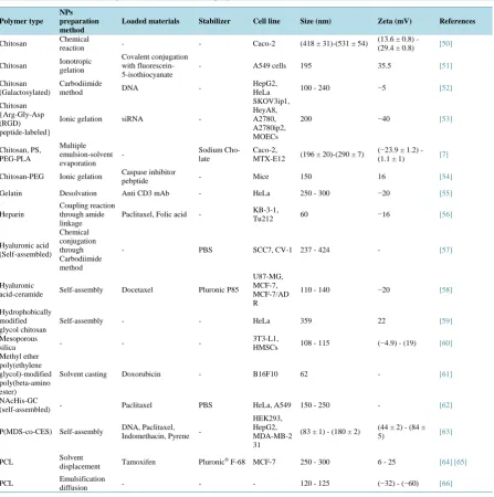
Cellular internalization or uptake is the most important physicochemical criteria prior to in vivo application.
Uptake of small molecules by any cells depends mainly on endocytosis among all other mechanisms (
Figure 1
).
Endocytosis is the bulk active transport process through lipid bilayer wrapping using energy in the form of ATP
to form required vesicles. Two main endocytosis mechanisms are reported as phagocytosis and pinocytosis [22].
Phagocytic cells (e.g. macrophages, neutrophils, dendritic cells, etc.) mediated cellular internalization is mostly
involved with engulfing the large particles (>1 µm) [23]. Adsorption or receptor dependent internalization is the
main mechanism of pinocytosis, which is mainly related to particle uptake by the cells through different
path-ways such as macro-pinocytosis, clathrin mediated, caveolin dependent or independent pinocytosis [3]. Size and
surface charge of polymeric NPs are likely the preliminary physicochemical variables, which govern the
endo-cytosis dependent cellular uptake. Besides, positive charge of the surface of polymeric NPs may endorse more
cellular attachment causing higher uptake either by endocytosis or by direct penetration, since cationic surface
of polymeric NPs interacts with anionic terminal of phospholipid, proteins and glycans on the cell surface due to
the electrostatic interactions [23].
An interesting experiment by Bhattacharjee et al. demonstrated the effects of size and surface charge of
fluo-rescent, monodisperse tri-block co-polymeric NPs based on cellular uptake through different endocytotic
path-ways [24]. They synthesized polymeric NPs (PNPs) with two different sizes (45 and 90 nm) and surface charges
such as neutral (PNP-OH, −4 mV), positive (PNP-NH
2, +22 mV) and negative (PNP-COOH, −19 mV) to
Figure 1.Relative sizes of NPs favorable for ingestion through various endocytotic pathways.
Inhibition of endocytic pathways was adopted to observe the role of endocytosis based cellular internalization of
polymeric NPs tracked by two mechanisms such as decreasing temperature to 4˚C of experimental unit and
ex-posing cells with 2-deoxyglucose (2-dOG) and sodium azide (NaN
3). Both inhibitory approaches showed
con-siderably lower uptake, which proved the higher uptake by positive charged polymeric NPs compared to that of
neutral to more negative charged polymeric NPs. Followed by the same strategy to block the clathrin and
caveo-lin mediated endocytosis, cells were exposed with hypertonic 450 mM sucrose solution and methyl-beta-cyclo-
dextran, respectively. Meeting the claimed fenestration sizes of these receptors dependent endocytosis, both
in-hibitions resulted with reduced uptake with smaller size after treating these cells with polymeric NPs, however
the uptake was varied with charge variations. For clathrin dependent endocytosis, uptake by both neutral and
negatively charged polymeric NPs was higher (65% and 75%, respectively) than positively charged polymeric
NPs (less than 38%), however an opposite result was found for caveolin dependent endocytosis.
On the other hand, Lai et al. investigated that polystyrene (PS) NPs with smaller size (>42 nm) were
success-fully internalized into HeLa cells following clathrin and caveolin independent endocytic pathways avoiding
en-dosomal or lysosomal accumulation [25]. Recent studies revealed that positively charged NPs uptake was
re-lated to energy dependent process such as proteins dynamin and F-actin but negatively charged NPs were not
dependent on dynamin proteins around the cell membrane [26]. Moreover, highly positively-charged NPs could
cause perforations in the cellular lipid bilayer to enter the cells by-passing endocytic pathways [23].
Another in vitro study for both fluorescent PS NPs and Coumarin-6 NPs in Caco-2 cells by Win et al. was
performed to assess the effects of different polymeric NPs size [27]. Raw Coumarin-6 could not increase the cell
uptake, however fluorescent PS NPs of 100 nm to 200 nm size showed the highest percentage of uptake. Smaller
particles (50 nm) showed the lowest uptake and particles as large as 1000 nm showed decrease in uptake, which
could be attributed to the uptake by other cellular mechanisms.
Optimization of antigen delivery to human dendritic cells (DCs; antigen presenting cells) is a challenge for
advanced vaccine delivery systems. To identify the effects of particle size and surface charge on human DCs, in
vitro cell uptake study has been investigated by Foged et al. in 2005 [28]. They designed the experiment based
on wide size ranges (0.1 µm to 4.5 µm) of fluorescent PS NPs with different surface charges (+12.4 to
−66.9
mV) after surface modification. Flow cytometric analysis of DCs after 24 hour incubation showed that lower
percentage of DCs had taken up 4.5 µm particles (30%); whereas the highest cellular uptake (60%) was
ob-Energy Independent
Pathway
Endocytosis
Energy Dependent Pathway
Pinocytosis
Macro-Pinocytosis Phagocytosis
Clathrin Dependent Pathway
Caveolin Dependent
Pathway
~50 nm ~80 nm ~120 nm 100 nm - 5 μm 0.5 nm - 10 μm Nanoparticle Size Range
Clathrin-Caveolin Independent
served for 0.5 µm and 0.1 µm sized particles. To optimize the charge dependent interactions, particles with two
sizes (0.1 and 1 µm) were modified by attaching variety poly amino acids/proteins covalently utilizing surface
amine and carboxyl groups. Sterically same positive and neutral charges particles were obtained using
polypep-tides poly-l-lysine (PLL) and poly-d-l-alanine (PA), respectively. After 24 hours incubation, only 10% cellular
uptake was observed with negatively charged 1 µm size particles, whereas positive charged particles were
ac-counted for 60% uptake. However, around 90% uptake was observed for lower size (0.1 µm) particles with
positive charge.
Prior to in vivo administration, it is essential to consider the compatibility, safety and biodegradability of the
particles with the human blood and cells. To investigate the efficiency of particle size and surface charge in
in
vitro cellular uptake and blood compatibility, recently Dash et al. employed chitosan/polyglutamic acid hollow
spheres to treat human umbilical vein endothelial cells (HUVECs) and human umbilical artery smooth muscle
cells (HUASMCs) [21]. Enhanced cellular uptake has been observed with 100 nm neutral charged (−4 mV) in
both cells such as 76% in HUVECs and 56% in HUASMCs compared to the other larger as well as pegylated
particles regardless of surface charge. However, negatively charged particles showed the least cell
internaliza-tion in both cases. To measure the effects of particles with erythrocytes of human blood, percentage of
hemoly-sis was accounted towards different sizes and charges of particles. All types of particles were partially associated
with very insignificant consequence on hemolysis (1% or less) without considering either size or surface charge.
But, highly anionic charged particles of smaller size resulted insignificant delayed clotting time and platelet
ac-tivation profile compared to larger particles and other types of charged particles.
Testing blood compatibility of polymeric NPs with human blood is another way for finding the probable
ad-verse effects, which may happen after in vivo administration. To rationalize the hemo-compatibility test, another
research group (Mayer et al.) employed PS NPs with variety of sizes and surface charges in different mediums
(such as cell culture medium with different FBS ratio, PBS) [29]. To assess the influence of polymeric NPs’ size
and surface charge on human blood, the aim of study was to monitor the adverse effects by measuring
comple-ment activation, induction of coagulation, thrombocyte activation, membrane integrity, granulocyte activation,
and hemolysis using flow cytometric analysis. Complement (C3a and C5a) levels detection is a consideration of
the body’s immune system activation. Cationic amidine PS particles were involved with high C3a generation
(150.8%). Irrespective to size and surface charge, no NPs were involved with prothrombin level induction.
CD62P/CD42b labelling was employed to investigate the thrombocyte activation, which was tested for both low
(0.5 mg/mL) and high (2 mg/mL) concentrations. But no thromocytic damage was observed, which were
con-firmed by no lactate-de-hydrogenase (LDH) release for any of the particles. The percentage of CD11b
expres-sion (marker for granulocyte activation) for particle’s different sizes and surface charges was reported in that
study. Increased percentage of hemolysis for all types of particles was reported using high concentration of
par-ticle treatment with human blood. However, larger parpar-ticles were found less hemolytic than smaller parpar-ticles,
and the most important point was that no influence was observed for negatively charged 160 nm size NPs on
erythrocytes of human blood by treating with lower concentration. Overall, positively charged larger particles
were involved with more hemolysis compared to negatively charged particles and the latter ones larger than 60
nm size appeared to be less hematotoxic than smaller particles. One interesting finding was; particles
resus-pended in cell culture medium with 10% fetal bovine serum (FBS) showed less negative zeta potential or about
to close to neutral charge compared to the particles resuspended in phosphate buffer saline (PBS). The presence
of salts and proteins in the dispersion cell medium might be accountable for neutralizing surface charge of
polymeric NPs.
Upon exposure of different types of PLGA NPs to different experimental media, Mura et al. also investigated
the possible size and zeta potential variations after resuspension of polymeric NPs in different media with time
dependent incubation up to 96 hours at 37˚C [30]. Three types of medium such as water, cell culture medium
plus 10% FBS and PBS have been considered for evaluation in this experiment. Among different media, water
and cell culture medium containing 10% FBS were not involved with significant variations in particle size
re-gardless of surface charge, however after incubation of PLGA/chitosan (CS) NOS in PBS the size was increased.
Furthermore, upon exposure to serum containing cell culture medium, PLGA/CS, PLGA/polyvinyl alcohol
(PVA), and PLGA/pluronic F-68 (PF-68) NPs did not show any noteworthy change in zeta potential values.
reaction [31] [32]. Cell viability responses due to NP treatment with higher concentration after 72 hours
incuba-tion demonstrated that only PLGA/PF68 NPs showed progressively decreased cell viability compared to other
types of NPs.
From other in vitro studies, it has also been found that NPs with 40 - 50 nm size range are involved with
maximum uptake [33] [34]. However, a recent experiment by Schadlich et al. revealed the effect of size for the
accumulation of near-infrared (NIR) fluorescent consisting PLA-PEG polymeric NPs in two tumor xenograft
models (HT29 colorectal carcinoma and A2780 ovarian carcinoma) utilizing in vivo fluorescence imaging
tech-nique [35]. NPs of 111 nm and 141 nm size showed higher biodistribution and accumulation in tumors
com-pared to the larger size (166 nm), which was due to rapid clearance of the larger particles by liver.
4. Effect of Particle Size and Surface Charge Based on
in Vivo
Studies
To explore the in vivo effects of specifically sized NPs with respect to surface charge, Kulkarni et al. injected
the fluorescent modified and unmodified PS NPs into Sprague–Dawley rats after physicochemical
characteriza-tion [36]. Modificacharacteriza-tion of PS NPs was performed by coating with D-α-tocopheryl polyethylene glycol succinate
or Vitamin E TPGS, which was able to switch the zeta potentials of different size NPs to less negative charge.
As previously known, circulating mononuclear phagocytic cells in the bloodstream are the key component of
reticuloendothelial system (RES). In addition, RES is also composed of matured cells such as macrophages
mainly available in lungs, liver and spleen [37]. Studies have shown that the NPs with the size range of 100 to
200 nm could be the optimum range in order to escape the RES recognition [27]. Due to rapid clearance from
systemic circulation, mostly uncoated NPs were distributed to those organs such as liver and spleen, where
mononuclear phagocytic system is located. Consequently, 100 and 200 nm size fluorescent PS-TPGS NPs
re-sulted in higher fluorescence concentration in blood plasma regardless of their surface charges. Liver and spleen
were the main target organs, where a substantial decrease in NP distribution was observed for all sizes of TPGS
modified PS NPs, since hydrophilic coated surface (stealth effect) possesses the ability to prevent the NPs from
RES capture.
NPs of 10 - 100 nm size is considered as mainly accepted range to design any NP formulation respective to
suitable clearance and biodistribution profile before any in vivo trial [3]. However, the upper range of particle
size is dependent on the interactions with body’s immune systems and the lower range is determined by the limit
of kidney filtration. Opsonization of larger particles by responsible proteins (e.g. plasma complement,
immu-noglobulins) in blood compartment is common to develop hypersensitivity response comparatively against
lar-ger foreign particles [44] [45]. On the other hand, smaller particles (<5.5 nm) have been found with rapid
clear-ance from the body by kidney’s glomerular filtration mechanism [46].
To explore the in vivo effects of different size of NPs, Liu
et al. prepared radioisotope labeled liposomes of
different sizes (30 - 400 nm) to inject into the mice models to observe the biodistribution in blood, liver, spleen,
and tumor [47]. After four hours of post administration, it was found that about 60% of 100 to 200 nm size
par-ticles were found in blood, but only 20% of injected parpar-ticles with size boundary (>250 nm or <50 nm) were
detected in blood. In liver, particle size with 100 nm was associated with 20% accumulation, whereas around 25%
distribution in liver was detected for larger particles. In spleen, 40% - 50% of the injected dose was detected for
larger size (>250 nm) but the percentage of detection was lower for the particle size range below 100 nm. In
2002, Levchenko et al. prepared liposomes of around 200 nm with variety of charged surfaces to evaluate the
tissue distribution in mice models [48]. The results from this study showed that the negatively charged
lipo-somes with zeta potential of around −40 mV were involved with higher clearance rate from the blood in
com-parison to liposomes with neutral zeta potential.
In addition, Yamamoto
et al. investigated the effect of surface charge of poly (ethylene glycol)-poly (D,
L-lactide) block copolymer micelles after injecting into male C57/BL6N mice through the tail vein [49]. They
prepared the micelles with both neutral (tyrosine) and negative (tyrosineglutamine) functionalities, which did
not show any significant variations in blood clearance kinetics. However, the negatively-charged micelles
dis-played a significant lower distribution in both liver and spleen after four hours of post intravenous injection.
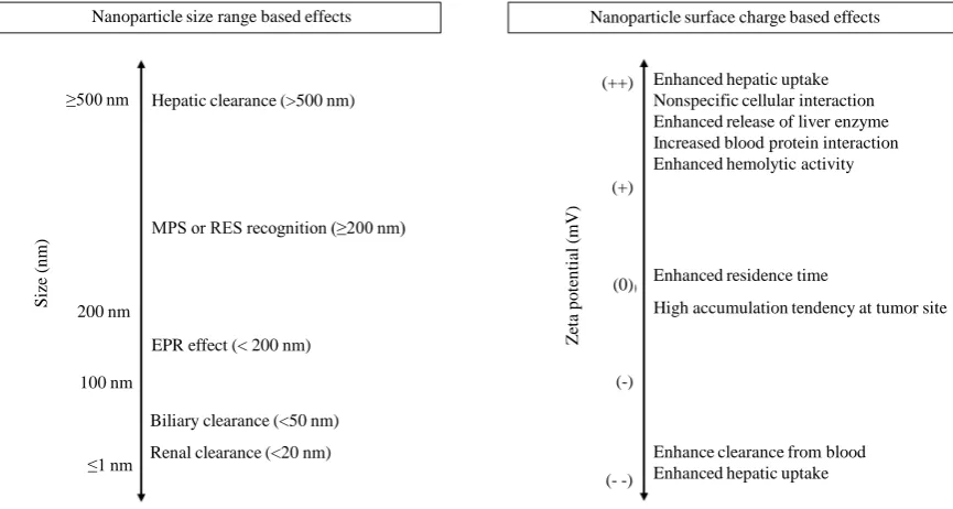
Overall effects of NPs size and surface charge could be summarized in
Figure 2
.
5. Conclusion
[image:10.595.96.529.465.696.2]In conclusion, polymeric NPs with size range from 10 to 200 nm might not only escape renal filtration and
bil-iary excretion but also accumulate in tumor utilizing EPR effects. Size range above 200 nm may be related to
rapid hepatic clearance and RES recognition. Pegylation strategy could be an ideal option to stealth the poly-
meric NPs for longer residing time during systemic circulation. After
in vivo administration of cationic
Figure 2.Relative biocompatibility of polymeric NPs based on the effects of size and surface charge. Abbreviation: MPS (Mononuclear Phagocyte System), RES (Reticuloendothelial System), EPR (Enhanced Permeability and Retention).
Nanoparticle size range based effects
Hepatic clearance (>500 nm) ≥500 nm
200 nm
100 nm
S
ize (
n
m
) MPS or RES recognition (≥200 nm)
EPR effect (< 200 nm)
Biliary clearance (<50 nm)
Renal clearance (<20 nm) ≤1 nm
Z
et
a p
o
te
n
tia
l (
m
V
)
Enhanced residence time
High accumulation tendency at tumor site Nanoparticle surface charge based effects
Enhanced hepatic uptake Nonspecific cellular interaction Enhanced release of liver enzyme Increased blood protein interaction Enhanced hemolytic activity
Enhance clearance from blood Enhanced hepatic uptake (- -)
polymeric NPs, non-specific interaction may occur with non-specific cells or opsonizing protein in blood
com-partment due to electrostatic bindings, which may involve unexpected cytotoxicity. In order to reduce such non-
specific surface reactivity or interaction, relatively less negatively charged anionic (almost neutral) polymeric
NPs with desired small size might be more rationale than cationic charged particles for a broad spectrum
bio-logical aspect. This review will help researchers to correlate the in vitro and
in vivo effects of polymeric NPs
based on particle size and charge. Further investigation and correlation of other physicochemical parameters
could be performed on polymeric NPs to understand their biological effects.
Acknowledgements
This work was funded by a research grant from the Canadian Breast Cancer Foundation (CBCF) and Natural
Sciences and Engineering Research (NSERC) Discovery Grant. The authors report no declarations of interest.
Conflict of Interest
The authors confirm that this article content has no conflict of interest.
References
[1] Grottkau, B.E., Cai, X., Wang, J., Yang, X. and Lin, Y. (2013) Polymeric Nanoparticles for a Drug Delivery System. Current Drug Metabolism, 14, 840-846. http://dx.doi.org/10.2174/138920021131400105
[2] He, C., Hu, Y., Yin, L., Tang, C. and Yin, C. (2010) Effects of Particle Size and Surface Charge on Cellular Uptake and Biodistribution of Polymeric Nanoparticles. Biomaterials, 31, 3657-3666.
http://dx.doi.org/10.1016/j.biomaterials.2010.01.065
[3] Wang, J., Byrne, J.D., Napier, M.E. and DeSimone, J.M. (2011) More Effective Nanomedicines through Particle De-sign. Small, 7, 1919-1931. http://dx.doi.org/10.1002/smll.201100442
[4] Kleber, J.W., Nash, J.F. and Lee, C.C. (1964) Synthetic Polymers as Potential Sustained-Release Coatings. Journal of Pharmaceutical Sciences, 53, 1519-1521. http://dx.doi.org/10.1002/jps.2600531219
[5] Singh, R. and Lillard Jr., J.W. (2009) Nanoparticle-Based Targeted Drug Delivery. Experimental and Molecular Pa-thology, 86, 215-223. http://dx.doi.org/10.1016/j.yexmp.2008.12.004
[6] Lamprecht, A., Schafer, U. and Lehr, C.M. (2001) Size-Dependent Bioadhesion of Micro- and Nanoparticulate Carri-ers to the Inflamed Colonic Mucosa. Pharmaceutical Research, 18, 788-793.
http://dx.doi.org/10.1023/A:1011032328064
[7] Behrens, I., Pena, A.I., Alonso, M.J. and Kissel, T. (2002) Comparative Uptake Studies of Bioadhesive and Non- Bioadhesive Nanoparticles in Human Intestinal Cell Lines and Rats: The Effect of Mucus on Particle Adsorption and Transport. Pharmaceutical Research, 19, 1185-1193. http://dx.doi.org/10.1023/A:1019854327540
[8] Galindo-Rodriguez, S.A., Allemann, E., Fessi, H. and Doelker, E. (2005) Polymeric Nanoparticles for Oral Delivery of Drugs and Vaccines: A Critical Evaluation of in Vivo Studies. Critical Reviews™ in Therapeutic Drug Carrier Systems,
22, 419-464. http://dx.doi.org/10.1615/CritRevTherDrugCarrierSyst.v22.i5.10
[9] Graf, A., McDowell, A. and Rades, T. (2009) Poly(alkylcyanoacrylate) Nanoparticles for Enhanced Delivery of Thera- peutics—Is There Real Potential? Expert Opinion on Drug Delivery, 6, 371-387.
[10] Vinogradov, S.B.E. and Kabanov, A. (1999) Poly(ethylene glycol)-polyethyleneimine NanoGel™ Particles: Novel Drug Delivery Systems for Antisense Oligonucleotides. Colloids and Surfaces B: Biointerfaces, 16, 291-304.
[11] Plapied, L., Duhem, N., Rieux, A.D. and Préat, V. (2011) Fate of Polymeric Nanocarriers for Oral Drug Delivery. Current Opinion in Colloid & Interface, 16, 228-237. http://dx.doi.org/10.1016/j.cocis.2010.12.005
[12] Ahlin, P., Kristl, J., Kristl, A. and Vrečer, F. (2002) Investigation of Polymeric Nanoparticles as Carriers of Enalaprilat for Oral Administration. International Journal of Pharmaceutics, 239, 113-120.
http://dx.doi.org/10.1016/S0378-5173(02)00076-5
[13] Kumari, A., Yadav, S.K. and Yadav, S.C. (2010) Biodegradable Polymeric Nanoparticles Based Drug Delivery Sys-tems. Colloids and Surfaces B: Biointerfaces, 75, 1-18. http://dx.doi.org/10.1016/j.colsurfb.2009.09.001
[14] Song, X., Zhao, X., Zhou, Y., Li, S. and Ma, Q. (2010) Pharmacokinetics and Disposition of Various Drug Loaded Biodegradable Poly(Lactide-Co-Glycolide) (PLGA) Nanoparticles. Current Drug Metabolism, 11, 859-869.
http://dx.doi.org/10.2174/138920010794479682
http://dx.doi.org/10.1208/s12249-010-9561-2
[16] Liu, Z., Jiao, Y., Wang, Y., Zhou, C. and Zhang, Z. (2008) Polysaccharides-Based Nanoparticles as Drug Delivery Systems. Advanced Drug Delivery Reviews, 60, 1650-1662. http://dx.doi.org/10.1016/j.addr.2008.09.001
[17] Hu, C.M., Fang, R.H., Luk, B.T. and Zhang, L. (2013) Polymeric Nanotherapeutics: Clinical Development and Ad-vances in Stealth Functionalization Strategies. Nanoscale, 6, 65-76. http://dx.doi.org/10.1039/C3NR05444F
[18] Kamaly, N., Xiao, Z., Valencia, P.M., Radovic-Moreno, A.F. and Farokhzad, O.C. (2012) Targeted Polymeric Thera-peutic Nanoparticles: Design, Development and Clinical Translation. Chemical Society Reviews, 41, 2971-3010.
http://dx.doi.org/10.1039/c2cs15344k
[19] Mundargi, R.C., Babu, V.R., Rangaswamy, V., Patel, P. and Aminabhavi, T.M. (2008) Nano/Micro Technologies for Delivering Macromolecular Therapeutics Using Poly(D,L-Lactide-co-Glycolide) and Its Derivatives. Journal of Con-trolled Release, 125, 193-209. http://dx.doi.org/10.1016/j.jconrel.2007.09.013
[20] Dinarvand, R., Sepehri, N., Manoochehri, S., Rouhani, H. and Atyabi, F. (2011) Polylactide-co-Glycolide Nanoparti-cles for Controlled Delivery of Anticancer Agents. International Journal of Nanomedicine, 6, 877-895.
http://dx.doi.org/10.2147/IJN.S18905
[21] Dash, B.C., Rethore, G., Monaghan, M., Fitzgerald, K., Gallagher, W. and Pandit, A. (2010) The Influence of Size and Charge of Chitosan/Polyglutamic Acid Hollow Spheres on Cellular Internalization, Viability and Blood Compatibility. Biomaterials, 31, 8188-8197. http://dx.doi.org/10.1016/j.biomaterials.2010.07.067
[22] Conner, S.D. and Schmid, S.L. (2003) Regulated Portals of Entry into the Cell. Nature, 422, 37-44.
http://dx.doi.org/10.1038/nature01451
[23] Zhao, F., Zhao, Y., Liu, Y., Chang, X., Chen, C. and Zhao, Y. (2011) Cellular Uptake, Intracellular Trafficking, and Cytotoxicity of Nanomaterials. Small, 7, 1322-1337. http://dx.doi.org/10.1002/smll.201100001
[24] Bhattacharjee, S., Ershov, D., Fytianos, K., van der Gucht, J., Alink, G.M., Rietjens, I.M., et al. (2012) Cytotoxicity and Cellular Uptake of Tri-Block Copolymer Nanoparticles with Different Size and Surface Characteristics. Particle and Fibre Toxicology, 9, 11. http://dx.doi.org/10.1186/1743-8977-9-11
[25] Lai, S.K., Hida, K., Man, S.T., Chen, C., Machamer, C., Schroer, T.A., et al. (2007) Privileged Delivery of Polymer Nanoparticles to the Perinuclear Region of Live Cells via a Non-Clathrin, Non-Degradative Pathway. Biomaterials, 28, 2876-2884. http://dx.doi.org/10.1016/j.biomaterials.2007.02.021
[26] Dausend, J., Musyanovych, A., Dass, M., Walther, P., Schrezenmeier, H., Landfester, K., et al. (2008) Uptake Mecha-nism of Oppositely Charged Fluorescent Nanoparticles in HeLa Cells. Macromolecular Bioscience, 8, 1135-1143.
http://dx.doi.org/10.1002/mabi.200800123
[27] Win, K.Y. and Feng, S.S. (2005) Effects of Particle Size and Surface Coating on Cellular Uptake of Polymeric Nanoparticles for Oral Delivery of Anticancer Drugs. Biomaterials, 26, 2713-2722.
http://dx.doi.org/10.1016/j.biomaterials.2004.07.050
[28] Foged, C., Brodin, B., Frokjaer, S. and Sundblad, A. (2005) Particle Size and Surface Charge Affect Particle Uptake by Human Dendritic Cells in an in Vitro Model. International Journal of Pharmaceutics, 298, 315-322.
http://dx.doi.org/10.1016/j.ijpharm.2005.03.035
[29] Mayer, A., Vadon, M., Rinner, B., Novak, A., Wintersteiger, R. and Frohlich, E. (2009) The Role of Nanoparticle Size in Hemocompatibility. Toxicology, 258, 139-147. http://dx.doi.org/10.1016/j.tox.2009.01.015
[30] Mura, S., Hillaireau, H., Nicolas, J., Le Droumaguet, B., Gueutin, C., Zanna, S., et al. (2011) Influence of Surface Charge on the Potential Toxicity of PLGA Nanoparticles towards Calu-3 Cells. International Journal of Nanomedicine,
6, 2591-2605.
[31] Shen, B.Q., Finkbeiner, W.E., Wine, J.J., Mrsny, R.J. and Widdicombe, J.H. (1994) Calu-3: A Human Airway Epithe-lial Cell Line That Shows cAMP-Dependent Cl-Secretion. American Journal of Physiology, 266, L493-L501. [32] da Paula, A.C., Ramalho, A.S., Farinha, C.M., Cheung, J., Maurisse, R., Gruenert, D.C., et al. (2005) Characterization
of Novel Airway Submucosal Gland Cell Models for Cystic Fibrosis Studies. Cellular Physiology and Biochemistry,
15, 251-262. http://dx.doi.org/10.1159/000087235
[33] Arap, W., Haedicke, W., Bernasconi, M., Kain, R., Rajotte, D., Krajewski, S., et al. (2002) Targeting the Prostate for Destruction through a Vascular Address. Proceedings of the National Academy of Sciences of the United States of America, 99, 1527-1531. http://dx.doi.org/10.1073/pnas.241655998
[34] Gratton, S.E., Ropp, P.A., Pohlhaus, P.D., Luft, J.C., Madden, V.J., Napier, M.E., et al. (2008) The Effect of Particle Design on Cellular Internalization Pathways. Proceedings of the National Academy of Sciences of the United States of America, 105, 11613-11618. http://dx.doi.org/10.1073/pnas.0801763105
8710-8720. http://dx.doi.org/10.1021/nn2026353
[36] Kulkarni, S.A. and Feng, S.S. (2013) Effects of Particle Size and Surface Modification on Cellular Uptake and Biodis-tribution of Polymeric Nanoparticles for Drug Delivery. Pharmaceutical Research, 30, 2512-2522.
http://dx.doi.org/10.1007/s11095-012-0958-3
[37] Hillery, A.M. and Florence, F.A. (1996) The Effect of Adsorbed Poloxamer 188 and 407 Surfactants on the Intestinal Uptake of 60-nm Polystyrene Particles after Oral Administration in the Rat. International Journal of Pharmaceutics,
132, 123-130. http://dx.doi.org/10.1016/0378-5173(95)04353-5
[38] Faraji, A.H. and Wipf, P. (2009) Nanoparticles in Cellular Drug Delivery. Bioorganic & Medicinal Chemistry, 17, 2950-2962. http://dx.doi.org/10.1016/j.bmc.2009.02.043
[39] Alexis, F., Pridgen, E., Molnar, L.K. and Farokhzad, O.C. (2008) Factors Affecting the Clearance and Biodistribution of Polymeric Nanoparticles. Molecular Pharmaceutics, 5, 505-515. http://dx.doi.org/10.1021/mp800051m
[40] Nigavekar, S.S., Sung, L.Y., Llanes, M., El-Jawahri, A., Lawrence, T.S., Becker, C.W., et al. (2004) 3H Dendrimer Nanoparticle Organ/Tumor Distribution. Pharmaceutical Research, 21, 476-483.
http://dx.doi.org/10.1023/B:PHAM.0000019302.26097.cc
[41] Liu, D., Mori, A. and Huang, L. (1992) Role of Liposome Size and RES Blockade in Controlling Biodistribution and Tumor Uptake of GM1-Containing Liposomes. Biochimica et Biophysica Acta, 1104, 95-101.
http://dx.doi.org/10.1016/0005-2736(92)90136-A
[42] Akiyama, Y., Mori, T., Katayama, Y. and Niidome, T. (2009) The Effects of PEG Grafting Level and Injection Dose on Gold Nanorod Biodistribution in the Tumor-Bearing Mice. Journal of Controlled Release, 139, 81-84.
http://dx.doi.org/10.1016/j.jconrel.2009.06.006
[43] Ishiwata, H., Suzuki, N., Ando, S., Kikuchi, H. and Kitagawa, T. (2000) Characteristics and Biodistribution of Cationic Liposomes and Their DNA Complexes. Journal of Controlled Release, 69, 139-148.
http://dx.doi.org/10.1016/S0168-3659(00)00293-5
[44] Vonarbourg, A., Passirani, C., Saulnier, P., Simard, P., Leroux, J.C. and Benoit, J.P. (2006) Evaluation of Pegylated Lipid Nanocapsules versus Complement System Activation and Macrophage Uptake. Journal of Biomedical Materials Research Part A, 78, 620-628. http://dx.doi.org/10.1002/jbm.a.30711
[45] Petros, R.A. and DeSimone, J.M. (2010) Strategies in the Design of Nanoparticles for Therapeutic Applications. Na-ture Reviews Drug Discovery, 9, 615-627. http://dx.doi.org/10.1038/nrd2591
[46] Choi, H.S., Liu, W., Misra, P., Tanaka, E., Zimmer, J.P., Itty Ipe, B., et al. (2007) Renal Clearance of Quantum Dots. Nature Biotechnology, 25, 1165-1170. http://dx.doi.org/10.1038/nbt1340
[47] Li, S.D. and Huang, L. (2008) Pharmacokinetics and Biodistribution of Nanoparticles. Molecular Pharmaceutics, 5, 496-504. http://dx.doi.org/10.1021/mp800049w
[48] Levchenko, T.S., Rammohan, R., Lukyanov, A.N., Whiteman, K.R. and Torchilin, V.P. (2002) Liposome Clearance in Mice: The Effect of a Separate and Combined Presence of Surface Charge and Polymer Coating. International Journal of Pharmaceutics, 240, 95-102. http://dx.doi.org/10.1016/S0378-5173(02)00129-1
[49] Yamamoto, Y., Nagasaki, Y., Kato, Y., Sugiyama, Y. and Kataoka, K. (2001) Long-Circulating Poly(Ethylene Gly-col)-Poly(D,L-Lactide) Block Copolymer Micelles with Modulated Surface Charge. Journal of Controlled Release, 77, 27-38. http://dx.doi.org/10.1016/S0168-3659(01)00451-5
[50] Ma, Z. and Lim, L.Y. (2003) Uptake of Chitosan and Associated Insulin in Caco-2 Cell Monolayers: A Comparison between Chitosan Molecules and Chitosan Nanoparticles. Pharmaceutical Research, 20, 1812-1819.
http://dx.doi.org/10.1023/B:PHAM.0000003379.76417.3e
[51] Huang, M., Ma, Z., Khor, E. and Lim, L.Y. (2002) Uptake of FITC-Chitosan Nanoparticles by A549 Cells. Pharma-ceutical Research, 19, 1488-1494. http://dx.doi.org/10.1023/A:1020404615898
[52] Kim, T.H., Park, I.K., Nah, J.W., Choi, Y.J. and Cho, C.S. (2004) Galactosylated Chitosan/DNA Nanoparticles Pre-pared Using Water-Soluble Chitosan as a Gene Carrier. Biomaterials, 25, 3783-3792.
http://dx.doi.org/10.1016/j.biomaterials.2003.10.063
[53] Han, H.D., Mangala, L.S., Lee, J.W., Shahzad, M.M., Kim, H.S., Shen, D., et al. (2010) Targeted Gene Silencing Us-ing RGD-Labeled Chitosan Nanoparticles. Clinical Cancer Research: An Official Journal of the American Association for Cancer Research, 16, 3910-3922.
[54] Aktas, Y., Yemisci, M., Andrieux, K., Gursoy, R.N., Alonso, M.J., Fernandez-Megia, E., et al. (2005) Development and Brain Delivery of Chitosan-PEG Nanoparticles Functionalized with the Monoclonal Antibody OX26. Bioconju-gate Chemistry, 16, 1503-1511. http://dx.doi.org/10.1021/bc050217o
Biomate-rials, 26, 2723-2732. http://dx.doi.org/10.1016/j.biomaterials.2004.07.047
[56] Wang, X., Li, J., Wang, Y., Cho, K.J., Kim, G., Gjyrezi, A., et al. (2009) HFT-T, a Targeting Nanoparticle, Enhances Specific Delivery of Paclitaxel to Folate Receptor-Positive Tumors. ACS Nano, 3, 3165-3174.
http://dx.doi.org/10.1021/nn900649v
[57] Choi, K.Y., Chung, H., Min, K.H., Yoon, H.Y., Kim, K., Park, J.H., et al. (2010) Self-Assembled Hyaluronic Acid Nanoparticles for Active Tumor Targeting. Biomaterials, 31, 106-114.
http://dx.doi.org/10.1016/j.biomaterials.2009.09.030
[58] Cho, H.J., Yoon, H.Y., Koo, H., Ko, S.H., Shim, J.S., Lee, J.H., et al. (2011) Self-Assembled Nanoparticles Based on Hyaluronic Acid-Ceramide (HA-CE) and Pluronic(R) for Tumor-Targeted Delivery of Docetaxel. Biomaterials, 32, 7181-7190. http://dx.doi.org/10.1016/j.biomaterials.2011.06.028
[59] Nam, H.Y., Kwon, S.M., Chung, H., Lee, S.Y., Kwon, S.H., Jeon, H., et al. (2009) Cellular Uptake Mechanism and Intracellular Fate of Hydrophobically Modified Glycol Chitosan Nanoparticles. Journal of Controlled Release, 135, 259-267.
[60] Chung, T.H., Wu, S.H., Yao, M., Lu, C.W., Lin, Y.S., Hung, Y., et al. (2007) The Effect of Surface Charge on the Up-take and Biological Function of Mesoporous Silica Nanoparticles in 3T3-L1 Cells and Human Mesenchymal Stem Cells. Biomaterials, 28, 2959-2966. http://dx.doi.org/10.1016/j.biomaterials.2007.03.006
[61] Ko, J., Park, K., Kim, Y.S., Kim, M.S., Han, J.K., Kim, K., et al. (2007) Tumoral Acidic Extracellular pH Targeting of pH-Responsive MPEG-Poly(Beta-Amino Ester) Block Copolymer Micelles for Cancer Therapy. Journal of Controlled Release, 123, 109-115.
[62] Park, J.S., Han, T.H., Lee, K.Y., Han, S.S., Hwang, J.J., Moon, D.H., et al. (2006) N-Acetyl Histidine-Conjugated Glycol Chitosan Self-Assembled Nanoparticles for Intracytoplasmic Delivery of Drugs: Endocytosis, Exocytosis and Drug Release. Journal of Controlled Release, 115, 37-45.
[63] Wang, Y., Gao, S., Ye, W.H., Yoon, H.S. and Yang, Y.Y. (2006) Co-Delivery of Drugs and DNA from Cationic Core- Shell Nanoparticles Self-Assembled from a Biodegradable Copolymer. Nature Materials, 5, 791-796.
http://dx.doi.org/10.1038/nmat1737
[64] Chawla, J.S. and Amiji, M.M. (2002) Biodegradable Poly(Epsilon-Caprolactone) Nanoparticles for Tumor-Targeted Delivery of taMoxifen. International Journal of Pharmaceutics, 249, 127-138.
http://dx.doi.org/10.1016/S0378-5173(02)00483-0
[65] Chawla, J.S. and Amiji, M.M. (2003) Cellular Uptake and Concentrations of Tamoxifen upon Administration in Poly(Epsilon-Caprolactone) Nanoparticles. AAPS PharmSci, 5, 28-34. http://dx.doi.org/10.1208/ps050103
[66] Cade, D., Ramus, E., Rinaudo, M., Auzely-Velty, R., Delair, T. and Hamaide, T. (2004) Tailoring of Bioresorbable Polymers for Elaboration of Sugar-Functionalized Nanoparticles. Biomacromolecules, 5, 922-927.
http://dx.doi.org/10.1021/bm034504b
[67] Wang, J., Tian, S., Petros, R.A., Napier, M.E. and Desimone, J.M. (2010) The Complex Role of Multivalency in Nanoparticles Targeting the Transferrin Receptor for Cancer Therapies. Journal of the American Chemical Society,
132, 11306-11313. http://dx.doi.org/10.1021/ja1043177
[68] Lee, H., Fonge, H., Hoang, B., Reilly, R.M. and Allen, C. (2010) The Effects of Particle Size and Molecular Targeting on the Intratumoral and Subcellular Distribution of Polymeric Nanoparticles. Molecular Pharmaceutics, 7, 1195-1208.
http://dx.doi.org/10.1021/mp100038h
[69] Xin, H., Jiang, X., Gu, J., Sha, X., Chen, L., Law, K., et al. (2011) Angiopep-Conjugated Poly(Ethylene Glycol)-co- Poly(Epsilon-Caprolactone) Nanoparticles as Dual-Targeting Drug Delivery System for Brain Glioma. Biomaterials,
32, 4293-4305. http://dx.doi.org/10.1016/j.biomaterials.2011.02.044
[70] Kim, H.R., Gil, S., Andrieux, K., Nicolas, V., Appel, M., Chacun, H., et al. (2007) Low-Density Lipoprotein Receptor- Mediated Endocytosis of PEGylated Nanoparticles in Rat Brain Endothelial Cells. Cellular and Molecular Life Sci- ences: CMLS, 64, 356-364. http://dx.doi.org/10.1007/s00018-007-6390-x
[71] Harush-Frenkel, O., Debotton, N., Benita, S. and Altschuler, Y. (2007) Targeting of Nanoparticles to the Clathrin-Me- diated Endocytic Pathway. Biochemical and Biophysical Research Communications, 353, 26-32.
http://dx.doi.org/10.1016/j.bbrc.2006.11.135
[72] Lu, W., Tan, Y.Z., Hu, K.L. and Jiang, X.G. (2005) Cationic Albumin Conjugated Pegylated Nanoparticle with Its Transcytosis Ability and Little Toxicity Against Blood-Brain Barrier. International Journal of Pharmaceutics, 295, 247-260. http://dx.doi.org/10.1016/j.ijpharm.2005.01.043
[73] Harush-Frenkel, O., Rozentur, E., Benita, S. and Altschuler, Y. (2008) Surface Charge of Nanoparticles Determines Their Endocytic and Transcytotic Pathway in Polarized MDCK Cells. Biomacromolecules, 9, 435-443.
http://dx.doi.org/10.1021/bm700535p
De-livery. ACS Nano, 4, 251-258. http://dx.doi.org/10.1021/nn9014032
[75] Danhier, F., Lecouturier, N., Vroman, B., Jerome, C., Marchand-Brynaert, J., Feron, O., et al. (2009) Paclitaxel- Loaded PEGylated PLGA-Based Nanoparticles: In Vitro and In Vivo Evaluation. Journal of Controlled Release, 133, 11-17.
[76] Mao, S., Germershaus, O., Fischer, D., Linn, T., Schnepf, R. and Kissel, T. (2005) Uptake and Transport of PEG- Graft-Trimethyl-Chitosan Copolymer-Insulin Nanocomplexes by Epithelial Cells. Pharmaceutical Research, 22, 2058- 2068. http://dx.doi.org/10.1007/s11095-005-8175-y
[77] Nukolova, N.V., Oberoi, H.S., Cohen, S.M., Kabanov, A.V. and Bronich, T.K. (2011) Folate-Decorated Nanogels for Targeted Therapy of Ovarian Cancer. Biomaterials, 32, 5417-5426.
http://dx.doi.org/10.1016/j.biomaterials.2011.04.006
[78] Cho, C.S., Cho, K.Y., Park, I.K., Kim, S.H., Sasagawa, T., Uchiyama, M., et al. (2001) Receptor-Mediated Delivery of All Trans-Retinoic Acid to Hepatocyte Using Poly(L-Lactic Acid) Nanoparticles Coated with Galactose-Carrying Polystyrene. Journal of Controlled Release, 77, 7-15.
[79] Banquy, X., Leclair, G., Rabanel, J.M., Argaw, A., Bouchard, J.F., Hildgen, P., et al. (2008) Selectins Ligand Deco-rated Drug Carriers for Activated Endothelial Cell Targeting. Bioconjugate Chemistry, 19, 2030-2039.
http://dx.doi.org/10.1021/bc800257m
[80] Nasongkla, N., Bey, E., Ren, J., Ai, H., Khemtong, C., Guthi, J.S., et al. (2006) Multifunctional Polymeric Micelles as Cancer-Targeted, MRI-Ultrasensitive Drug Delivery Systems. Nano Letters, 6, 2427-2430.
http://dx.doi.org/10.1021/nl061412u
[81] Prabha, S. and Labhasetwar, V. (2004) Critical Determinants in PLGA/PLA Nanoparticle-Mediated Gene Expression. Pharmaceutical Research, 21, 354-364. http://dx.doi.org/10.1023/B:PHAM.0000016250.56402.99
[82] Musumeci, T., Ventura, C.A., Giannone, I., Ruozi, B., Montenegro, L., Pignatello, R., et al. (2006) PLA/PLGA Nanoparticles for Sustained Release of Docetaxel. International Journal of Pharmaceutics, 325, 172-179.
http://dx.doi.org/10.1016/j.ijpharm.2006.06.023
[83] Prabha, S., Zhou, W.Z., Panyam, J. and Labhasetwar, V. (2002) Size-Dependency of Nanoparticle-Mediated Gene Transfection: Studies with Fractionated Nanoparticles. International Journal of Pharmaceutics, 244, 105-115.
http://dx.doi.org/10.1016/S0378-5173(02)00315-0
[84] Sahoo, S.K., Panyam, J., Prabha, S. and Labhasetwar, V. (2002) Residual Polyvinyl Alcohol Associated with Poly (D,L-Lactide-co-Glycolide) Nanoparticles Affects Their Physical Properties and Cellular Uptake. Journal of Con-trolled Release, 82, 105-114.
[85] Sahoo, S.K. and Labhasetwar, V. (2005) Enhanced Antiproliferative Activity of Transferrin-Conjugated Paclitaxel- Loaded Nanoparticles Is Mediated via Sustained Intracellular Drug Retention. Molecular Pharmaceutics, 2, 373-383.
http://dx.doi.org/10.1021/mp050032z
[86] Obermajer, N., Kocbek, P., Repnik, U., Kuznik, A., Cegnar, M., Kristl, J., et al. (2007) Immunonanoparticles—An Ef-fective Tool to Impair Harmful Proteolysis in Invasive Breast Tumor Cells. The FEBS Journal, 274, 4416-4427.
http://dx.doi.org/10.1111/j.1742-4658.2007.05971.x
[87] Davda, J. and Labhasetwar, V. (2002) Characterization of Nanoparticle Uptake by Endothelial Cells. International Journal of Pharmaceutics, 233, 51-59. http://dx.doi.org/10.1016/S0378-5173(01)00923-1
[88] Mo, Y. and Lim, L.Y. (2005) Preparation and in Vitro Anticancer Activity of Wheat Germ Agglutinin (WGA)-Conju- gated PLGA Nanoparticles Loaded with Paclitaxel and Isopropyl Myristate. Journal of Controlled Release, 107, 30-42. [89] Mo, Y. and Lim, L.Y. (2005) Paclitaxel-Loaded PLGA Nanoparticles: Potentiation of Anticancer Activity by Surface
Conjugation with Wheat Germ Agglutinin. Journal of Controlled Release, 108, 244-262.
[90] Mo, Y. and Lim, L.Y. (2004) Mechanistic Study of the Uptake of Wheat Germ Agglutinin-Conjugated PLGA Nanoparticles by A549 Cells. Journal of Pharmaceutical Sciences, 93, 20-28. http://dx.doi.org/10.1002/jps.10507
[91] McCarron, P.A., Marouf, W.M., Quinn, D.J., Fay, F., Burden, R.E., Olwill, S.A., et al. (2008) Antibody Targeting of Camptothecin-Loaded PLGA Nanoparticles to Tumor Cells. Bioconjugate Chemistry, 19, 1561-1569.
http://dx.doi.org/10.1021/bc800057g
[92] Tosi, G., Fano, R.A., Bondioli, L., Badiali, L., Benassi, R., Rivasi, F., et al. (2011) Investigation on Mechanisms of Glycopeptide Nanoparticles for Drug Delivery across the Blood-Brain Barrier. Nanomedicine, 6, 423-436.
http://dx.doi.org/10.2217/nnm.11.11
[93] Fonseca, C., Simoes, S. and Gaspar, R. (2002) Paclitaxel-Loaded PLGA Nanoparticles: Preparation, Physicochemical Characterization and in Vitro Anti-Tumoral Activity. Journal of Controlled Release, 83, 273-286.
[95] Esmaeili, F., Dinarvand, R., Ghahremani, M.H., Ostad, S.N., Esmaily, H. and Atyabi, F. (2010) Cellular Cytotoxicity and In-Vivo Biodistribution of Docetaxel Poly(Lactide-co-Glycolide) Nanoparticles. Anti-Cancer Drugs, 21, 43-52.
http://dx.doi.org/10.1097/CAD.0b013e328331f934
[96] Betancourt, T., Brown, B. and Brannon-Peppas, L. (2007) Doxorubicin-Loaded PLGA Nanoparticles by Nanoprecipi-tation: Preparation, Characterization and in Vitro Evaluation. Nanomedicine, 2, 219-232.
http://dx.doi.org/10.2217/17435889.2.2.219
[97] Manchanda, R., Fernandez-Fernandez, A., Nagesetti, A. and McGoron, A.J. (2010) Preparation and Characterization of a Polymeric (PLGA) Nanoparticulate Drug Delivery System with Simultaneous Incorporation of Chemotherapeutic and Thermo-Optical Agents. Colloids and Surfaces B: Biointerfaces, 75, 260-267.
http://dx.doi.org/10.1016/j.colsurfb.2009.08.043
[98] Yallapu, M.M., Gupta, B.K., Jaggi, M. and Chauhan, S.C. (2010) Fabrication of Curcumin Encapsulated PLGA Nanoparticles for Improved Therapeutic Effects in Metastatic Cancer Cells. Journal of Colloid and Interface Science,
351, 19-29. http://dx.doi.org/10.1016/j.jcis.2010.05.022
[99] Yan, F., Zhang, C., Zheng, Y., Mei, L., Tang, L., Song, C., et al. (2010) The Effect of Poloxamer 188 on Nanoparticle Morphology, Size, Cancer Cell Uptake, and Cytotoxicity. Nanomedicine, 6, 170-178.
http://dx.doi.org/10.1016/j.nano.2009.05.004
[100]Song, X.R., Cai, Z., Zheng, Y., He, G., Cui, F.Y., Gong, D.Q., et al. (2009) Reversion of Multidrug Resistance by Co-Encapsulation of Vincristine and Verapamil in PLGA Nanoparticles. European Journal of Pharmaceutical Sci-ences: Official Journal of the European Federation for Pharmaceutical Sciences, 37, 300-305.
[101]Zeisser-Labouebe, M., Lange, N., Gurny, R. and Delie, F. (2006) Hypericin-Loaded Nanoparticles for the Photody-namic Treatment of Ovarian Cancer. International Journal of Pharmaceutics, 326, 174-181.
http://dx.doi.org/10.1016/j.ijpharm.2006.07.012
[102]Shah, N., Chaudhari, K., Dantuluri, P., Murthy, R.S. and Das, S. (2009) Paclitaxel-Loaded PLGA Nanoparticles Sur-face Modified with Transferrin and Pluronic((R))P85, an in Vitro Cell Line and in Vivo Biodistribution Studies on Rat Model. Journal of Drug Targeting, 17, 533-542. http://dx.doi.org/10.1080/10611860903046628
[103]Win, K.Y. and Feng, S.S. (2006) In Vitro and in Vivo Studies on Vitamin E TPGS-Emulsified Poly(D,L-Lactic-co- Glycolic Acid) Nanoparticles for Paclitaxel Formulation. Biomaterials, 27, 2285-2291.
http://dx.doi.org/10.1016/j.biomaterials.2005.11.008
[104]Dong, Y. and Feng, S.S. (2007) Poly(D,L-Lactide-co-Glycolide) (PLGA) Nanoparticles Prepared by High Pressure Homogenization for Paclitaxel Chemotherapy. International Journal of Pharmaceutics, 342, 208-214.
http://dx.doi.org/10.1016/j.ijpharm.2007.04.031
[105]Bondioli, L., Costantino, L., Ballestrazzi, A., Lucchesi, D., Boraschi, D., Pellati, F., et al. (2010) PLGA Nanoparticles Surface Decorated with the Sialic Acid, N-Acetylneuraminic Acid. Biomaterials, 31, 3395-3403.
http://dx.doi.org/10.1016/j.biomaterials.2010.01.049
[106]Zheng, Y., Yu, B., Weecharangsan, W., Piao, L., Darby, M., Mao, Y., et al. (2010) Transferrin-Conjugated Lipid- Coated PLGA Nanoparticles for Targeted Delivery of Aromatase Inhibitor 7alpha-APTADD to Breast Cancer Cells. International Journal of Pharmaceutics, 390, 234-241. http://dx.doi.org/10.1016/j.ijpharm.2010.02.008
[107]Kou, G., Gao, J., Wang, H., Chen, H., Li, B., Zhang, D., et al. (2007) Preparation and Characterization of Paclitaxel- Loaded PLGA Nanoparticles Coated with Cationic SM5-1 Single-Chain Antibody. Journal of Biochemistry and Mo-lecular Biology, 40, 731-739. http://dx.doi.org/10.5483/BMBRep.2007.40.5.731
[108]Kocbek, P., Obermajer, N., Cegnar, M., Kos, J. and Kristl, J. (2007) Targeting Cancer Cells Using PLGA Nanoparti-cles Surface Modified with Monoclonal Antibody. Journal of Controlled Release, 120, 18-26.
[109]Acharya, S., Dilnawaz, F. and Sahoo, S.K. (2009) Targeted Epidermal Growth Factor Receptor Nanoparticle Biocon-jugates for Breast Cancer Therapy. Biomaterials, 30, 5737-5750. http://dx.doi.org/10.1016/j.biomaterials.2009.07.008
[110]Sun, B., Ranganathan, B. and Feng, S.S. (2008) Multifunctional Poly(D,L-Lactide-co-Glycolide)/Montmorillonite (PLGA/MMT) Nanoparticles Decorated by Trastuzumab for Targeted Chemotherapy of Breast Cancer. Biomaterials,
29, 475-486. http://dx.doi.org/10.1016/j.biomaterials.2007.09.038
[111]Gao, J., Kou, G., Wang, H., Chen, H., Li, B., Lu, Y., et al. (2009) PE38KDEL-Loaded Anti-HER2 Nanoparticles In-hibit Breast Tumor Progression with Reduced Toxicity and Immunogenicity. Breast Cancer Research and Treatment,
115, 29-41. http://dx.doi.org/10.1007/s10549-008-0043-0
[112]Bicho, A., Peca, I.N., Roque, A.C. and Cardoso, M.M. (2010) Anti-CD8 Conjugated Nanoparticles to Target Mam-malian Cells Expressing CD8. International Journal of Pharmaceutics, 399, 80-86.
http://dx.doi.org/10.1016/j.ijpharm.2010.08.005