CVID-associated TACI mutations affect
autoreactive B cell selection and activation
Neil Romberg, … , Charlotte Cunningham-Rundles, Eric
Meffre
J Clin Invest.
2013;
123(10)
:4283-4293.
https://doi.org/10.1172/JCI69854
.
Common variable immune deficiency (CVID) is an assorted group of primary diseases that
clinically manifest with antibody deficiency, infection susceptibility, and autoimmunity.
Heterozygous mutations in the gene encoding the tumor necrosis factor receptor
superfamily member TACI are associated with CVID and autoimmune manifestations,
whereas two mutated alleles prevent autoimmunity. To assess how the number of
TACI
mutations affects B cell activation and tolerance checkpoints, we analyzed healthy
individuals and CVID patients carrying one or two
TACI
mutations. We found that TACI
interacts with the cleaved, mature forms of TLR7 and TLR9 and plays an important role
during B cell activation and the central removal of autoreactive B cells in healthy donors and
CVID patients. However, only subjects with a single
TACI
mutation displayed a breached
immune tolerance and secreted antinuclear antibodies (ANAs). These antibodies were
associated with the presence of circulating B cell lymphoma 6–expressing T follicular
helper (Tfh) cells, likely stimulating autoreactive B cells. Thus,
TACI
mutations may favor
CVID by altering B cell activation with coincident impairment of central B cell tolerance,
whereas residual B cell responsiveness in patients with one, but not two,
TACI
mutations
enables autoimmune complications.
Research Article
Autoimmunity
Find the latest version:
CVID-associated
TACI
mutations affect
autoreactive B cell selection and activation
Neil Romberg,1 Nicolas Chamberlain,2 David Saadoun,3 Maurizio Gentile,4 Tuure Kinnunen,2 Yen Shing Ng,2 Manmeet Virdee,1 Laurence Menard,2 Tineke Cantaert,2 Henner Morbach,2 Rima Rachid,5 Natalia Martinez-Pomar,6 Nuria Matamoros,6 Raif Geha,5 Bodo Grimbacher,7Andrea Cerutti,4,8 Charlotte Cunningham-Rundles,8 and Eric Meffre2
1Department of Pediatrics, Division of Allergy and Clinical Immunology, and 2Department of Immunobiology, Yale University School of Medicine,
New Haven, Connecticut, USA. 3Immunology, Immunopathology, Immunotherapy Laboratory, Department of Internal Medicine,
Groupe Hospitalier Pitié-Salpétrière and Université Pierre et Marie Curie, Paris, France. 4Institut Municipal d’Investigació Mèdica-Hospital del Mar,
Barcelona, Spain. 5Division of Immunology, Children’s Hospital, Department of Pediatrics, Harvard Medical School, Boston, Massachusetts, USA. 6Servei d’Immunología, Hospital Universitari Son Espases, Palma de Mallorca, Spain.
7Center of Chronic Immunodeficiency (CCI), University Medical Center Freiburg and the University of Freiburg, Freiburg, Germany. 8Department of Medicine, Mount Sinai Medical Center, New York, New York, USA.
Common variable immune deficiency (CVID) is an assorted group of primary diseases that clinically
man-ifest with antibody deficiency, infection susceptibility, and autoimmunity. Heterozygous mutations in the
gene encoding the tumor necrosis factor receptor superfamily member TACI are associated with CVID and
autoimmune manifestations, whereas two mutated alleles prevent autoimmunity. To assess how the number
of TACI
mutations affects B cell activation and tolerance checkpoints, we analyzed healthy individuals and
CVID patients carrying one or two TACI mutations. We found that TACI interacts with the cleaved, mature
forms of TLR7 and TLR9 and plays an important role during B cell activation and the central removal of
autoreactive B cells in healthy donors and CVID patients. However, only subjects with a single TACI
muta-tion displayed a breached immune tolerance and secreted antinuclear antibodies (ANAs). These antibodies
were associated with the presence of circulating B cell lymphoma 6–expressing T follicular helper (Tfh) cells,
likely stimulating autoreactive B cells. Thus, TACI mutations may favor CVID by altering B cell activation
with coincident impairment of central B cell tolerance, whereas residual B cell responsiveness in patients with
one, but not two, TACI mutations enables autoimmune complications.
Introduction
Common variable immune deficiency (CVID) is a heterogeneous group of primary diseases that manifest with antibody deficiency, susceptibility to infections, and autoimmunity (1). Since its iden-tification as a clinical entity, CVID has been principally described as a B cell disorder (2). B cell receptor (BCR) and TLR7 and TLR9 responses are found altered in most CVID patients (3–5). However, CVID patients often have additional immunological abnormali-ties not apparently attributable to B cells, such as Treg deficiency, a finding associated with clinical autoimmunity (6, 7). Although the majority of CVID cases have no identifiable pathogenic muta-tion, 7%–10% of patients bear a mutation in the TNFRSF13B gene encoding TACI, a tumor necrosis factor receptor superfamily member expressed on B cells (8, 9). TACI can bind two ligands, a proliferation-inducing ligand (APRIL) and B cell activation factor (BAFF), both of which were found elevated in the serum of CVID patients (10–12). Interestingly, elevated serum BAFF concentra-tions in mice have been reported to interfere with the removal of autoreactive B cells (13, 14). TACI mutations in CVID patients are typically found in the heterozygous state, suggesting either that
TACI mutations exert a dominant-negative effect on the unmu-tated allele, or that defects induced by TACI mutations result from haploinsufficiency (15–17). Yet, the lack of disease in the majority of carriers with TACI mutations and their puzzling relative com-monness (approximately 1%) in the general population cast doubt
on their role in the pathogenesis of immune deficiency (18). When associated with CVID, a single TACI mutation predicts the devel-opment of autoantibody-mediated autoimmune disease, whereas patients with two mutated alleles are mostly spared clinical auto-immune conditions, suggesting a complex role for TACI in main-taining B cell tolerance (19, 20).
In healthy controls, most autoreactivity is purged from the rep-ertoire at two distinct B cell tolerance checkpoints (21). The first checkpoint occurs centrally in the bone marrow and is dependent upon B cell intrinsic factors including the BCR and TLR signaling pathways that mediate binding to self-antigens (22–25). In contrast, regulation of the peripheral B cell tolerance checkpoint involves Tregs and potentially plasma BAFF concentrations (26–28). To determine the impact of TACI mutations on the establish-ment of human B cell tolerance, we cloned and expressed in vitro recombinant antibodies from single new emigrant/translational and mature naive B cells from subjects with or without CVID car-rying one or two TACI mutation(s). We found that TACI muta-tions impaired the removal of autoreactive B cells at the central B cell tolerance checkpoint by imposing BCR and TLR defects in a dose-dependent manner in all subjects, regardless of CVID sta-tus. In contrast, only healthy individuals, and not CVID patients, were capable of mitigating central B cell tolerance defects with an effective peripheral B cell tolerance checkpoint, which does not rely on functional TACI. Finally, we report that secreted antinu-clear antibodies (ANAs) are common in CVID patients with one
TACI mutation and correlate with the presence of circulating T follicular helper (Tfh) cells as well as a high incidence of autoim-Conflict of interest: The authors have declared that no conflict of interest exists.
munity, whereas subjects with two TACI mutations who are mostly protected from autoimmunity were completely devoid of ANAs and circulating Tfh cells.
Results
Central B cell tolerance is defective in all subjects with TACI mutations. Central B cell tolerance is responsible for the removal of most polyreactive and antinuclear B cells (21). To determine whether this checkpoint is affected by TACI mutations, we cloned anti-bodies expressed by single CD10++CD21loIgMhiCD27–CD20+ new emigrant/transitional B cells from four representative individuals from the following three subject groups: healthy donors with one
TACI mutation and CVID patients with one or two TACI muta-tions. We found a significant increase in the frequency of polyre-active clones in new emigrant/transitional B cells from all indi-viduals with TACI mutations, which represented 20.0%–26.1% in either healthy individuals or CVID patients with a single muta-tion, compared with 5.3%–11.5% in healthy controls without mutations (Figure 1, A and B, and Supplemental Tables 1–18; sup-plemental material available online with this article; doi:10.1172/ JCI69854DS1). Polyreactive B cells were even more prevalent in patients with two TACI mutations comprising 28.5%–39.9% of their new emigrant/transitional B cells and were also frequent in CVID patients without TACI mutations as previously reported (Figure 1, A and B, and ref. 29). This increase in autoreactive clones in patients with two TACI mutations compared with subjects with a single TACI mutation was further evidenced by the significantly increased frequency of both HEp-2–reactive and nuclear-reactive new emigrant/transitional B cells in these subjects (Figure 1, B–D). Hence, TACI mutations interfere in a gene-dosage manner with the establishment of central B cell tolerance in all individuals regard-less of their CVID status.
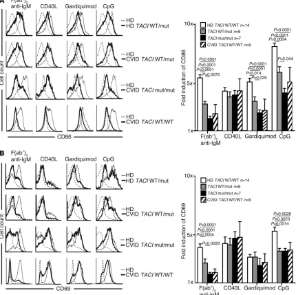
B cell activation after BCR, TLR7, and TLR9 stimulation is TACI gene dosage dependent. The high frequency of ANA-expressing new emigrant B cells in subjects with TACI mutations is reminiscent of IRAK4- and MYD88-deficient patients who display similar features, suggesting defective TLR functions in the presence of
mutated TACI molecules (23). We therefore assessed the in vitro activation of naive B cells from individuals with and without TACI
mutations; the cells were stimulated for 2 days through BCR, CD40, TLR7, or TLR9 (Figure 2). We found that independent of disease status, naive B cells carrying a single TACI mutation dis-played a diminished induction of B cell activation markers CD86 and CD69 when stimulated with F(ab′)2 anti-human IgM, which triggers BCRs, guardiquimod (TLR7 agonist), or CpG (TLR9 ago-nist). By contrast, B cells with two TACI mutations were nearly unresponsive to such stimulation as also evidenced by the defec-tive induction of the coactivation molecule ICOSL (Figure 2A and Supplemental Figure 1A). Similarly to adenosine deaminase (ADA) inhibition (25), TACI mutations imposed selective, not global, TLR7- and TLR9-dependent B cell activation defects because the induction of CD25 by TLR agonists was unaffected in B cells car-rying TACI mutations (Figure 2B and Supplemental Figure 1B). In addition, B cells with TACI mutations did not suffer from an intrinsic inability to upregulate CD86 because this molecule was normally induced after CD40 triggering (Figure 2A). Moreover, we observed B cell activation defects in CVID patients without TACI
mutations that were similar to the defects in B cells carrying TACI
mutations but with overall greater variability (Figure 2, A and B, Supplemental Figure 1, A and B, and refs. 3, 4). Thus, TACI plays an important and selective role in mediating BCR, TLR7, and TLR9 functions during B cell activation.
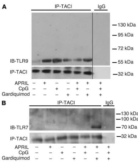
TACI interacts with TLR7 and TLR9 in B cells. The identification of TACI-dependent TLR7 and TLR9 functions in B cells and the rapid induction of TACI on the B cell surface after activation by these TLRs suggest an interplay between TACI, TLR7, and TLR9, since each uses MyD88 for intracellular signaling (25, 30, 31). To explore the potential interaction between TACI and these TLRs, we performed coprecipitation experiments using healthy donor splenic B cells under different stimulating conditions (Figure 3). We found that TACI coprecipitated with TLR9 and that the inter-action was enhanced upon activation of these receptors by their respective ligands APRIL and CpG, but less so by the TLR7 ligand gardiquimod (Figure 3A). TACI did not associate with the 120-kDa unactivated full-length TLR9, but associated only with its cleaved 65-kDa mature form created after the binding of its ligand, DNA (32). Hence, TACI only interacts with TLR9 after activation (Figure 3A). Coprecipitation of TACI and mature TLR9 was also observed in the human IgD+ E2E B cell line under similar activating condi-tions (Supplemental Figure 2). In contrast to TLR9, the interaction between TACI and TLR7 was only detected upon simultaneous acti-vation by APRIL and gardiquimod, respectively (Figure 3B). Once more, TACI did not bind to the 120-kDa unactivated full-length TLR7, but was found to bind only to its cleaved 75-kDa mature form, thereby demonstrating that the association of TACI with TLR7 follows TLR7 activation by its ligand (Figure 3B and ref. 33).
A role for TACI in potentiating TLR7 and TLR9 functions in B cells was further revealed by B cell activation experiments using TACI and TLR costimulations. Indeed, TACI triggering by its ligand APRIL enhanced TLR7- or TLR9-induced B cell prolifera-tion and IgM secreprolifera-tion in vitro, whereas APRIL alone did not elicit any responses (Supplemental Figure 3). The specificity of the syn-ergy between TACI and TLR7 or TLR9 was demonstrated by the addition of TACI-Ig, a decoy receptor for soluble APRIL and BAFF that blocked the enhanced B cell responses induced by APRIL and TLR agonists (Supplemental Figure 3 and ref. 33). The in vivo acti-vation of TACI likely relies upon the production of its ligands in Figure 1
Defective central B cell tolerance checkpoint in individuals carrying
TACI mutation(s). (A) Recombinant antibodies derived from new
emi-grant/transitional B cells from representative individuals were tested by ELISA for reactivity against dsDNA, insulin, and LPS (21). Antibodies were considered polyreactive when they reacted against all three anti-gens. Dashed lines show ED38 antibody–positive control, and solid lines show binding for each cloned recombinant antibody. Horizontal lines define the cutoff OD405 for positive reactivity. For each individual, the frequency of polyreactive and nonpolyreactive clones is summa-rized in pie charts, with the total number of antibodies tested indicated in the center. (B) The frequency of polyreactive new
emigrant/transi-tional B cells increased in all individuals carrying TACI mutation(s) and most CVID patients without TACI mutations, whereas the frequency of HEp-2–reactive new emigrant/transitional B cells only increased in patients with two mutated TACI alleles. (C) Autoreactive antibodies
expressed by new emigrant/transitional B cells from individuals carrying
TACI mutation(s) included clones with various nuclear staining patterns on HEp-2 cells. Original magnification, ×40. (D) The frequency of
anti-nuclear new emigrant/transitional B cells in individuals carrying TACI
research article
trans by non-B cells, including dendritic cells, since B cells did not seem to secrete substantial amounts of BAFF or APRIL (Supple-mental Figure 4 and refs. 34, 35). Hence, we identified ligand-de-pendent interactions between TACI and endosomal receptors TLR7 and TLR9, which further reveal that TACI plays an impor-tant role in mediating human B cell activation by these TLRs.
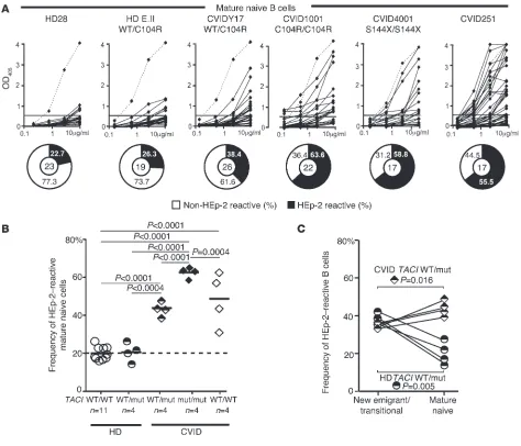
Peripheral B cell tolerance is defective in CVID patients. A second B cell tolerance checkpoint is responsible for the elimination of additional autoreactive B cells before they enter the CD10– C-D21+IgM+CD27–CD20+ mature naive B cell compartment (21). We
assessed the impact of TACI mutations on peripheral B cell toler-ance by testing HEp-2 cell lysates for the reactivity of recombinant antibodies cloned from mature naive B cells from healthy subjects and CVID patients with or without TACI mutations (Supplemen-tal Tables 19–35). We observed a significantly elevated frequency of HEp-2 reactive mature naive B cells in all tested CVID patients with one TACI mutation (38.5%–47.6%; P < 0.001), those with two
TACI mutations (58.5%–65%; P < 0.001), and those without TACI
mutations (28.6%–56%; P < 0.001) compared with healthy controls (16.0%–26.3%), demonstrating an impaired peripheral B cell toler-Figure 2
TACI mutation(s) result in selective defects for in vitro naive B cell activation. Purified naive B cells from representative individuals with and without
TACI mutation(s) (thick lines) displayed decreased CD86 (A) but mostly normal CD69 (B) induction compared with healthy controls (thin lines)
after 48 hours of stimulation with F(ab′)2 anti-IgM, CpG, and gardiquimod, but not CD40L. CVID patients without TACI mutation(s) (thick lines)
[image:5.585.78.508.88.516.2]ance checkpoint in these patients (Figure 4, A and B). Peripheral B cell tolerance defects were further evidenced by higher frequen-cies of polyreactive mature naive clones in CVID patients compared with healthy controls, with the highest frequency being observed in B cells from subjects with two TACI mutations that frequently displayed antinuclear reactivity (Supplemental Figure 5 and Supple-mental Figure 6, A and B). In contrast, between the new emigrant/ transitional and mature naive B cell developmental stages, healthy donors with one TACI mutation showed a significant reduction in both HEp-2–reactive (37.7% to 19.3%; P = 0.005) and polyreac-tive (24.9% to 18.2%; P = 0.046) clones (Figure 4 and Supplemental Figure 6B). We conclude that a single TACI mutation does not affect the peripheral B cell tolerance checkpoint, which is functional in all healthy subjects but defective in CVID patients.
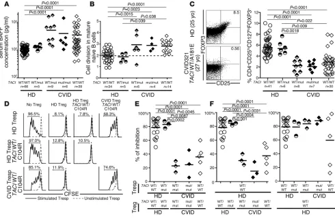
Peripheral B cell tolerance defects in CVID patients correlate with ele-vated plasma BAFF concentrations and decreased Treg frequencies. Defects in peripheral B cell tolerance have been correlated with elevated plasma BAFF concentrations and lower Treg frequencies, both features that have already been reported in CVID (7, 11, 12, 26–28). We found 4- to 5-fold increased elevations in plasma BAFF concentrations in CVID patients, contrasting with normal BAFF concentrations in healthy donors with a single TACI mutation (Figure 5A). Using a quantitative PCR–based assay for enumerat-ing κ-deletion recombination excision circles (KRECs) that esti-mates the number of divisions undergone by a B cell population (36), we found that mature naive B cells from CVID patients with and without TACI mutations displayed significantly increased homeostatic proliferation, with an average of 3.0 divisions in vivo compared with 1.9 divisions in B cells from healthy donors both with and without TACI mutations (Figure 5B). Both the defec-tive peripheral B cell tolerance checkpoint and the dysregulated homeostatic B cell proliferation observed in CVID patients were characteristic of FOXP3-deficient patients who do not have func-tional Tregs, suggesting a similarly altered Treg compartment in
CVID (28). Indeed, we found that the frequency of CD4+CD25hi CD127loFOXP3+ Tregs was decreased in most CVID patients, inde-pendent of the presence and/or number of TACI mutations, but not in healthy donors with a single TACI mutation (Figure 5C). Impaired Treg function in CVID patients was further suggested by their altered Ki67+CD45RO+CD25lo phenotype, also reminiscent of nonfunctional Tregs from FOXP3-deficient IPEX patients (Sup-plemental Figure 7, A and B, and ref. 28). Our direct assessment of Treg suppressive function in vitro demonstrated the impaired abil-ity of Tregs from CVID patients with and without TACI mutations to inhibit the proliferation of CFSE-labeled CD3+CD4+CD25– autologous and heterologous T responder (Tresp) cells (Figure 5, D and E). In contrast, Tregs from healthy donors with a single
TACI mutation were as suppressive as Tregs from controls with no TACI mutation, further demonstrating that TACI mutations are not inducing Treg defects per se (Figure 5, D–F). Thus, TACI
mutations are not responsible for the elevated plasma BAFF con-centrations, the low Treg frequencies, or the poor Treg suppressive function associated with the majority of CVID patients displaying a defective peripheral B cell tolerance checkpoint.
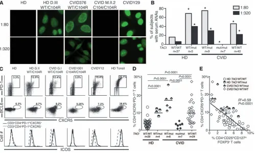
Circulating ANAs and BCL6+ Tfh cells are associated with a single TACI
mutation. CVID patients with a single TACI mutation are prone to developing autoimmune syndromes, whereas carrying two mutations seems to prevent autoimmunity (19, 20). To further investigate this apparent discrepancy, we assessed the plasma of healthy donors and CVID patients with or without TACI muta-tion(s) for IgG ANAs. In agreement with previous reports, 16.2% of healthy donors were found positive for low-titer (1:80 dilution) plasma ANAs, with very few displaying high ANA titers (5.4% at a dilution of 1:320) (37). In contrast, most CVID patients with one
[image:6.585.49.275.78.343.2]TACI mutation displayed low-titer plasma ANAs (1:80 dilution), and 30% of them had detectable high-titer ANAs (1:320) (Figure 6, A and B). We found that 40% of healthy donors carrying a sin-gle TACI mutation also frequently harbored significant high-titer Figure 3
TACI interacts with activated/cleaved TLR7 and TLR9. TACI associ-ates with activated/cleaved 65-kDa TLR9 (A) or 75-kDa TLR7 (B) after
the receptors triggered or cotriggered with their respective ligands. Immunoprecipitations were performed using human splenic B cells stimulated or not by the TACI ligand APRIL, TLR9 ligand CpG, and/or TLR7 ligand gardiquimod, as indicated below the blots. The thin black vertical line in A signifies the absence of a single lane from an
research article
ANAs, thereby suggesting a role for TACI in preventing autoanti-body secretion (Figure 6, A and B). Remarkably, ANAs were unde-tectable even at a low 1:80 titer in all seven CVID patients with two TACI mutations, revealing that the absence of functional TACI blocks autoreactive B cell activation (Figure 6, A and B).
Since the generation of IgG ANAs is considered T cell dependent, we analyzed effector T cell subsets potentially contributing to auto-antibody production, such as CXCR5+PD-1hiCD4+ circulating Tfh cells (38, 39). These T cells are rare in the peripheral blood of con-trol healthy donors without a TACI mutation and in CVID patients with two TACI mutations, comprising 3.7% and 2.1% of their CD4+ T cells, respectively (Figure 6, C and D). In contrast, we found a high average frequency of CXCR5+PD-1hiCD4+ T cells in healthy donors and CVID patients with a single TACI mutation as well as in some
[image:7.585.55.529.81.487.2]CVID patients without TACI mutations (healthy donor [HD] TACI WT/mut: 10.1%; CVID TACI WT/mut: 17.1%; and CVID: 9.9% of CD4+ cells) (Figure 6, C and D). These cells resembled tonsilar Tfh cells, which are normally localized in GCs, since they expressed intracellular BCL6, the master transcriptional regulator responsible for Tfh differentiation, as well as the coactivation molecules ICOS and CD40L (Figure 6C and data not shown) (40). Tfh cells were generally most prevalent in the peripheral blood of subjects who had the lowest frequencies of circulating Tregs, whereas in subjects with two TACI mutations, Tfh cells were nearly absent (Figure 6E). In addition, several cytokines secreted by Tfh cells, including IL-4 and IL-10, but not IL-21, were found at elevated concentrations in the plasma of CVID patients but not in that of healthy donors with one TACI mutation (Supplemental Figure 8). Hence, a single Figure 4
A defective peripheral B cell tolerance checkpoint in CVID patients. (A) Recombinant antibodies derived from mature naive B cells from subjects
or CVID patients with or without TACI mutation(s) were tested by ELISA for anti–HEp-2 cell reactivity. Dashed lines show the ED38-positive control (21). Horizontal lines define the cutoff OD405 for positive reactivity. For each individual, the frequencies of HEp-2–reactive and non–HEp-2–reactive clones are summarized in pie charts, with the number of antibodies tested indicated in the center. (B) The frequency of HEp-2–reactive mature
naive B cells was higher in all CVID patients than was observed in healthy donors with or without one TACI mutation. (C) The frequency of
TACI mutation correlates with an increased frequency of circulating CD4+CXCR5+PD-1hiICOShiBCL6+ Tfh cells and the production of high-titer IgG ANAs, thereby revealing an additional, more distal B cell tolerance breach often associated with autoimmune syn-dromes. In contrast, a second TACI mutation resulting in a lack of functional TACI counteracts the effects of a single TACI mutation, creating a scenario of scant circulating Tfh cells, absent ANAs, and a reduced susceptibility to autoantibody-mediated disease.
Discussion
We report herein several roles played by TACI mutations in the development of CVID. Previously, the association between TACI
mutation(s) and CVID was unclear because many people carrying
TACI mutations are healthy and without infectious susceptibility or decreased serum immunoglobulins (18, 41). However, we found that all individuals with TACI mutations failed to remove devel-oping autoreactive B cells in the marrow. This defective central B cell tolerance checkpoint is the first evidence of the full
pene-trance of TACI mutations in B cell development. This effect is not influenced by the nature of the mutation itself or the CVID status of the carrier. Indeed, healthy carriers with a single C104R
TACI mutation abrogating ligand binding displayed abnormalities similar to those in CVID patients carrying the A181E mutation affecting the TACI transmembrane domain. Likewise, patients homozygous for the allele encoding the C104R mutation showed profound B cell defects and elevated autoreactive B cell frequen-cies similar to those in the S144X/S144X individual in whom no TACI protein is expressed (8). Hence, various mutated TACI alleles encode similarly nonfunctional molecules. It remains to be deter-mined whether these mutations result in the expression of interfer-ing dominant-negative products or whether their impact on B cell activation and central selection is solely due to haploinsufficiency.
[image:8.585.51.541.84.405.2]How do TACI mutations affect the removal of developing autoreactive B cells? Previous studies have suggested that the regulation of central B cell tolerance is intrinsic to B cells and involves the sensing of self-antigens through binding with BCRs Figure 5
Increased plasma BAFF concentrations, homeostatic naive B cell proliferation, and an impaired Treg compartment are features common to CVID patients with and without TACI mutations. TACI mutations did not affect plasma BAFF concentrations (pg/ml) measured by ELISA (A) or mature
naive B cell expansion detected by KREC analysis (B). Both were greater in CVID patients compared with healthy controls. (C–F) CVID status,
but not TACI mutations, affected Treg frequency and function. (C) Dot plots represent CD4+ gated CD25hiFOXP3+ T cells of a healthy donor control
and an age-matched CVID patient with one TACI mutation. Scatter plots reveal that decreased CD4+CD25hiCD127loFOXP3+ Treg frequency only
correlated with CVID. yo, year-old. (D) Representative histograms of Treg-mediated suppression of autologous and heterologous CFSE-labeled
research article
and potentially TLRs (23). Our data from subjects with TACI
mutations further support such a model, as TACI is expressed primarily by B cells and regulates BCR, TLR7, and TLR9 func-tions. Indeed, B cells carrying a single TACI mutated allele display decreased CD86 upregulation in vitro when BCR, TLR7, or TLR9 are triggered, whereas B cells with two mutated TACI alleles fail to upregulate this costimulatory molecule. Interestingly, defec-tive CD86 induction after BCR and TLR9 stimulation as well as impaired TLR responses are features already reported for B cells from CVID patients and may represent one mechanism by which
TACI mutations favor the development of CVID (5, 42). TACI- deficient patients as well as subjects with a single TACI mutation share a similar defective central B cell tolerance checkpoint that is also characterized by a failure to remove antinuclear clones. The presence of antinuclear clones in the new emigrant/tran-sitional B cell compartment is reminiscent of IRAK4-deficient, MYD88-deficient, and ADA-deficient severe combined immune deficiency patients who have revealed the IRAK4/MYD88 and ADA pathways to be essential for the counterselection of such reactivity (23, 25). Since TLR7 and TLR9 bind nucleic acids and require IRAK4, MYD88, ADA, and TACI to function in B cells, these receptors may therefore play an important role in
establish-ing central tolerance against RNA- and DNA-containestablish-ing antigens. In addition, TACI’s intracellular domain binds MYD88 (30), and our data show that TACI can interact with both TLR7 and TLR9 upon coengagement of these receptors. Hence, polyreactive and antinuclear BCRs that bind self-antigens in the bone marrow may internalize and deliver them to endosomal compartments. TLR7 and TLR9 may then recognize RNA- and DNA-containing complexes, leading to TLR proteolytic activation and interac-tion with TACI, which in turn may allow the amplificainterac-tion of IRAK4/MYD88–dependent signals to mediate central tolerance mechanisms. We conclude that TACI plays an essential role in the establishment of central B cell tolerance in humans.
[image:9.585.45.544.83.382.2]TACI mutations potentially favor CVID development by impairing B cell activation. However, many individuals carrying a TACI muta-tion do not develop CVID (18, 41). By comparing such subjects with CVID patients with TACI mutation(s), we found that disease correlated with an abnormal peripheral B cell tolerance checkpoint independently of TACI mutations. Indeed, all asymptomatic individ-uals with a single TACI mutation managed to prevent the accumula-tion of autoreactive clones in their mature naive B cell compartment. This demonstrates that peripheral B cell tolerance can be established despite a defective central B cell tolerance checkpoint. The increased Figure 6
Heterozygous TACI mutations correlate with secreted ANAs and circulating Tfh cells. (A and B) ANAs with diverse nuclear staining patterns at low
(1:80) and high (1:320) dilutions were common in individuals with one TACI mutation, but were absent in patients with two mutated alleles. *P < 0.05 (statistical differences compared with healthy controls by χ2 testing). (C and D) Expanded CD4+PD-1hiCXCR5+ Tfh cells in the peripheral blood of
individuals with one, but not two, TACI mutations and some CVID patients without TACI mutations. In addition to PD-1 and CXCR5 expression, Tfh cells in carriers of a single TACI mutation were further evidenced by increased intracellular BCL6 expression in and increased cell surface ICOS expression on CD4+PD-1hiCXCR5+ T cells (solid line in lower panel of C) compared with CD4+PD-1loCXCR5– T cells (dashed line in lower panel
of C). (E) The frequency of circulating Tfh cells was inversely correlated with the frequency of circulating Tregs in enrolled subjects with or without
the development of an SLE-like autoimmune condition in TACI- deficient mice with the absence of autoimmune manifestations in CVID patients with two TACI mutations (54). On the other hand, as in Taci–/– mice, Tfh cells are enriched in CVID patients with a
single TACI mutation and likely contribute to the activation of autoreactive B cells through cell-cell interactions and the produc-tion of cytokines such as IL-4 and IL-10, leading to ANA secreproduc-tion. It is unknown at this point why Tfh cell frequency is increased in CVID patients with a single TACI mutation or without TACI
mutations, but their low Treg numbers and poor Treg suppres-sive function may fail to downregulate Tfh production in GCs (55, 56). Nevertheless, increased Tfh cells may favor autoreactive B cell activation in CVID patients and contribute to the develop-ment of autoimmune conditions characteristic of these patients.
In summary, TACI mutations favor CVID development by altering B cell activation after BCR, TLR7, and TLR9 stimula-tion. However, additional genetic or environmental factors are required for the development of CVID. Since TACI regulates the function of BCR, TLR7, and TLR9, which are likely involved in sensing self-antigens in the marrow, subjects with TACI muta-tion(s) not only suffer from B cell activation defects but also from an impaired removal of developing autoreactive B cells. B cells from CVID patients with a single TACI mutation may respond to self-antigen stimulation and receive additional T cell help due to the presence of Tfh cells, leading to autoantibody secretion and an increased risk for autoimmune syndromes. In contrast, autoreactive B cells from subjects with two mutated
TACI alleles lack the functional TACI necessary for B cell acti-vation, resulting in diminished immune and autoimmune humoral responses.
Methods
Patients. We obtained peripheral blood from 21 individuals with TACI
mutations including 9 CVID patients with one TACI mutation and
7 immune-deficient patients with two TACI mutations, 6 with CVID,
and 1 with hypogammaglobulinemia (Supplemental Table 1). Patient A.II.1, a TACI S144X homozygote who has a complete absence of mea-surable TACI-specific mRNA and protein, has no increased suscep-tibility to infections and does not require Ig replacement therapy, yet is nonetheless hypogammaglobulinemic (serum IgG 410 mg/dl) (8). Although not a classical CVID phenotype, this patient was grouped
here with other CVID patients carrying two TACI mutations to
sim-plify subject categorization. Healthy relatives of these patients made up
the remaining 5 subjects; each carried one TACI mutation. TACI alleles
present in our study cohort included common missense variants asso-ciated with CVID (C104R and A181E) as well as rare mutations such as insertions (204insA, 571insG) and truncations (S144X and S194X). For purposes of comparison, we obtained peripheral blood samples
from 50 healthy controls and 50 CVID patients without TACI
muta-tions. The study groups were controlled for age and sex. In addition, healthy controls analyzed for their autoreactive B cell frequencies were previously reported and included a 50-year-old male of mixed European descent previously splenectomized after accidental blunt-force trauma (HD28) (25). We also included HD29, the 29-year-old healthy brother
of CVID patient CVID332. All subjects were genotyped at the TACI gene
loci by Sanger sequencing using published primer sets (exons 1–4) (8).
For exon 5, the following primer pair was used: 5′
-CCTAGTGCAGGGC-CAGGCCTG-3′ and 5′-CCGACCTCCTGCTCTATCT-3′. All samples
were collected in accordance with Yale University’s IRB-approved protocol HIC0906005336.
frequency of autoreactive mature naive B cells in patients with auto-immune diseases such as rheumatoid arthritis, lupus erythema-tosus, and type 1 diabetes — all of whom display defective central B cell tolerance — is therefore likely due to a defective peripheral B cell tolerance checkpoint and not just an accumulation in the periph-ery of autoreactive clones produced by the marrow (24, 43, 44). The defect in peripheral tolerance of CVID patients is associated with an impaired Treg compartment, which plays an important role in reg-ulating this checkpoint (28). In addition, decreased Treg functions may also lead to increased proliferative events in the mature naive B cell compartment of CVID patients compared with healthy donors with or without TACI mutations (28). The elevated BAFF concentra-tion in the plasma of CVID patients may also favor B cell homeo-static expansion and provide survival signals to autoreactive clones, thereby preventing their elimination in the periphery (14, 26–28, 45). Hence, one TACI mutation is not sufficient to induce the develop-ment of CVID and is not responsible for the defective peripheral B cell tolerance checkpoint observed in CVID patients. It is unclear at this point whether another susceptibility gene is responsible for CVID development, potentially affecting Treg function, or whether nongenetic environmental factors are more important in the development of this disease.
It was previously reported that CVID patients with a single TACI
mutation are prone to autoimmune cytopenias, whereas patients devoid of functional TACI are protected from autoimmunity (20). This apparent discrepancy was initially puzzling, as we found that subjects with two mutated TACI alleles had increased frequencies of autoreactive clones in all naive B cell compartments compared with CVID patients with a single TACI mutation. However, in the absence of functional TACI, this increase in autoreactive B cells is accompanied by profound impairments of B cell activation after BCR, TLR7, and TLR9 triggering that are significantly worse than in subjects with a single TACI mutation. Moreover, TLRs play an essential role in the development of autoimmunity (46). Hence, severe TLR function impairment in TACI-deficient individuals is likely to be protective against autoimmunity despite profoundly defective autoreactive B cell counterselection, leaving these indi-viduals with an immunodeficient phenotype. In agreement with this hypothesis, we could detect ANAs in the plasma of CVID patients and asymptomatic individuals with a single TACI muta-tion, but not in subjects with two mutated TACI alleles.
research article
KREC assay. The ratio of KREC joints (signal joint) to the Jκ-Cκ recom-bination genomic joints (coding joint) was determined as previously described (36). In brief, genomic DNA was isolated from sorted B cell frac-tions by lysing cell pellets in 10 mM Tris-HCl, pH 8.0, containing 100 μg/ml proteinase K (Roche), incubating for 1 hour at 56°C, and heat inactivating the enzyme at 95°C for 10 minutes. Two separate RT-PCR reactions were performed, one reaction to amplify the signal joint and another to amplify the coding joint, as previously detailed. The number of cell divisions was calculated by subtracting the cycle threshold of the PCR detecting the cod-ing joint from that detectcod-ing the signal joint.
In vitro Treg suppression assay. CD4+ T cells were enriched using
the EasySep Human CD4+T cell enrichment kit (STEMCELL
Tech-nologies). CD4+CD25hiCD127lo/– Tregs were sorted by flow
cytom-etry, whereas CD3+CD4+CD25– Tresp cells were obtained after
the depletion of CD25+ cells with anti-human CD25 microbeads
(Miltenyi Biotec) and then labeled with CellTrace CFSE
(Invi-voGen) at 5 μM. Treg and Tresp cells were cocultured at a 1:1 ratio
in the presence of beads loaded with anti-CD2, anti-CD3, and anti-CD28 (Treg suppression inspector human; Miltenyi Biotec). On days 3.5 to 4.5, cocultures were stained for viability with the LIVE/DEAD kit (Invitrogen), and proliferation of the viable Tresp was analyzed by CFSE dilution.
Cytokine detection. Plasma BAFF concentrations were determined by ELISA according to the manufacturer’s instructions (R&D Systems). Cytokines (IL-4, IL-10) in plasma were measured with the High Sensitiv-ity Human Cytokine Magnetic Bead Kit (Millipore) using a Luminex 200. IL-21 was measured by ELISA with the Human IL-21 ELISA Ready-Set-Go Kit (eBioscience).
Statistics. Statistical analysis was performed using GraphPad Prism soft-ware, version 5.0 (GraphPad Software). Differences between groups of research subjects were analyzed for statistical significance with unpaired
or paired (when within groups), two-tailed Student’s t tests except for
plasma indirect immunofluorescence assay comparisons that used χ2
testing. For these statistical tests, α was set to α = 0.05; accordingly, a
P value less than or equal to 0.05 was considered significant. The
rela-tionship between Tfh cells and Tregs was statistically analyzed using lin-ear regression analysis.
Acknowledgments
We thank J. Bussel, F. Kantor, P. Askenase, C. Randolf, J. Sproviero, F. Hsu, C. Price, and O. Pagovich for providing blood samples from CVID patients, and S. Rudchenko, L. Devine, and C. Wang for cell sorting. This work was supported by grants from the NIH-NIAID (AI071087, AI082713, and AI095848, to E. Meffre, and AI061093, to E. Meffre, A. Cerutti, and C. Cunningham-Rundles); and the NIH-NICHD (K12HD0141401-10, to N. Romberg); and by a CIS/ Talecris Fellowship Award (to N. Romberg). T. Kinnunen was supported by the Sigrid Juselius Foundation, the Finnish Med-ical Foundation, and the Saastamoinen Foundation. L. Menard was supported by NIH-NIAID grant T32 AI089704. T. Cantaert received support from the Rubicon program, Netherlands Orga-nization for Scientific Research, and H. Morbach received support from the German Research Foundation (DFG, MO 2160/2-1).
Received for publication March 14, 2013, and accepted in revised form July 25, 2013.
Address correspondence to: Eric Meffre, Yale University School of Medicine, 300 George Street, New Haven, Connecticut 06511, USA. Phone: 203.737.4535; Fax: 203.785.7903; E-mail: [email protected].
Cell staining and sorting, cDNA, RT-PCR, antibody production, ELISAs, and indirect fluorescence assays. Peripheral B cells were purified from the periph-eral blood of research subjects by positive selection using CD20 magnetic
beads (Miltenyi Biotec). Single CD19+CD21loCD10+IgMhiCD27– new
emi-grant/transitional and CD19+CD21+CD10–IgM+CD27– peripheral mature
naive B cells from patients and control donors were sorted on a FACSVan-tage cell sorter (BD) into 96-well PCR plates. RT-PCR reactions, primer sequences, cloning strategy, expression vectors, recombinant antibody expression, recombinant antibody purification, and recombinant antibody reactivity determination were as previously described (21). For indirect immunofluorescence assays, HEp-2 cell–coated slides (Bion Enterprises) were incubated in a moist chamber at room temperature with patient plasma samples at 1:80 and 1:320 dilutions in PBS according to the manufacturer’s instructions. FITC-conjugated goat anti-human IgG was used as a detection reagent. The following antibodies were used for flow cytometric stainings: CD19 APC-Cy7, CD27 PerCP-Cy5.5, CD10 PE-Cy7, IgM FITC, CD21 APC, CD25 FITC, CD69 PE, CD80 FITC, CD86 PE, CD4 APC-Cy7, CD25 PE-Cy7, CD127 PerCP-Cy5.5, CD45RO Pacific Blue, TACI PE, CD303 FITC, CD14 Pacific Blue, CD4 APC-Cy7, CXCR5 PerCP-Cy5.5, PD-1 PE-Cy7, ICOSL PE, CD25 PE, and CD40 APC (all from Biolegend), CD3 eFluor 605NC, BCL6 PE (both from eBioscience), and CD21 BD Horizon V450 (BD). Intracellular staining for FOXP3 Alexa Fluor 488 (clone PCH101; eBioscience) was per-formed using the FOXP3/Transcription Factor Staining Buffer Set (eBiosci-ence) in accordance with the manufacturer’s instructions.
B cell activation and proliferation. Naive B cells were enriched from the blood of healthy donors by negative selection using the Naive B Cell Isolation Kit II (Miltenyi Biotec) and plated for 48 hours at 150,000–200,000 cells per well in a 96-well plate in RPMI 10% FBS and either 2.5 μg/ml polyclonal F(ab′)2 rabbit
anti-human IgM (Jackson ImmunoResearch), multimeric soluble
recombi-nant human CD40L 1.0 μg/ml (Alexis Biochemicals), 0.5 μg/ml CpG (TLR9
agonist; Invitrogen), or 1.0 μg/ml gardiquimod (TLR7 agonist; InvivoGen). In
proliferation and antibody secretion experiments, total human splenic B cells isolated by negative selection using EasySep were stimulated for 5 days with CpG and gardiquimod in the presence or absence of 500 ng/ml APRIL (R&D
Systems). APRIL was blocked with 30 μg/ml of TACI-Ig (Ancell). Mouse IgG1
with irrelevant binding activity was used as a control for TACI-Ig. Cell prolif-eration was assessed by CFSE staining using the CellTrace CFSE Cell Prolif-eration Kit (Invitrogen) according to the manufacturer’s instructions. Super-natants were collected and analyzed to test the presence of BAFF and APRIL using a soluble human BAFF ELISA Kit (Adipogen) and a LEGEND MAX Human APRIL/TNFSF13 ELISA Kit (BioLegend). IgM secretion was detected by ELISA. In brief, M96-Nunc ELISA plates were coated with anti-human IgM (SouthernBiotech). After blocking with 1% BSA in PBS, samples were added to the plate overnight. Purified anti IgM-HRP antibody (Cappel) and TMB sub-strate solution (BD) were used according to the manufacturer’s instructions.
Coimmunoprecipitation experiments. FACS-sorted human CD19+ splenic
B cells were treated with 0.2 μg/ml recombinant human APRIL (R&D
Sys-tems), 0.5 μg/ml CpG (Invitrogen), and/or 1.0 μg/ml gardiquimod
(Invi-voGen) for 2 hours. For each condition, 107 cells were used. Cells were
homogenized and solubilized in lysis buffer (50 mM Tris-HCl, pH 7.5, 150 mM NaCl, 1 mM EDTA, 1 mM EGTA, 0.5% Triton, 0.2 mg/ml BSA, and protease inhibitors) using a Dounce homogenizer. An equal amount of each protein lysate was incubated with anti-TACI biotinylated antibody (Abcam)
overnight at 4°C, followed by incubation with 50 μl of
1. Al-Herz W, et al. Primary immunodeficiency dis-eases: an update on the classification from the international union of immunological societies expert committee for primary immunodeficiency. Front Immunol. 2011;2:54.
2. Sanford JP, Favour CB, Tribeman MS. Absence of serum gamma globulins in an adult. N Engl J Med. 1954; 250(24):1027–1029.
3. Bryant A, Calver NC, Toubi E, Webster AD, Farrant J. Classification of patients with common variable immunodeficiency by B cell secretion of IgM and IgG in response to anti-IgM and interleukin-2. Clin Immunol Immunopathol. 1990;56(2):239–248. 4. Yu JE, et al. Toll-like receptor 7 and 9 defects in
common variable immunodeficiency. J Allergy Clin Immunol. 2009;124(2):349–356.
5. Denz A, Eibel H, Illges H, Kienzle G, Schlesier M, Peter HH. Impaired up-regulation of CD86 in B cells of “type A” common variable immu-nodeficiency patients. Eur J Immunol. 2000; 30(4):1069–1077.
6. Horn J, et al. Decrease in phenotypic regulatory T cells in subsets of patients with common vari-able immunodeficiency. Clin Exp Immunol. 2009; 156(3):446–454.
7. Arumugakani G, Wood PM, Carter CR. Fre-quency of Treg cells is reduced in CVID patients with autoimmunity and sple-nomegaly and is associated with expanded CD21lo B lymphocytes. J Clin Immunol. 2010; 30(2):292–300.
8. Salzer U, et al. Mutations in TNFRSF13B encod-ing TACI are associated with common variable immunodeficiency in humans. Nat Genet. 2005; 37(8):820–828.
9. Castigli E, et al. TACI is mutant in common vari-able immunodeficiency and IgA deficiency. Nat Genet. 2005;37(8):829–834.
10. Wu Y, et al. Tumor necrosis factor (TNF) receptor superfamily member TACI is a high affinity recep-tor for TNF family members APRIL and BLyS. J Biol Chem. 2000;275(45):35478–35485.
11. Knight AK, Radigan L, Marron T, Langs A, Zhang L, Cunningham-Rundles C. High serum levels of BAFF, APRIL, and TACI in common variable immu-nodeficiency. Clin Immunol. 2007;124(2):182–189. 12. Kreuzaler M, et al. Soluble BAFF levels inversely
correlate with peripheral B cell numbers and the expression of BAFF receptors. J Immunol. 2012; 188(1):497–503.
13. Lesley R, et al. Reduced competitiveness of autoan-tigen-engaged B cells due to increased dependence on BAFF. Immunity. 2004;20(4):441–453. 14. Thien M, et al. Excess BAFF rescues self-reactive
B cells from peripheral deletion and allows them to enter forbidden follicular and marginal zone niches. Immunity. 2004;20(6):785–798.
15. Garibyan L, Lobito AA, Siegel RM, Call ME, Wucherpfennig KW, Geha RS. Dominant-negative effect of the heterozygous C104R TACI mutation in common variable immunodeficiency (CVID). J Clin Invest. 2007;117(6):1550–1557.
16. Chinen J, et al. Transmembrane activator and CAML interactor (TACI) haploinsufficiency results in B-cell dysfunction in patients with Smith- Magenis syndrome. J Allergy Clin Immunol. 2011; 127(6):1579–1586.
17. Lee JJ, et al. The C104R mutant impairs the function of transmembrane activator and calcium modulator and cyclophilin ligand interactor (TACI) through haploinsufficiency. J Allergy Clin Immunol. 2010; 126(6):e1234–e1232.
18. Pan-Hammarstrom Q, et al. Reexamining the role of TACI coding variants in common variable
immunodeficiency and selective IgA deficiency. Nat Genet. 2007;39(4):429–430.
19. Zhang L, et al. Transmembrane activator and calcium-modulating cyclophilin ligand inter-actor mutations in common variable immuno-deficiency: clinical and immunologic outcomes in heterozygotes. J Allergy Clin Immunol. 2007; 120(5):1178–1185.
20. Salzer U, et al. Relevance of biallelic versus monoal-lelic TNFRSF13B mutations in distinguishing dis-ease-causing from risk-increasing TNFRSF13B vari-ants in antibody deficiency syndromes. Blood. 2009; 113(9):1967–1976.
21. Wardemann H, Yurasov S, Schaefer A, Young JW, Meffre E, Nussenzweig MC. Predominant autoan-tibody production by early human B cell precur-sors. Science. 2003;301(5638):1374–1377. 22. Ng Y-S, Wardemann H, Chelnis J,
Cunningham-Run-dles C, Meffre E. Bruton’s tyrosine kinase (Btk) is essential for human B cell tolerance. J Exp Med. 2004; 200(7):927–934.
23. Isnardi I, et al. IRAK-4- and MyD88-dependent pathways are essential for the removal of develop-ing autoreactive B cells in humans. Immunity. 2008; 29(5):746–757.
24. Menard L, et al. The PTPN22 allele encoding an R620W variant interferes with the removal of developing autoreactive B cells in humans. J Clin Invest. 2011;121(9):3635–3644.
25. Sauer AV, Morbach H, Brigida I, Ng YS, Aiuti A, Meffre E. Defective B cell tolerance in adenosine deaminase deficiency is corrected by gene therapy. J Clin Invest. 2012;122(6):2141–2152.
26. Herve M, et al. CD40 ligand and MHC class II expression are essential for human peripheral B cell tolerance. J Exp Med. 2007;204(7):1583–1593. 27. Meyers G, et al. Activation-induced cytidine
deaminase (AID) is required for B-cell toler-ance in humans. Proc Natl Acad Sci U S A. 2011; 108(28):11554–11559.
28. Kinnunen T, et al. Accumulation of peripheral auto-reactive B cells in the absence of functional human regulatory T cells. Blood. 2013;121(9):1595–1603. 29. Romberg N, Ng YS, Cunningham-Rundles C,
Meffre E. Common variable immunodeficiency patients with increased CD21-/lo B cells suffer from altered receptor editing and defective central B cell tolerance. Blood. 2011;118(22):5977–5978. 30. He B, et al. The transmembrane activator TACI
trig-gers immunoglobulin class switching by activating B cells through the adaptor MyD88. Nat Immunol. 2010; 11(9):836–845.
31. Kawai T, Adachi O, Ogawa T, Takeda K, Akira S. Unresponsiveness of MyD88-deficient mice to endotoxin. Immunity. 1999;11(1):115–122. 32. Ewald SE, et al. The ectodomain of Toll-like
recep-tor 9 is cleaved to generate a functional receprecep-tor. Nature. 2008;456(7222):658–662.
33. Ewald SE, Engel A, Lee J, Wang M, Bogyo M, Barton GM. Nucleic acid recognition by Toll-like receptors is coupled to stepwise processing by cathepsins and asparagine endopeptidase. J Exp Med. 2011; 208(4):643–651.
34. Moore PA, et al. BLyS: member of the tumor necrosis factor family and B lymphocyte stimulator. Science. 1999;285(5425):260–263.
35. Dillon SR, Gross JA, Ansell SM, Novak AJ. An APRIL to remember: novel TNF ligands as therapeutic tar-gets. Nat Rev Drug Discov. 2006;5(3):235–246. 36. van Zelm MC, Szczepanski T, van der Burg M, van
Dongen JJ. Replication history of B lymphocytes reveals homeostatic proliferation and extensive antigen-induced B cell expansion. J Exp Med. 2007; 204(3):645–655.
37. Tan EM, et al. Range of antinuclear antibodies in “healthy” individuals. Arthritis Rheum. 1997; 40(9):1601–1611.
38. Mietzner B, et al. Autoreactive IgG memory antibodies in patients with systemic lupus ery-thematosus arise from nonreactive and polyre-active precursors. Proc Natl Acad Sci U S A. 2008; 105(28):9727–9732.
39. Morita R, et al. Human blood CXCR5(+)CD4(+)
T cells are counterparts of T follicular cells and contain specific subsets that differentially support antibody secretion. Immunity. 2011;34(1):108–121. 40. Nurieva RI, et al. Bcl6 mediates the develop-ment of T follicular helper cells. Science. 2009; 325(5943):1001–1005.
41. Martinez-Gallo M, Radigan L, Almejún MB, Martínez-Pomar N, Matamoros N, Cunning-ham-Rundles C. TACI mutations and impaired B-cell function in subjects with CVID and healthy heterozygotes. J Allergy Clin Immunol. 2013;131(2):468–476.
42. Cunningham-Rundles C, Radigan L, Knight AK, Zhang L, Bauer L, Nakazawa A. TLR9 activation is defective in common variable immune deficiency. J Immunol. 2006;176(3):1978–1987.
43. Samuels J, Ng YS, Coupillaud C, Paget D, Meffre E. Human B cell tolerance and its failure in rheuma-toid arthritis. Ann N Y Acad Sci. 2005;1062:116–126. 44. Yurasov S, et al. Defective B cell tolerance checkpoints in systemic lupus erythematosus. J Exp Med. 2005; 201(5):703–711.
45. Mackay F, et al. Mice transgenic for BAFF develop lymphocytic disorders along with autoimmune manifestations. J Exp Med. 1999;190(11):1697–1710. 46. Sadanaga A, et al. Protection against autoimmune nephritis in MyD88-deficient MRL/lpr mice. Arthritis Rheum. 2007;56(5):1618–1628.
47. Grimbacher B, et al. Homozygous loss of ICOS is associated with adult-onset common variable immu-nodeficiency. Nat Immunol. 2003;4(3):261–268. 48. Bossaller L, et al. ICOS deficiency is associated with
a severe reduction of CXCR5+CD4 germinal center Th cells. J Immunol. 2006;177(7):4927–4932. 49. Warnatz K, et al. Human ICOS deficiency
abro-gates the germinal center reaction and provides a monogenic model for common variable immuno-deficiency. Blood. 2006;107(8):3045–3052. 50. Borriello F, et al. B7-1 B7-2 have overlapping,
criti-cal roles in immunoglobulin class switching germi-nal center formation. Immunity. 1997;6(3):303–313. 51. Mak TW, et al. Costimulation through the induc-ible costimulator ligand is essential for both T helper and B cell functions in T cell-dependent B cell responses. Nat Immunol. 2003;4(8):765–772. 52. Salek-Ardakani S, et al. B cell-specific expression
of B7-2 is required for follicular Th cell function in response to vaccinia virus. J Immunol. 2011; 186(9):5294–5303.
53. Ou X, Xu S, Lam KP. Deficiency in TNFRSF13B (TACI) expands T-follicular helper germinal center B cells via increased ICOS-ligand expression but impairs plasma cell survival. Proc Natl Acad Sci U S A. 2012; 109(38):15401–15406.
54. Seshasayee D, Valdez P, Yan M, Dixit VM, Tumas D, Grewal IS. Loss of TACI causes fatal lymphop-roliferation and autoimmunity, establishing TACI as an inhibitory BLyS receptor. Immunity. 2003; 18(2):279–288.
55. Chung Y, et al. Follicular regulatory T cells express-ing Foxp3 and Bcl-6 suppress germinal center reac-tions. Nat Med. 2011;17(8):983–988.