Onset of autoimmune lymphoproliferative
syndrome (ALPS) in humans as a consequence
of genetic defect accumulation
Aude Magerus-Chatinet, … , Alain Fischer, Frédéric
Rieux-Laucat
J Clin Invest. 2011;121(1):106-112. https://doi.org/10.1172/JCI43752.
Autoimmune diseases develop in approximately 5% of humans. They can arise when
self-tolerance checkpoints of the immune system are bypassed as a consequence of inherited
mutations of key genes involved in lymphocyte activation, survival, or death. For example,
autoimmune lymphoproliferative syndrome (ALPS) results from defects in self-tolerance
checkpoints as a consequence of mutations in the death receptor–encoding gene TNF
receptor superfamily, member 6 (TNFRSF6; also known as FAS). However, some mutation
carriers remain asymptomatic throughout life. We have now demonstrated in 7 ALPS
patients that the disease develops as a consequence of an inherited TNFRSF6
heterozygous mutation combined with a somatic genetic event in the second TNFRSF6
allele. Analysis of the patients’ CD4
–CD8
–(double negative) T cells — accumulation of
which is a hallmark of ALPS — revealed that in these cells, 3 patients had somatic
mutations in their second TNFRSF6 allele, while 4 patients had loss of heterozygosity by
telomeric uniparental disomy of chromosome 10. This observation provides the molecular
bases of a nonmalignant autoimmune disease development in humans and may shed light
on the mechanism underlying the occurrence of other autoimmune diseases.
Research Article
Find the latest version:
Onset of autoimmune lymphoproliferative
syndrome (ALPS) in humans as a
consequence of genetic defect accumulation
Aude Magerus-Chatinet,1 Bénédicte Neven,1,2 Marie-Claude Stolzenberg,1 Cécile Daussy,1
Peter D. Arkwright,3 Nina Lanzarotti,1 Catherine Schaffner,1 Sophie Cluet-Dennetiere,4
Filomeen Haerynck,5 Gérard Michel,6 Christine Bole-Feysot,7 Mohammed Zarhrate,7
Isabelle Radford-Weiss,8 Serge P. Romana,8 Capucine Picard,2,9,10
Alain Fischer,1,2,9 and Frédéric Rieux-Laucat1,2
1INSERM U768, Université Paris Descartes, Hôpital Necker-Enfants Malades, Paris, France. 2Unité d’Immunologie et d’Hématologie Pédiatrique,
Assistance Publique des Hôpitaux de Paris, Paris, France. 3Royal Manchester Children’s Hospital, University of Manchester, Manchester, United Kingdom. 4Service d’Hématologie du Centre Hospitalier de Compiègne, Compiègne, France. 5University Hospital Ghent, Belgium. 6Service d’Hématologie Pédiatrique,
Hôpital des Enfants de La Timone, Marseille, France. 7Genomic Core Facility, Imagine Fondation, Hôpital Necker-Enfants Malades, Paris, France. 8Service de Cytogénétique, Hôpital Necker-Enfants Malades, Paris, France. 9Centre d’Etude des Déficits Immunitaires,
Assistance Publique des Hôpitaux de Paris, Paris, France. 10INSERM U980, Université Paris Descartes, Paris, France.
Autoimmune diseases develop in approximately 5% of humans. They can arise when self-tolerance checkpoints
of the immune system are bypassed as a consequence of inherited mutations of key genes involved in
lympho-cyte activation, survival, or death. For example, autoimmune lymphoproliferative syndrome (ALPS) results
from defects in self-tolerance checkpoints as a consequence of mutations in the death receptor–encoding gene
TNF receptor superfamily, member 6 (
TNFRSF6
; also known as
FAS
). However, some mutation carriers remain
asymptomatic throughout life. We have now demonstrated in 7 ALPS patients that the disease develops as a
consequence of an inherited
TNFRSF6
heterozygous mutation combined with a somatic genetic event in the
second
TNFRSF6
allele. Analysis of the patients’ CD4
–CD8
–(double negative) T cells — accumulation of which
is a hallmark of ALPS — revealed that in these cells, 3 patients had somatic mutations in their second
TNFRSF6
allele, while 4 patients had loss of heterozygosity by telomeric uniparental disomy of chromosome 10. This
observation provides the molecular bases of a nonmalignant autoimmune disease development in humans
and may shed light on the mechanism underlying the occurrence of other autoimmune diseases.
Introduction
Autoimmune lymphoproliferative syndrome (ALPS, also known as lymphoproliferative syndrome with autoimmunity or Canale-Smith syndrome) (1) is a human disease characterized by benign lymphoproliferation (splenomegaly and/or diffuse lymphadenop-athy), and elevated serum immunoglobulins (IgG and IgA), plasma IL-10 levels (2, 3), and FAS-L levels. (4), accumulation in the blood and lymphoid organs of CD4–CD8–TCRαβ+ T cells (also referred
to as double-negative [DN] T cells), susceptibility to malignant conditions (5), and autoimmune manifestations (6, 7).
Mutations in the TNF receptor superfamily, member 6 (TNFRSF6) gene (encoding FAS, also known as the CD95 receptor) are the most common cause of ALPS (8, 9). Patients carrying a homozy-gous TNFRSF6 mutation have an early-onset severe phenotype, whereas subjects with heterozygous TNFRSF6 mutations present ALPS of variable intensity. T cells from patients with homozygous or heterozygous TNFRSF6 mutations, respectively, exhibit a com-plete or partial functional impairment in an in vitro FAS-induced apoptosis assay. Moreover, somatic heterozygous mutations in
TNFRSF6 in patients with a clinical ALPS phenotype were recently described (10, 11). In our experimental conditions, activated T cells showed normal sensitivity to FAS-induced apoptosis in vitro (11). The inability to detect an apoptosis defect was due to
spontane-ous in vitro apoptosis of the mutant T cells. However, more than 80% of these patients’ DN T cells were mutated. These patients dis-played mosaic expression of a somatic TNFRSF6 mutation, which provided the affected cells a selective advantage and accounted for the observed lymphoproliferation and autoimmunity.
However, heterozygous germline TNFRSF6 mutations are not always associated with clinical expression (partial clinical pen-etrance), since some mutation-positive relatives (MPRs) remain asymptomatic despite an in vitro FAS-induced apoptosis impair-ment on their T cells. This observation led us to postulate that a second event is necessary for disease expression in patients with
TNFRSF6 mutations when an incomplete penetrance is observed. Here, we report that the combination of a germline TNFRSF6
mutation and a somatic event impairing the second TNFRSF6
allele can account for the onset of clinical phenotype in ALPS.
Results
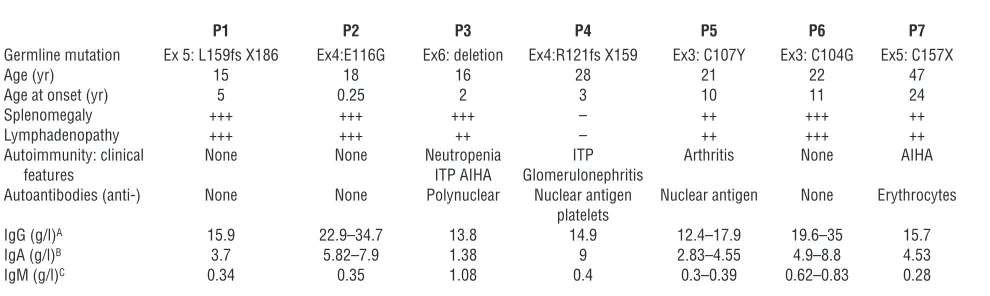
Biological characteristics of the 7 ALPS patients and their asymptomatic relatives. Within the cohort of ALPS patients with heterozygous
TNFRSF6 mutation studied in our institution (n = 87), 16% (n = 14) of the germline mutations affected the extracellular domain (ECD) of TNFRSF6. The clinical penetrance has been found to be much lower for heterozygous ECD mutations (30% of carriers developed ALPS) than for heterozygous intracellular domain (ICD) mutations (80% of carriers developed ALPS) (12, 13). This observation suggests that additional events are necessary for ALPS disease expression in Conflict of interest: The authors have declared that no conflict of interest exists.
research article
The Journal of Clinical Investigation http://www.jci.org Volume 121 Number 1 January 2011 107
carriers of TNFRSF6 mutations with incomplete clinical penetrance. In order to search for these additional events, we analyzed the avail-able T cells from 7 patients with the typical clinical symptoms of ALPS associated with monoallelic mutations affecting the ECD of
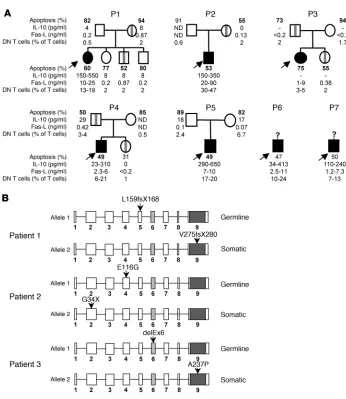
TNFRSF6 (Table 1) and compared with asymptomatic family car-riers in 4 cases. We first quantified ALPS markers: the percentage of DN T cells, plasma FAS-L, and IL-10 concentrations (2, 4, 14) (Figure 1A). In strict agreement with the clinical phenotype, all 7 patients presented markedly elevated numbers of DN T cells and plasma FAS-L and IL-10 concentrations, whereas their asymptom-atic MPRs carrying the same TNFRSF6 mutations exhibited normal DN T cell percentages and plasma IL-10 concentration and little or no increase in the plasma FAS-L concentration.
For all 7 patients, FAS-mediated T cell apoptosis was lower than for controls (Figure 1A). When simultaneously tested, we found that FAS-mediated apoptosis of patients’ T cells and their asymp-tomatic MPRs had similarly low apoptosis levels; this contrasted with the results for nonmutated relatives (Figure 1A). Since the magnitude of the FAS-mediated apoptosis defect was similar in symptomatic and asymptomatic carriers of TNFRSF6 mutations, we postulated that any additional causative events did not directly have an impact on the FAS-induced apoptosis as assayed in vitro. This result was reminiscent of ALPS cases carrying somatic
TNFRSF6 mutations but which were associated with normal FAS-mediated T cell apoptosis in vitro (11).
Somatic mutations of TNFRSF6 in 3 patients with heterozygous germline mutations. Based on the literature description of ALPS sufferers carry-ing a somatic TNFRSF6 mutation, which was predominantly found in DN T cells, we sequenced the TNFRSF6 gene in sorted DN T cells from each of the 7 patients. In addition to the previously identi-fied germline TNFRSF6 mutation, we found a second heterozygous
TNFRSF6 mutation in the DN T cells from patients 1, 2, and 3 (Fig-ure 1B). In patient 1, a base insertion in exon 9 had created a prema-ture stop codon at position 280. In patient 2, a nonsense mutation was found in exon 2 (G34X). In patient 3, a missence mutation was found in exon 9 (A237P). These somatic heterozygous TNFRSF6
mutations were present in the second allele, since the cloned PCR products from cDNA amplified from these patients’ DN T cells exhibited only 1 of the 2 TNFRSF6 mutations (data not shown).
The proportion of cells carrying the somatic mutation was estab-lished in several lineages for patients 1 and 2. In patient 1, the second mutation was found only in DN T cells and not in either single-positive T cells or epithelial cells (sensitivity threshold: 2.5% of cells) (Supplemental Table 1; supplemental material available online with this article; doi:10.1172/JCI43752DS1). It was there-fore impossible to determine at which stage of lymphocyte differen-tiation the mutation had occurred. In patient 2, the mutation was found in 7%–35% of T cells, B cells, NK cells, and monocytes but also in buccal epithelial cells — indicating that the somatic mutation had occurred very early in embryogenesis. In the other 4 patients,
TNFRSF6 sequencing did not highlight any second heterozygous mutation but revealed only the mutated allele in DN T cells (not shown), suggesting a somatic loss of the wild-type allele.
Loss of the wild-type TNFRSF6 allele in 4 patients. In order to identify the molecular basis of the loss of the wild-type allele in DN T cells from patients 4 to 7, we checked for single-nucleotide polymor-phisms at the TNFRSF6 locus in DNA from DN T cells and from CD4+ T cells. In all 4 patients, DNA from the CD4+ T cells
dis-played heterozygous polymorphic microsatellite markers, whereas DNA from the DN T cells displayed homozygous markers (Figure 2A). We then used serial microsatellite typing along chromosome 10q in CD4+ T cell and DN T cell DNA to determine the location
of the mutations. For patients 5 and 6, we honed in on a centro-meric breakpoint region at about 1.3 kb upstream of the TNFRSF6
locus (Figure 2A). Loss of heterozygosity (LOH) was observed down to the more distal telomeric marker, indicating that about 45 Mb of parental DNA was missing. The conserved heterozygos-ity observed in the CD4+ T cells suggested that very few (if any)
of these cells carried the same chromosome aberration. Our data indicated that DN T cells exhibited telomeric uniparental disomy (UPD) or partial loss of the long arm of chromosome 10. The same analysis performed on the DN T cells from patient 4 and patient 7 also revealed a mosaic LOH encompassing 60 Mb or more, respec-tively (Figure 2A). This phenomenon was not observed in DN T cells from 3 ALPS patients carrying ICD mutations of TNFRSF6
[image:3.585.52.547.107.255.2]with incomplete clinical penetrance or from healthy carriers from families 1 to 4 (data not shown). Array-based comparative genom-ic hybridization in patients 4, 6, and 7 (Figure 2B) and FISH in
Table 1
Clinical features of 7 ALPS-FAS patients
P1 P2 P3 P4 P5 P6 P7
Germline mutation Ex 5: L159fs X186 Ex4:E116G Ex6: deletion Ex4:R121fs X159 Ex3: C107Y Ex3: C104G Ex5: C157X Age (yr) 15 18 16 28 21 22 47 Age at onset (yr) 5 0.25 2 3 10 11 24 Splenomegaly +++ +++ +++ – ++ +++ ++ Lymphadenopathy +++ +++ ++ – ++ +++ ++ Autoimmunity: clinical None None Neutropenia ITP Arthritis None AIHA features ITP AIHA Glomerulonephritis
Autoantibodies (anti-) None None Polynuclear Nuclear antigen Nuclear antigen None Erythrocytes platelets
IgG (g/l)A 15.9 22.9–34.7 13.8 14.9 12.4–17.9 19.6–35 15.7
IgA (g/l)B 3.7 5.82–7.9 1.38 9 2.83–4.55 4.9–8.8 4.53
IgM (g/l)C 0.34 0.35 1.08 0.4 0.3–0.39 0.62–0.83 0.28
AControl values, 9.2–14.8 g/l. BControl values, 1.42–2.62 g/l. CControl values, 0.88–1.84 g/l. +, enlargement of the spleen not exceeding half the distance
patients 6 and 7 (data not shown) confirmed the LOH and con-servation of the same gene copy number. The size of the chromo-somal segment affected by the LOH in patient 4 was about 75 Mb. These results demonstrate that (a) the wild-type allele had been lost and (b) the mutated allele had been duplicated from the esti-mated breakpoint down to the end of chromosome 10.
Measurement of FAS expression in cells with 2 disrupted TNFRSF6 alleles. In order to determine the impact of these additional
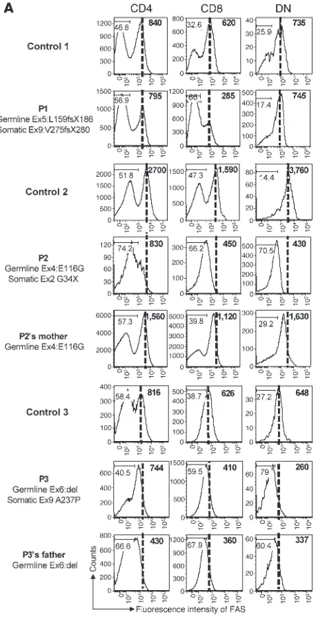
TNFRSF6 mutations or chromosome aberration, we analyzed FAS protein expression in CD4, CD8, and DN T cells from the 7 patients, 3 asymptomatic MPRs (patient 2’s mother, patient 3’s father, and patient 4’s father), and 5 healthy controls (Figure 3). First, we found that FAS expression in single-positive (CD4 or CD8) T cells was slightly lower than control values in all patients or relatives carrying a low-penetrance ECD TNFRSF6 mutation (except for patient 1); this suggests that the germline TNFRSF6
mutation in allele 1 led to defective expression in patients 2 to 7. Further analysis indicated that the great majority of these patients’ DN T cells did not express FAS. Interestingly, DN T cells from patients 2, 3, and 4 expressed less FAS than those from patient 2’s mother, patient 3’s father, and patient 4’s father, respectively (Figure 3, A and B). The low FAS expression in these cells was not due to an activation defect, since they expressed high amounts of
human leukocyte antigen class II molecules (Supplemental Fig-ure 1). Strikingly, no decrease of FAS expression was observed in 3 patients carrying germline ICD TNFRSF6 mutations associated with low penetrance. Accordingly, no somatic mutation has been detected in these patients. These results indicated that the somatic event in DN T cells is mostly associated with reduced FAS expres-sion via loss of the wild-type TNFRSF6 allele.
Discussion
[image:4.585.41.387.76.475.2]Severe ALPS always occurs when both TNFRSF6 alleles are mutated in the germline in humans (9, 15, 16) or mice (17). In contrast, if the FAS defect is partial (as in the case of a heterozygous mutation, for example), the human disease develops in 70% of carriers (13). The partial clinical penetrance of heterozygous TNFRSF6 mutations in ALPS suggests that in addition to the germline TNFRSF6 mutation, a “second hit” is required for disease onset. Here, we identified somat-ic genetsomat-ic events affecting the second TNFRSF6 allele in 7 patients carrying a germline heterozygous mutation with low clinical pen-etrance. In these patients, a small proportion of lymphocytes lacked functional FAS — leading to uncontrolled proliferation and autoim-munity. Missence or nonsense somatic mutations were identified in 3 patients, whereas somatic UPD (duplication of the mutant allele with loss of the wild-type allele) were found in the other 4 patients.
Figure 1
Family tree of 7 ALPS patients with
germline heterozygous TNFSFR6
mutations and additional somatic muta-tions in patients 1, 2, and 3. (A) Family trees of 7 ALPS patients with a germ-line ECD TNFRSF6 mutation. Asymp-tomatic mutated relatives are indicated by vertical gray bars. Subjects with ALPS are marked in black (arrows). The range of values determined for the 4 ALPS markers, i.e., apoptosis of acti-vated T cells (control > 75%), plasma IL-10 concentration (control < 20 pg/ ml), plasma FAS-L concentration (con-trol < 0.2 ng/ml), and the percentage of DN T cells (< 2% of TCRαβ cells) are shown for these patients and their rela-tives. Apoptosis values depicted in bold were obtained in an assay performed at the same time for patients’ cells and relatives’ cells. P1, patient 1. (B) Localization of the 2 mutations in the
research article
The Journal of Clinical Investigation http://www.jci.org Volume 121 Number 1 January 2011 109
Interestingly, we identified somatic events in 7 patients carrying germline heterozygous TNFRSF6 mutations affecting the ECD of FAS. The latter are known to frequently induce a loss of FAS expres-sion at the cell membrane and therefore haploinsufficiency (18). This was confirmed by analyzing cells from the MPRs, in which the only germline mutation was present. Expression of half the normal level of FAS receptor is sufficient to trigger apoptosis and might preclude the onset of ALPS and account for the low clinical
[image:5.585.59.535.81.556.2]penetrance seen with ECD TNFRSF6 mutations. The additional somatic event led to a clear, profound expression defect in a small proportion of single-positive T cells and in the majority of DN T cells from all but 1 patient. In this latter case, patient 1 carried 2 missence mutations (one germline and one somatic), which were predicted to be nonfunctional. The subsequent in vivo functional defect could thus be the consequence of either the lack of FAS expression at the cell membrane or the lack of apoptosis signal.
Figure 2
Figure 3
Impact of TNFRSF6 mutations on FAS protein expression by lym-phocyte subsets. FAS expression by CD4, CD8, and DN T cells was determined by FACS analysis (A) for patients 1 to 3 carrying a germ-line TNFRSF6 mutation and a somatic TNFRSF6 mutation in the sec-ond allele; (B) for patients 4 to 7 carrying a germline heterozygous
research article
The Journal of Clinical Investigation http://www.jci.org Volume 121 Number 1 January 2011 111
This could result from a lack of multimerization of the receptor or the lack of death-inducing signaling complex (DISC) recruitment by the residual FAS molecules. Consequently, the double-mutant lymphocytes could not be killed by FAS-L. A similar situation has been observed in patients carrying inherited homozygous or compound heterozygous mutations (9, 15). The lymphoprolifera-tion observed in the latter patients indicated that a complete FAS defect led to uncontrolled proliferation of T and (to a lesser extent) B cells. In contrast, most missence mutations affecting the ICD of TNFRSF6 are known to exert a transdominant negative effect, since the mutant protein is likely to trimerize with wild-type FAS. Assuming that efficient DISC formation and subsequent apopto-sis triggering require wild-type FAS trimers only, it was estimated that 1 out of 8 trimers would be functional. The ensuing apoptosis defect may be sufficient to account for the onset of ALPS. Accord-ingly, the clinical penetrance of the ICD missence TNFRSF6 muta-tion was complete in most families (19). Moreover, no addimuta-tional defect in the TNFRSF6 gene could be found in 3 tested patients with ICD mutations. It is noteworthy that previously described cases of ALPS caused by somatic heterozygous mutations (10, 11) were mostly associated with ICD TNFRSF6 mutations. This obser-vation suggested that somatic ICD TNFRSF6 mutations markedly impair FAS function and provide the mutant cells with a selective advantage in vivo. Moreover, the patients carrying somatic ICD mutations developed clinical disease, despite the low proportion (10%–20%) of mutant lymphocytes. A similar picture was seen in our current cases, since the somatic events were seen in a minor fraction of single-positive T cells and in a major fraction of DN T cells. This observation supports the concept whereby the sec-ond hit (somatic mutation or UPD) together with the germline ECD-mutated TNFRSF6 gene, profoundly impair FAS function and provide the double-mutant T cells with a selective advantage in vivo. The double-mutant cells died spontaneously in vitro and so could not be tested. Consequently, in vitro expansion of the patients’ T cells led to accumulation of the germline mutants only. Accordingly, the magnitude of the FAS-induced apoptosis defect was similar in T cells from the patients and their asymptomatic mutated relatives, respectively.
The combination of germline and somatic mutations in the same gene has been reported in cancers with a hereditary predisposi-tion, such as retinoblastoma (20). Similarly, missence mutations of TNFRSF6 outside the death domain are associated with LOH in non-Hodgkin lymphomas (21). The LOH observed in 4 of our patients with germline TNFRSF6 ECD mutations resulted from loss of the wild-type allele and concomitant duplication of the mutant one, i.e., telomeric UPD. This type of mechanism is usually observed in genetic disorders of development, where it can be involved in trisomy or monosomy rescue during embryonic development (22). The telomeric UPD observed in our ALPS cases might reflect a recombination hotspot on the 10q chromosome (as described in the 10q syndrome; ref. 23), but more likely reflect the selective advantage provided by the resulting homozygous TNFRSF6 muta-tions. These events may occur during embryogenesis (patient 2) or later in life (since the onset of clinical manifestations was delayed in patients 5, 6, and 7). Implication of modifier genes in the onset of ALPS was previously postulated. The present findings highlight the role of somatic events affecting the second TNFRSF6 allele as modi-fier in ALPS expression. However, we could not strictly exclude that these somatic FAS events result from other, yet undefined, germline mutation in the actual “ modifier” genes.
Lymphocyte homeostasis and self tolerance are maintained by several checkpoints, including antigen receptor editing, modula-tion, anergy, and central or peripheral clonal deletion of self-reactive lymphocytes (24). Variants of proteins involved in cell activation, survival, proliferation, and death may contribute to the emergence of autoimmunity (25). For instance, variants of the genes for HLA molecules (selecting for self-reactive lymphocytes), PTPN22 (regu-lating Antigen receptor signaling), and CTLA-4 (inhibiting T cell proliferation) are risk factors in the occurrence of various autoim-mune diseases. It has been postulated that in view of the moderate functional impact of these variants, additional events are required to bypass the tolerance checkpoints. This model accounts for the rela-tively long time to onset of autoimmune disease in most patients. Our identification of a combined germline TNFRSF6 mutation (with incomplete clinical penetrance) and a somatic event affecting the second TNFRSF6 allele now demonstrates that a hit on each of the 2 alleles of the same gene can lead to lymphoproliferation and autoimmunity. On the basis of this observation, one can pos-tulate that combinations of gene variants and somatic mutations in genes involved in cell death (such as Bim, Bid, or caspases) or in the regulation of cell proliferation (such as PD-1 or CTLA-4) could be involved in the emergence of autoimmune disease via the selec-tion of self-reactive T and B lymphocytes. This hypothesis could be tested in models of autoimmunity in which self-reactive T and/or B cell clones can be identified. Somatic mutations occur relatively frequently (i.e., 10–6 per gene per cell division) (26). Consequently,
in the context of the high-rate cell division seen in both lymphoid precursors and mature lymphocytes, a role for somatic mutations in triggering autoimmune disease onset should be considered.
In conclusion, our present findings validate the “2-hit” hypoth-esis in the setting of a nonmalignant disease and highlight the pos-sible impact of somatic mutations on the onset of other autoim-mune diseases in humans.
Methods
Study population. Seven patients presenting with typical ALPS manifesta-tions (including autoimmune manifestamanifesta-tions in 3 cases) and carrying germline heterozygous TNFRSF6 mutations affecting the ECD of the protein were enrolled in this study (Table 1). Relatives of patients 1 to 5 (including 7 asymptomatic MPRs) as well as 3 patients carrying a germline heterozygous ICD TNFRSF6 mutation (D269fsX279; G253D; K296fsX279) were also investigated as controls. The study protocol was approved by the local investigational review board (CPP ile de France II, Ministry of Research, Paris France). Informed consent was obtained from the patients or their families prior to study entry.
Immunofluorescence and cell sorting. Using flow cytometry, the percentage of DN T cells was defined as the percentage of TCR-αβ–positive cells that lacked CD4 and CD8 expression. Thawed peripheral blood mononuclear cells were incubated with a cocktail of 2.5 μl of anti–TCR PAN α/β PC5 antibody (IP26A; Immunotech; Beckman Coulter), 5 μl of anti–CD4-APC antibody (SK3), 10 μl of anti–CD8-V500 antibody (RPAT8), 10 μl of anti–CD95-PE antibody (DX2), and 10 μl of anti–HLA-DP-DQ-DR-FITC (TU39) (BD Biosciences) per 4 million cells. Acquisition and cell sorting were performed with an ARIA I or ARIA II cytometer (BD Biosciences), and data were analyzed using Flowjo software (TreeStar Inc.).
Plasma IL-10 and FAS-ligand and apoptosis assays. Plasma IL-10 and FAS-ligand levels and apoptosis were assayed as previously described (4).
DNA cloning. PCR products were cloned using TOPO TA cloning kit (Invitrogen) following the manufacturer’s instructions. The sequences of at least 80 clones were then determined by capillary electrophoresis using Applied Biosystems DNA sequencing kits and a 3130xl Genetic Analyzer (Applied Biosystems).
Genotype analysis. All analyzed microsatellite markers flanked the TNFRSF6 locus or genes located on the 10q chromosome. Primer sequenc-es were retrieved from the Genome Database (http://www.ncbi.nlm.nih. gov). FISH experiments were performed with BAC RP11-399O19 (a probe spanning the TNFRSF6 locus) as previously described (27). Analysis of copy number variations and LOH was performed using Whole-Genome Cytoge-netics 2.7M DNA chips (Affymetrix). The results were visualized using the Chromosome Analysis Suite (version 1.0.1; Affymetrix).
Acknowledgments
This work was funded by grants from INSERM, the Agence Natio-nale de la Recherche (ANR) (grant no. 08-GENO-015-01, E-rare 2007
[epinostics]), the Association pour la Recherche contre le Cancer (ARC), and the European STREP (autorome). A. Magerus-Chati-net was supported by a postdoctoral fellowship from ARC and the European STREP. B. Neven was supported by a doctoral fellowship from INSERM and the Fondation pour la Recherche Médicale. The authors thank Françoise Bourgue Vion-Dury, Corinne Cordier, Vir-ginie Grandin, Chantal Harre, Corinne Jacques, Nathalie Lambert, Alexandra Léger, Stephanie Ndaga, Fabienne Poulain, Solenn Pruvost, and Magali Salmon; and M. Jerôme Megret for technical support.
Received for publication May 19, 2010, and accepted in revised form October 6, 2010.
Address correspondence to: Frédéric Rieux-Laucat, INSERM U768, Hôpital Necker-Enfants Malades, 149 Rue de Sèvres, F-75015 Paris, France. Phone: 33.0.144.495.082; Fax: 33.0.142.730.640; E-mail: [email protected].
1. Canale VC, Smith CH. Chronic lymphoadenopa-thy simulating malignant lymphoma. J Pediatr. 1967;70(6):891–899.
2. Lopatin U, et al. Increases in circulating and phoid tissue interleukin-10 in autoimmune lym-phoproliferative syndrome are associated with disease expression. Blood. 2001;97(10):3161–3170. 3. Ohga S, et al. Dominant expression of interleukin
10 but not interferon gamma in CD4(–) CD8(–) alphabetaT cells of autoimmune lymphoprolifera-tive syndrome. Br J Haematol. 2002;119(2):535–538. 4. Magerus-Chatinet A, et al. FAS-L, IL-10, and dou-ble-negative CD4- CD8- TCR alpha/beta+ T cells are reliable markers of autoimmune lymphoprolif-erative syndrome (ALPS) associated with FAS loss of function. Blood. 2009;113(13):3027–3030. 5. Straus SE, et al. The development of lymphomas in
families with autoimmune lymphoproliferative syn-drome with germline Fas mutations and defective lymphocyte apoptosis. Blood. 2001;98(1):194–200. 6. Rieux-Laucat F, Le Deist F, Fischer A. Autoimmune
lymphoproliferative syndromes:genetic defects of apoptosis pathways. Cell Death and Differentiation. 2003;10(1):124–133.
7. Sneller MC, et al. Clincal, immunologic, and genet-ic features of an autoimmune lymphoproliferative syndrome associated with abnormal lymphocyte apoptosis. Blood. 1997;89(4):1341–1348. 8. Fisher GH, et al. Dominant interfering Fas gene
muta-tions impair apoptosis in a human autoimmune lym-phoproliferative syndrome. Cell. 1995;81(6):935–946. 9. Rieux-Laucat F, et al. Mutations in Fas associated with human lymphoproliferative syndrome and
autoimmunity. Science. 1995;268(5215):1347–1349. 10. Dowdell KC, et al. Somatic FAS mutations are
common in patients with genetically undefined autoimmune lymphoproliferative syndrome. Blood. 2010;115(25):5164–5169.
11. Holzelova E, et al. Autoimmune lymphoprolifera-tive syndrome with somatic Fas mutations. N Engl J Med. 2004;351(14):1409–1418.
12. Vaishnaw AK, Orlinick JR, Chu JL, Krammer PH, Chao MV, Elkon KB. The molecular basis for apop-totic defects in patients with CD95 (Fas/Apo-1) mutations. J Clin Invest. 1999;103(3):355–363. 13. Rieux-Laucat F, et al. Lymphoproliferative
syn-drome with autoimmunity: A possible genetic basis for dominant expression of the clinical manifesta-tions. Blood. 1999;94(8):2575–2582.
14. Bleesing JJ, et al. TcR-alpha/beta(+) CD4(–)CD8(–) T cells in humans with the autoimmune lympho-proliferative syndrome express a novel CD45 iso-form that is analogous to murine B220 and rep-resents a marker of altered O-glycan biosynthesis.
Clin Immunol. 2001;100(3):314–324.
15. Bettinardi A, et al. Missence mutations in the Fas gene resulting in autoimmune lymphoproliferative syndrome: a molecular and immunological analy-sis. Blood. 1997;89(3):902–909.
16. Kasahara Y, et al. Novel Fas (CD95/APO-1) muta-tions in infants with a lymphoproliferative disor-der. Int Immunol. 1998;10(2):195–202.
17. Watanabe D, Suda T, Hashimoto H, Nagata S. Con-stitutive activation of the Fas ligand gene in mouse lymphoproliferative disorders. EMBO J. 1995; 14(1):12–18.
18. Roesler J, et al. Haploinsufficiency, rather than the effect of an excessive production of soluble CD95 (CD95{Delta}TM), is the basis for ALPS Ia in a fam-ily with duplicated 3′ splice site AG in CD95 intron 5 on one allele. Blood. 2005;106(5):1652–1659. 19. Jackson CE, et al. Autoimmune lymphoproliferative
syndrome with defective Fas: genotype influences penetrance. Am J Hum Genet. 1999;64(4):1002–1014. 20. Rushlow D, et al. Detection of mosaic RB1 muta-tions in families with retinoblastoma. Hum Mutat. 2009;30(5):842–851.
21. Gronbaek K, et al. Somatic Fas mutations in non-Hodgkin’s lymphoma: association with extra-nodal disease and autoimmunity. Blood. 1998; 92(9):3018–3024.
22. Conlin LK, et al. Mechanisms of mosaicism, chime-rism and uniparental disomy identified by single nucleotide polymorphism array analysis. Hum Mol Genet. 2010;19(7):1263–1275.
23. Yatsenko SA, et al. Identification of critical regions for clinical features of distal 10q deletion syn-drome. Clin Genet. 2009;76(1):54–62.
24. Goodnow CC. Multistep pathogenesis of autoim-mune disease. Cell. 2007;130(1):25–35.
25. Zenewicz LA, Abraham C, Flavell RA, Cho JH. Unraveling the genetics of autoimmunity. Cell. 2010;140(6):791–797.
26. Araten DJ, et al. A quantitative measurement of the human somatic mutation rate. Cancer Res. 2005;65(18):8111–8117.