Amendment history:
Corrigendum
(March 2015)
Complement as a multifaceted modulator of
kidney transplant injury
Paolo Cravedi, Peter S. Heeger
J Clin Invest.
2014;
124(6)
:2348-2354.
https://doi.org/10.1172/JCI72273
.
Improvements in clinical care and immunosuppressive medications have positively affected
outcomes following kidney transplantation, but graft survival remains suboptimal, with
half-lives of approximately 11 years. Late graft loss results from a confluence of processes
initiated by ischemia-reperfusion injury and compounded by effector mechanisms of
uncontrolled alloreactive T cells and anti-HLA antibodies. When combined with
immunosuppressant toxicity, post-transplant diabetes and hypertension, and recurrent
disease, among other factors, the result is interstitial fibrosis, tubular atrophy, and graft
failure. Emerging evidence over the last decade unexpectedly identified the complement
cascade as a common thread in this process. Complement activation and function affects
allograft injury at essentially every step. These fundamental new insights, summarized
herein, provide the foundation for testing the efficacy of various complement antagonists to
improve kidney transplant function and long-term graft survival.
Review Series
Find the latest version:
Complement as a multifaceted modulator of
kidney transplant injury
Paolo Cravedi and Peter S. Heeger
Department of Medicine, Recanati Miller Transplant Institute and Immunology Institute, Icahn School of Medicine at Mount Sinai, New York, New York, USA.
Improvements in clinical care and immunosuppressive medications have positively affected outcomes following
kidney transplantation, but graft survival remains suboptimal, with half-lives of approximately 11 years. Late graft
loss results from a confluence of processes initiated by ischemia-reperfusion injury and compounded by effector
mechanisms of uncontrolled alloreactive T cells and anti-HLA antibodies. When combined with
immunosuppres-sant toxicity, post-transplant diabetes and hypertension, and recurrent disease, among other factors, the result
is interstitial fibrosis, tubular atrophy, and graft failure. Emerging evidence over the last decade unexpectedly
identified the complement cascade as a common thread in this process. Complement activation and function
affects allograft injury at essentially every step. These fundamental new insights, summarized herein, provide the
foundation for testing the efficacy of various complement antagonists to improve kidney transplant function and
long-term graft survival.
Introduction
Kidney transplantation is the treatment of choice for end-stage kidney disease. Advances in immunosuppression, HLA matching, and improvements in medical care have reduced acute rejection rates to less than 10%, but long-term graft survival remains sub-optimal, as kidney transplant half-lives are only 8 to 11 years (1). As a consequence, the extension of allograft survival is one current focus of transplant research. An improved understanding of the pathophysiology of acute and chronic allograft injury will likely guide development and implementation of novel therapies capa-ble of prolonging kidney transplant survival.
Current concepts are that kidney transplant injury is initiated by surgical trauma and the requisite associated ischemia-reperfusion (IR) injury, the latter being particularly detrimental in recipients of deceased donor allografts. IR rates following transplantation are increasing (2), and kidney graft survival is worse in recipients with significant IR injury (3). Donor-reactive T cells and HLA anti-bodies are established mediators of transplant injury and must be controlled by appropriate immunosuppression. Inadequate immunosuppression predisposes to acute cellular rejection and antidonor HLA alloantibody formation, both of which are asso-ciated with progressive interstitial fibrosis and tubular atrophy (IF/TA) and shortened graft survival (4–6). In contrast, excessive immunosuppression not only predisposes to infectious compli-cations, but is associated with multiple toxicities including direct kidney damage (7) and indirect effects of drug-induced diabetes (8) and hypertension (9).
Emerging evidence over the past 15 years supports the concept that the complement cascade, traditionally considered a compo-nent of innate immunity, unexpectedly regulates kidney IR injury, T cell and humoral alloimmunity that underlie transplant rejec-tion, and progressive kidney injury that results in late graft failure. This body of literature, to be reviewed herein, suggests that comple-ment components produced by the liver as well as by kidney cells and immune cells play crucial roles as pathogenic mediators of
transplant rejection in animals and humans. The data support the need for further efficacy testing of targeting complement and/or its receptors to improve kidney transplant outcome in humans.
Overview of the complement cascade
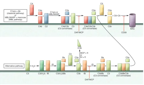
The complement system is composed of over 30 soluble and mem-brane-bound proteins that are activated as a cascade by three initi-ating pathways: (a) the lectin pathway triggered by carbohydrates present on bacteria surface, (b) the classical pathway triggered by cross-linking, cell-bound subclasses of IgG and IgM antibodies, and (c) the alternative pathway that undergoes spontaneous acti-vation on cell surfaces (Figure 1 and ref. 10).
The three pathways converge to form C3 convertases, multim-eric protein complexes with enzymatic activity (10). Cleavage of C3 yields C3a and C3b, the latter of which triggers formation of the C5 convertase. Subsequent C5 cleavage initiates formation of the membrane attack complex (MAC, C5b-9) on the target cells. In addition to the MAC, soluble and surface-bound split products, including C3a, C3b, iC3b, C3dg, and C5a, mediate inflammation by directly lysing target cells, serving as chemoattractants, func-tioning as opsonins, and activating innate immune cells such as macrophages and neutrophils (11).
Complement activation is tightly regulated (Figure 1) so as to prevent bystander damage to self cells (11). This regulation is accomplished through the expression of membrane-bound and soluble complement–regulating proteins. Decay accelerating fac-tor (DAF or CD55) is a glycophosphatidylinositol-anchored, mem-brane-bound complement regulator that accelerates the decay of cell surface–assembled C3 convertases. DAF limits downstream complement activation and restricts production of the afore-mentioned cleavage products (12). Notably, DAF only functions intrinsically, limiting complement activation on the cell surface upon which it is expressed, but not on proximally located patho-gens, which lack DAF expression. Human CD46 (murine homolog Crry), also known as membrane cofactor protein (MCP), has simi-lar decay-accelerating function, but also exhibits cofactor activity. In conjunction with soluble factor I, this membrane-bound regula-tor inactivates C3b to iC3b, thereby preventing re-formation of the C3 convertase. Other examples of complement regulators include
Conflict of interest: Peter S. Heeger is a recipient of a research grant from Alexion Pharmaceuticals.
review series
CD59 (protectin), a cell surface–expressed regulator that inhibits formation of the MAC at the C9 step; factor H, a soluble comple-ment regulatory protein that exhibits both decay-accelerating and cofactor activity; and CR1, which limits amplification of the com-plement cascade and MAC formation by inhibiting C3 convertases. The majority of the complement components circulating in the blood (systemic compartment) are produced by the liver. Comple-ment proteins are also generated by tissue-resident (e.g., tubular cells in the kidney, ref. 13) and migratory/immune cells, including T cells and APCs (14). Theoretically, non–liver cell–derived com-plement could activate and function locally without affecting sys-temic complement activation.
Complement and IR injury
The pathophysiology of post-transplant IR injury and delayed graft function (DGF) has been reviewed elsewhere in detail (15). Briefly, IR injury results from tissue hypoxia, mitochondrial dam-age, and ATP depletion, followed by the generation of free oxy-gen radicals upon reperfusion, which initially damage endothe-lium. Ensuing inflammation driven by TLR signaling and locally secreted cytokines, chemokines, and complement amplify the inflammation, resulting in tubular injury and kidney dysfunction.
Early evidence implicating a role for complement in IR injury derived from mouse models in which complement deposition and loss of membrane-bound complement regulators were described during kidney IR injury and overexpression of Crry ameliorated IR injury (16, 17). IR injury was later shown to be dampened in complement-depleted mice (18, 19) and in C3-deficient (16, 19), factor B-deficient (20) or C5-deficient (21) mice, while mice deficient in DAF (22) or in Crry and factor H (23, 24) (in which restraint on complement activation is lifted) were more suscep-tible to IR injury. Strikingly, IR injury studies performed in ani-mals following transplantation of WT or C3-deficient kidneys into syngeneic WT or C3-deficient recipients showed that donor kidney–derived C3, and not systemic recipient C3, is the predom-inant mediator of IR injury (25).
Peng et al. (26) used C3a receptor– (C3aR-), C5aR-, and C3aR/ C5aR-deficient mice and models of renal IR injury to demonstrate that deficiency of either or both of these receptors protected mice from injury. The C3aR/C5aR- and C5aR-deficient mice were most protected. Studies performed in BM chimeras showed that the absence of C3aR and C5aR on either renal tubular epithelial cells or circulating leukocytes attenuated renal IR injury, indicating that expression of C3aR and C5aR on both renal cells and
circulat-Figure 1
Schematic representation of the complement cascade. C1q,r,s cross-linking of antibodies activates the classical pathway. Mannose-associated serine proteases (MASPs) bind to mannose motifs expressed on bacteria to activate complement via the MBL pathway. Subsequent cleavage and assembly of C4 and then C2 form the C4bC2b C3 convertase. C3 spontaneously binds to H2O, forming C3(H2O), which binds to cell
[image:3.585.46.545.75.369.2]ing leukocytes contributes to the pathogenesis of renal IR injury. In these studies, protection from injury was associated with less cellular infiltration and lower mRNA levels of kidney injury mol-ecule-1, proinflammatory mediators, and adhesion molecules in post-ischemic kidneys. One specific mechanism is that C3a pro-duced in response to IR injury drives renal tubular epithelial cell production of chemokines (27). Together, the animal model data support the conclusion that IR injury upregulates production of complement components by kidney endothelial and tubular cells as well as by infiltrating immune cells. Local activation through the alternative pathway yields C3a and C5a, which amplify local inflammation and injury through autocrine and paracrine liga-tions with their receptors expressed on cells in the graft.
A role for complement activation in human IR injury was eval-uated by de Vries et al. (28). These investigators detected soluble C5b-9 following reperfusion of deceased donor but not living donor kidneys. Whole genome expression profiling of 53 human renal allograft protocol biopsies obtained at implantation con-firmed significantly higher expression levels of complement genes in deceased donor kidneys (29). Extending these findings prior to surgical removal of the donor organ, van Werkhoven et al. found that brain death initiates systemic complement activation, upreg-ulates C5aR expression in renal tubular cells (30), and is associated with induction of intrarenal inflammatory cytokines. The authors hypothesized that complement activation that occurs in the donor accounts in part for the poorer outcomes of grafts harvested from deceased compared with living donors.
The recognition that complement participates in the pathogene-sis of post-transplant IR injury has prompted investigators to test whether complement inhibitors are effective therapies. An analog of the human complement-regulatory protein CD35 (CR1; blocks complement activation at the C3 convertase step) has been con-jugated to a myristoylated peptidyl tail, such that when admin-istered by intravenous perfusion of the harvested organ ex vivo it will self-insert into the lipid bilayer of the EC membranes (31). Patel et al. used this approach to show that the reagent was effec-tive in preventing post-transplant kidney IR injury in a rat model (32). The human reagent mirococept (APT070) is currently being studied in a clinical trial for prevention of DGF (31). Eculizumab (Soliris; Alexion Pharmaceuticals Inc.), a humanized anti-C5 mAb that is FDA approved to treat paroxysmal nocturnal hemoglobi-nuria is also being tested for efficacy in preventing post-transplant DGF (NCT01403389; NCT01919346).
The aforementioned findings indicating that brain death is asso-ciated with complement activation in the donor kidney prior to organ removal raise the intriguing concept that complement inhi-bition in the donor could be an effective prophylactic therapy to prevent IR injury. Innovative study designs involving randomized treatment of organ donors and collaborations between organ pro-curement agencies and multiple transplant centers will need to be developed to test this possibility.
Complement and alloreactive T cells
Complement and effector T cells. Building upon the paradigm-shifting observation published in 2002 that WT mice do not reject allog-rafts from C3-deficient donors (33), work from several groups, including ours, uncovered an unexpected role for complement as a regulator of T cell immunity. During cognate interactions between T cells and APCs, both partners upregulate and secrete alternative pathway complement components C3, fB, and fD, as well as the
common pathway protein C5, and upregulate surface expression of C3aR and C5aR (34, 35). These changes are a consequence of costimulatory molecule signaling via CD28/CD80/CD86 and CD154/CD40 (35), which simultaneously and transiently reduce cell surface–expressed DAF (thereby lifting restraint on comple-ment activation). Locally produced C3a and C5a bind to their receptors and function as autocrine and paracrine stimulators of the T cell and the APC (34, 35). Signaling via these GPCRs in T cells activates PI3Kγ and induces phosphorylation of the intracellular signaling molecule AKT (14, 35), upregulating the antiapoptotic protein Bcl2 and downregulating expression of the proapoptotic molecule Fas. Together, these complement-dependent mechanisms enhance T cell proliferation and diminish T cell apoptosis (14). The evidence also indicates that C3aR and C5aR signaling is required for T cell homeostasis, as T cells deficient in both receptors spon-taneously undergo accelerated cell death in vitro and in vivo (35).
On DCs and macrophages, C3a/C3aR and C5a/C5aR ligations induce upregulation and release of innate cytokines (e.g., IL-12, IL-23) and costimulatory molecules (e.g., CD80, CD86) (35–38). APCs deficient in C5aR/C3aR or C3 produce less IL-12, express lower levels of CD80, and are weaker T cell stimulators than WT APCs, while DCs and macrophages obtained from mice genetically deficient in DAF produce more IL-12 and induce stronger T cell responses than cells from WT animals (35–38). Independent of the phenotype of the APC, T cells deficient in C3aR and C5aR signal-ing respond poorly to WT and Daf1–/– APCs and undergo
acceler-ated cell death (14, 35).
Alloresponses are dampened in WT chimeric mouse recipients of C3–/– BM, despite normal serum complement. In contrast,
C3-deficient chimeric hosts repopulated with WT BM exhibited normal T cell alloreactivity (14, 39). Analogously, BM chimeras produced using C5aR-deficient donors or recipients confirmed that T cell immunity is dependent on C5aR expression on BM- derived cells (14, 39).
Complement and T cell–mediated rejection. Studies performed in transplant models revealed WT mice reject Daf1–/– heart allografts
with accelerated kinetics (39), and that the accelerated rejection is due to a complement-dependent augmentation of antidonor T cell immunity. Collaborative work additionally showed that donor or recipient DAF deficiency accelerates skin graft rejection (34) and overcomes the immune privilege of the eye by enhancing patho-genic T cell alloimmunity induced by normally toleropatho-genic corneal transplants, resulting in rapid corneal rejection (40).
CD8+ T cells, the principal mediators of solid organ transplant
rejection, require helper signals derived from CD4+ T cells in
order to become optimal pathogenic effector cells (CD4-defi-cient mice do not reject cardiac allografts; ref. 41). Current con-cepts regarding how CD4+ T cells help alloreactive CD8+ T cells
are that during cognate TCR/APC interactions, CD154 expressed on CD4+ T cells transmits activating signals to APCs through
ligation with CD40 (42, 43). This in turn upregulates costimula-tory molecule (CD80/86) and MHC expression on the APC and induces proinflammatory cytokines (e.g., IL-12), which together facilitate optimal CD8+ T cell activation, expansion,
differentia-tion, and survival. Building upon previous findings linking com-plement to T cell activation, our research group provided exper-imental evidence, using in vivo transplant models, that immune cell–derived complement is a crucial molecular intermediary underlying how CD4 cells provide help to alloreactive CD8+ T
review series
Local complement production also influences effector CD8+
T cell responses to allogeneic vascular ECs (45). Experiments performed using in vitro culture systems and in vivo heart trans-plantation models showed that complement is produced by ECs and regulates T cell function and expansion (46). The effects of EC-derived complement are transmitted in part through C5aR signaling on T cells (45).
C5a/C5aR interactions also modulate T cell–dependent kidney transplant rejection in rodents (47). Together with the findings that anti-C5 mAb synergizes with CTLA4-Ig to prevent T cell priming, limits T cell trafficking to an allograft, and prolongs transplant survival in mice (48), the body of work supports the conclusion that complement is a physiologically important regu-lator of pathogenic T cell immunity that causes allograft rejection in animal models.
Important confirmatory human experiments published in 2013 showed that C3a and C5a are generated during in vitro cultures of human T cells responding to allogeneic DCs (49). Both part-ners express the receptors for C3a and C5a (50–53), and C3aR and C5aR antagonists inhibit human T cell proliferation (49). Recombinant C3a/C5a promote human CD4+ T cell expansion,
bypassing the inhibitory effects of CTLA4-Ig, and inducing AKT phosphorylation. Lowering human DC C3a/C5a production by siRNA knockdown of DC-expressed C3 reduces human T cell alloresponses. Conversely, downregulating DC expression of DAF increases immune cell C3a/C5a and augments human alloreac-tive T cell proliferation, identifying APCs as the dominant com-plement source (49). In vitro studies by Zhou et al. also showed that different subsets of human DCs produce complement, and that C5aR/C3aR signaling regulates DC activation and function (54). Pharmacological C5aR blockade reduced human anti-mouse graft-versus-host disease (GVHD) scores and inhibited T cell responses in NOD/SCID/γcnull mouse recipients of human
periph-eral blood mononuclear cells, verifying that the C5aR-dependent effects on human T cells apply in vivo (49).
In further support of the clinical relevance of local complement in human transplantation, the quantity of RNA message for alter-native pathway complement components and complement recep-tors, including C5aR and C3aR, is higher in human allograft biop-sies with histologic evidence of rejection compared to non-injured control tissue (47, 55).
Complement and Tregs. Tregs are instrumental for tolerance induc-tion and maintenance (56) in rodents and are associated with improved long-term transplant outcomes in humans (56, 57). Data published in 2013 indicated that complement also regulates Treg induction, function, and stability (58, 59). Our laboratory demonstrated that peripheral murine natural Tregs (nTregs) express C3aR and C5aR, and that signaling through these recep-tors inhibits Treg function (60). Genetic and pharmacologic block-ade of C3aR/C5aR signal transduction in nTregs augments their in vitro and in vivo suppressive activity, abrogates autoimmune colitis, and prolongs allogeneic skin graft survival. Mechanisms involve C3a/C5a-induced phosphorylation of AKT and, as a conse-quence, phosphorylation of the transcription factor Foxo1, which results in lowered nTreg Foxp3 expression. Two additional sets of data showed that genetic deficiency or pharmacologic blockade of C3aR/C5aR signaling augments murine induced Treg (iTreg) generation, stabilizes Foxp3 expression, resists iTreg conversion to IFN-γ/TNF-α–producing effector T cells, and as a consequence, limits the clinical expression of GVHD (58, 59).
Immune cell–derived complement also modulates human Treg generation and function (58, 61). Pharmacologic antagonists to human C3aR and C5aR augment in vitro generation and stabil-ity of human iTreg from naive precursors. In NOD/SCID/γcnull
mouse recipients of human peripheral blood mononuclear cells, we showed that pharmacologic C5aR blockade enhances human iTreg generation and stability and results in better disease protec-tion (61). These clinically relevant translaprotec-tional findings provide proof of concept that C3a/C3aR and C5a/C5aR ligations are viable targets for facilitating iTreg-mediated transplant tolerance.
Building upon previously published evidence that coengage-ment of the TCR and the complecoengage-ment regulator CD46 promote regulatory IL-10 production (62), these new results underscore the crucial role of complement in modulating the balance between pathogenic and protective adaptive T cell responses. Whether com-plement antagonists can therapeutically control T cell alloreactiv-ity while simultaneously promoting Treg induction, function, and stability in transplant patients remains to be determined. Anti-C5 mAb and C5aR antagonists are currently being tested in humans for other indications, providing opportunities to assess their effects on human alloreactive T cells in vivo (NCT01363388).
Complement and antibody-mediated transplant injury
Donor reactive anti-HLA antibodies are generally accepted as pathogenic mediators of acute and chronic transplant injury (63). It has been known for decades that complement depletion impairs antibody production (64). One mechanism involves anti-gen-bound C3dg (a C3b cleavage product; see Figure 1) binding to B cell–expressed complement receptor 2 (CD21), which facilitates antigen presentation to B cells and lowers the threshold for B cell activation (65, 66). Isotype switching in B cells that results in IgG production is dependent upon T follicular helper (TFH) cell/B cell
interactions (reviewed in ref. 67), transmitted in part by IL-21/ IL-21 receptor, and CD40/CD154, ligations. As outlined above, T cell immunity is regulated by locally produced complement, indicating that complement could impact antibody production indirectly through regulating T cell immunity. To our knowledge, there are no data addressing whether complement specifically alters induction, function, or stability of TFH cells. Regardless of
the specific mechanism, C3-deficient mice fail to produce high- affinity IgG responses against major histocompatibility antigens in skin grafts (68), demonstrating the relevance of complement’s influence over antibody formation in response to transplant anti-gens. Whether complement blockade impacts anti-HLA antibody formation in human transplant recipients has not been tested.
ther-apeutic approach to humans, the anti-human C5 mAb eculizumab plus plasma exchange reduced the incidence of antibody-mediated rejection in 26 sensitized kidney transplant recipients compared with a historical control group of 51 patients treated with a plasma exchange–based protocol alone (72). Anti-C5 mAb also successfully reversed established antibody-mediated rejection (73). In light of the association between anti-HLA antibodies and chronic antibody-medi-ated graft rejection, ongoing studies are testing the efficacy of eculi-zumab in preventing graft loss in kidney transplant recipients with donor-specific antibodies (NCT01327573).
From a diagnostic/biomarker standpoint, investigators hypothe-sized that detection of serum anti-HLA antibodies capable of bind-ing C1q would enhance the prognostic utility of serum alloantibody analysis in kidney transplantation (74, 75). The latest iteration of the single antigen bead technology currently used for detecting anti-HLA antibodies (76) additionally identifies those antibodies
that bind C1q. A 2013 population-based study of 1,016 kidney transplant recipients suggested that, among patients with anti-HLA antibodies, those that were C1q+ had the worst graft survival (77).
Colvin and colleagues pioneered the use of C4d staining in graft tissue as a diagnostic test for antibody-mediated allograft rejection (AMR) (78). C4d is an activation byproduct of classical pathway complement activation and its detection in graft tissue is more sensitive than detection of immunoglobulin. As a consequence, C4d staining has become a valuable tool for diagnosing AMR. Importantly, diagnostic sensitivity depends on staining method-ology and cases of C4d– AMR have been described (79).
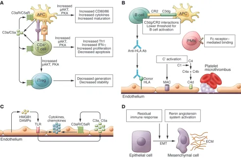
Complement and kidney graft injury and fibrosis
[image:6.585.45.536.83.404.2]Mechanisms of late graft failure are complex and involve immune and non-immune mechanisms (80), but late graft loss routinely results in pathological evidence of progressive glomerulosclerosis,
Figure 2
review series
IF/TA, and arteriosclerosis (81, 82). The functional and structural changes of chronic renal allograft failure share similarities with those observed in other forms of chronic progressive kidney dis-ease, in which decline of functioning nephron mass has been con-sidered the key event (83).
Unexpectedly, several pieces of evidence suggest that intragraft complement activation contributes to this progressive kidney injury. C3–/– kidney isografts transplanted into WT recipients were
protected from toxin-induced tubular damage, proteinuria, and progressive renal failure, despite the presence of abundant circu-lating C3 (84). Follow-up work showed that C3 is implicated in the activation of the renin-angiotensin system and in the epitheli-al-to-mesenchymal transition (85, 86), supporting the concept that synthesis of complement components by renal epithelial cells is one critical mediator of tubular damage in proteinuria-associated renal disease. As proteinuria is one hallmark of kidney graft dysfunction (87), these results raise the possibility that kidney-derived comple-ment participates in the developcomple-ment of IF/TA after transplant.
Indirect associative evidence linking complement to progressive transplant injury derives from studies of complement gene morphisms and transplant outcomes in humans. Specific C5 poly-morphisms in both the donor and the recipient have been asso-ciated with worse late graft function, but interestingly not with the risk of acute rejection (88). Although controversial, some addi-tional evidence suggests that donor kidney expression of a specific polymorphic variant of C3 is associated with worse post-trans-plant outcomes (89, 90). In further support of the concept that intragraft complement production modulates progressive kidney injury, proteomic studies of kidney allograft tissue by the Salomon
group demonstrated strong associations between IF/TA and the alternative pathway, but not the classical pathway, complement components (91). An ongoing study of chronic anti-C5 mAb ther-apy in kidney transplant recipients (NCT01327573) could poten-tially provide further insight into the role of complement as a mediator of progressive graft dysfunction and IF/TA.
Conclusions
The complement system is now firmly established as a pervasive, multifaceted mediator of transplant injury in animal models, and studies from multiple groups have largely confirmed that these newly recognized mechanisms apply to human transplant recipients (Figure 2). This success of translational immunol-ogy, along with the development of pharmacologic agents that block human complement components and receptors (92, 93), now allows for testing of the intriguing concept that targeting complement in kidney transplant recipients will improve graft survival and patient health.
Acknowledgments
This work was supported by NIH grants AI43578 and AI071185 awarded to P.S. Heeger. P. Cravedi is a recipient of American Heart Association grant 12POST12050294. The authors thank Jeremy Leventhal for his comments on the manuscript.
Address correspondence to: Peter S. Heeger, Icahn School of Medicine at Mount Sinai, Box 1243, One Gustave L. Levy Plaza, New York, New York 10029, USA. Phone: 212.241.6324; Fax: 212.987.0389; E-mail: [email protected].
1. Lodhi SA, Lamb KE, Meier-Kriesche HU. Solid organ allograft survival improvement in the United States: the long-term does not mirror the dramatic short-term success. Am J Transplant. 2011; 11(6):1226–1235.
2. Tapiawala SN, et al. Delayed graft function and the risk for death with a functioning graft. J Am Soc Nephrol. 2010;21(1):153–161.
3. Khalkhali HR, Ghafari A, Hajizadeh E, Kazemne-jad A. Risk factors of long-term graft loss in renal transplant recipients with chronic allograft dys-function. Exp Clin Transplant. 2010;8(4):277–282. 4. El Ters M, et al. Kidney allograft survival after
acute rejection, the value of follow-up biopsies.
Am J Transplant. 2013;13(9):2334–2341. 5. Smith JD, et al. De novo donor HLA-specific
anti-bodies after heart transplantation are an indepen-dent predictor of poor patient survival. Am J Trans-plant. 2011;11(2):312–319.
6. Terasaki PI, Ozawa M. Predicting kidney graft failure by HLA antibodies: a prospective trial. Am J Transplant. 2004;4(3):438–443.
7. Gaston RS. Chronic calcineurin inhibitor nephro-toxicity: reflections on an evolving paradigm. Clin J Am Soc Nephrol. 2009;4(12):2029–2034.
8. Kaposztas Z, Gyurus E, Kahan BD. New-onset dia-betes after renal transplantation: diagnosis, inci-dence, risk factors, impact on outcomes, and novel implications. Transplant Proc. 2011;43(5):1375–1394. 9. Luft FC. How calcineurin inhibitors cause hyperten-sion. Nephrol Dial Transplant. 2012;27(2):473–475. 10. Walport MJ. Complement. First of two parts.
N Engl J Med. 2001;344(14):1058–1066.
11. Ricklin D, Hajishengallis G, Yang K, Lambris JD. Complement: a key system for immune sur-veillance and homeostasis. Nat Immunol. 2010; 11(9):785–797.
12. Medof ME, Kinoshita T, Nussenzweig V. Inhibi-tion of complement activaInhibi-tion on the surface of
cells after incorporation of decay-accelerating fac-tor (DAF) into their membranes. J Exp Med. 1984; 160(5):1558–1578.
13. Peake PW, O’Grady S, Pussell BA, Charlesworth JA. C3a is made by proximal tubular HK-2 cells and activates them via the C3a receptor. Kidney Int. 1999; 56(5):1729–1736.
14. Lalli PN, Strainic MG, Yang M, Lin F, Medof ME, Heeger PS. Locally produced C5a binds to T cell-expressed C5aR to enhance effector T-cell expan-sion by limiting antigen-induced apoptosis. Blood. 2008;112(5):1759–1766.
15. Siedlecki A, Irish W, Brennan DC. Delayed graft function in the kidney transplant. Am J Transplant. 2011;11(11):2279–2296.
16. Zhou W, et al. Predominant role for C5b-9 in renal ischemia/reperfusion injury. J Clin Invest. 2000; 105(10):1363–1371.
17. Thurman JM, et al. Altered renal tubular expres-sion of the complement inhibitor Crry permits complement activation after ischemia/reperfusion.
J Clin Invest. 2006;116(2):357–368.
18. Stein JH, Osgood RW, Barnes JL, Reineck HJ, Pinck-ard RN, McManus LM. The role of complement in the pathogenesis of postischemic acute renal fail-ure. Miner Electrolyte Metab. 1985;11(4):256–261. 19. Park P, et al. Inhibiting the complement system does
not reduce injury in renal ischemia reperfusion.
J Am Soc Nephrol. 2001;12(7):1383–1390. 20. Thurman JM, Ljubanovic D, Edelstein CL, Gilkeson
GS, Holers VM. Lack of a functional alternative com-plement pathway ameliorates ischemic acute renal failure in mice. J Immunol. 2003;170(3):1517–1523. 21. De Vries B, Matthijsen RA, Wolfs TG, Van Bijnen AA,
Heeringa P, Buurman WA. Inhibition of complement factor C5 protects against renal ischemia-reperfusion injury: inhibition of late apoptosis and inflammation.
Transplantation. 2003;75(3):375–382.
22. Yamada K, Miwa T, Liu J, Nangaku M, Song WC.
Critical protection from renal ischemia reperfu-sion injury by CD55 and CD59. J Immunol. 2004; 172(6):3869–3875.
23. Renner B, et al. Binding of factor H to tubular epi-thelial cells limits interstitial complement activation in ischemic injury. Kidney Int. 2011;80(2):165–173. 24. Renner B, et al. The complement inhibitors Crry
and factor H are critical for preventing autologous complement activation on renal tubular epithelial cells. J Immunol. 2010;185(5):3086–3094. 25. Farrar CA, Zhou W, Lin T, Sacks SH. Local
extravas-cular pool of C3 is a determinant of postischemic acute renal failure. FASEB J. 2006;20(2):217–226. 26. Peng Q, et al. C3a and C5a promote renal
ische-mia-reperfusion injury. J Am Soc Nephrol. 2012; 23(9):1474–1485.
27. Thurman JM. Triggers of inflammation after renal ischemia/reperfusion. Clin Immunol. 2007; 123(1):7–13.
28. de Vries DK, et al. Acute but transient release of terminal complement complex after reperfusion in clinical kidney transplantation. Transplantation. 2013;95(6):816–820.
29. Naesens M, et al. Expression of complement com-ponents differs between kidney allografts from living and deceased donors. J Am Soc Nephrol. 2009; 20(8):1839–1851.
30. van Werkhoven MB, et al. Complement mediated renal inflammation induced by donor brain death: role of renal C5a-C5aR interaction. Am J Transplant. 2013;13(4):875–882.
31. Sacks SH, Zhou W. The role of complement in the early immune response to transplantation. Nat Rev Immunol. 2012;12(6):431–442.
33. Pratt JR, Basheer SA, Sacks SH. Local synthesis of complement component C3 regulates acute renal transplant rejection. Nat Med. 2002;8(6):582–587. 34. Heeger PS, et al. Decay-accelerating factor
modu-lates induction of T cell immunity. J Exp Med. 2005; 201(10):1523–1530.
35. Strainic MG, et al. Locally produced complement fragments C5a and C3a provide both costimu-latory and survival signals to naive CD4+ T cells.
Immunity. 2008;28(3):425–435.
36. Lalli PN, Strainic MG, Lin F, Medof ME, Heeger PS. Decay accelerating factor can control T cell differentiation into IFN-γ-producing effector cells via regulating local C5a-induced IL-12 production.
J Immunol. 2007;179(9):5793–5802.
37. Li K, et al. Cyclic AMP plays a critical role in C3a-re-ceptor-mediated regulation of dendritic cells in antigen uptake and T-cell stimulation. Blood. 2008; 112(13):5084–5094.
38. Peng Q, et al. Dendritic cell function in allostimu-lation is modulated by C5aR signaling. J Immunol. 2009;183(10):6058–6068.
39. Pavlov V, et al. Donor deficiency of decay-accelerat-ing factor accelerates murine T cell-mediated cardiac allograft rejection. J Immunol. 2008;181(7):4580–4589. 40. Esposito A, et al. Decay accelerating factor is essen-tial for successful corneal engraftment. Am J Trans-plant. 2010;10(3):527–534.
41. Krieger NR, Yin DP, Fathman CG. CD4+ but not
CD8+ cells are essential for allorejection. J Exp Med. 1996;184(5):2013–2018.
42. Ridge JP, Di Rosa F, Matzinger P. A conditioned dendritic cell can be a temporal bridge between a CD4+ T-helper and a T-killer cell. Nature. 1998;
393(6684):474–478.
43. Schoenberger SP, Toes RE, van der Voort EI, Offringa R, Melief CJ. T-cell help for cytotoxic T lymphocytes is mediated by CD40-CD40L interac-tions. Nature. 1998;393(6684):480–483.
44. Vieyra M, et al. Complement regulates CD4 T-cell help to CD8 T cells required for murine allograft rejection. Am J Pathol. 2011;179(2):766–774. 45. Raedler H, Yang M, Lalli PN, Medof ME, Heeger
PS. Primed CD8(+) T-cell responses to allogeneic endothelial cells are controlled by local complement activation. Am J Transplant. 2009;9(8):1784–1795. 46. Jane-Wit D, et al. Alloantibody and complement
promote T cell-mediated cardiac allograft vascu-lopathy through noncanonical nuclear factor-κB signaling in endothelial cells. Circulation. 2013; 128(23):2504–2516.
47. Gueler F, et al. Complement 5a receptor inhibition improves renal allograft survival. J Am Soc Nephrol. 2008;19(12):2302–2312.
48. Raedler H, et al. Anti-complement component C5 mAb synergizes with CTLA4Ig to inhibit alloreac-tive T cells and prolong cardiac allograft survival in mice. Am J Transplant. 2011;11(7):1397–1406. 49. Cravedi P, Leventhal J, Lakhani P, Ward SC,
Dono-van MJ, Heeger PS. Immune cell-derived C3a and C5a costimulate human T cell alloimmunity. Am J Transplant. 2013;13(10):2530–2539.
50. Li K, et al. Functional modulation of human monocytes derived DCs by anaphylatoxins C3a and C5a. Immunobiology. 2012;217(1):65–73. 51. Werfel T, et al. Activated human T lymphocytes
express a functional C3a receptor. J Immunol. 2000; 165(11):6599–6605.
52. Nataf S, Davoust N, Ames RS, Barnum SR. Human T cells express the C5a receptor and are chemoat-tracted to C5a. J Immunol. 1999;162(7):4018–4023. 53. Nataf S, Levison SW, Barnum SR. Expression of the
anaphylatoxin C5a receptor in the oligodendrocyte lineage. Brain Res. 2001;894(2):321–326. 54. Li K, et al. Expression of complement components,
receptors and regulators by human dendritic cells.
Mol Immunol. 2011;48(9–10):1121–1127. 55. Keslar K, Rodriguez ER, Tan CD, Starling RC, Heeger
PS. Complement gene expression in human cardiac allograft biopsies as a correlate of histologic grade of injury. Transplantation. 2008;86(9):1319–1321. 56. Roncarolo MG, Battaglia M. Regulatory T-cell
immunotherapy for tolerance to self antigens and alloantigens in humans. Nat Rev Immunol. 2007; 7(8):585–598.
57. Burrell BE, Nakayama Y, Xu J, Brinkman CC, Bromberg JS. Regulatory T cell induction, migra-tion, and function in transplantation. J Immunol. 2012;189(10):4705–4711.
58. Strainic MG, Shevach EM, An F, Lin F, Medof ME. Absence of signaling into CD4(+) cells via C3aR and C5aR enables autoinductive TGF-beta1 signal-ing and induction of Foxp3(+) regulatory T cells.
Nat Immunol. 2013;14(2):162–171.
59. van der Touw W, Cravedi P, Kwan WH, Paz-Artal E, Merad M, Heeger PS. Cutting edge: Receptors for C3a and C5a modulate stability of alloantigen- reactive induced regulatory T cells. J Immunol. 2013; 190(12):5921–5925.
60. Kwan WH, van der Touw W, Paz-Artal E, Li MO, Heeger PS. Signaling through C5a receptor and C3a receptor diminishes function of murine natural reg-ulatory T cells. J Exp Med. 2013;210(2):257–268. 61. van der Touw W, Paolo C, Wing K, Heeger PS.
Blocking C3a/C5a-receptor signaling promotes induction and stability of murine and human alloreactive regulatory T cells. In: American Trans-plant Congress Meeting; May 18–22, 2013; Seattle, Washington, USA. Abstract 162.
62. Cardone J, et al. Complement regulator CD46 tem-porally regulates cytokine production by conven-tional and unconvenconven-tional T cells. Nat Immunol. 2010;11(9):862–871.
63. Lachmann N, et al. Anti-human leukocyte antigen and donor-specific antibodies detected by luminex posttransplant serve as biomarkers for chronic rejection of renal allografts. Transplantation. 2009;87(10):1505–1513.
64. Pepys MB. Role of complement in induction of antibody production in vivo. Effect of cobra factor and other C3-reactive agents on thymus-dependent and thymus-independent antibody responses. J Exp Med. 1974;140(1):126–145.
65. Fang Y, Xu C, Fu YX, Holers VM, Molina H. Expres-sion of complement receptors 1 and 2 on follicular dendritic cells is necessary for the generation of a strong antigen-specific IgG response. J Immunol. 1998;160(11):5273–5279.
66. Dempsey PW, Allison ME, Akkaraju S, Goodnow CC, Fearon DT. C3d of complement as a molecular adjuvant: bridging innate and acquired immunity.
Science. 1996;271(5247):348–350.
67. Tangye SG, Ma CS, Brink R, Deenick EK. The good, the bad and the ugly — TFH cells in human health and disease. Nat Rev Immunol. 2013;13(6):412–426. 68. Marsh JE, Farmer CK, Jurcevic S, Wang Y, Carroll MC, Sacks SH. The allogeneic T and B cell response is strongly dependent on complement components C3 and C4. Transplantation. 2001;72(7):1310–1318. 69. Baldwin WM, Baldwin WM 3rd, Valujskikh A,
Fairchild RL. Antibody-mediated rejection: emer-gence of animal models to answer clinical ques-tions. Am J Transplant. 2010;10(5):1135–1142. 70. Patel R, Terasaki PI. Significance of the positive
crossmatch test in kidney transplantation. N Engl J Med. 1969;280(14):735–739.
71. Wang H, et al. Inhibition of terminal complement components in presensitized transplant recipients prevents antibody-mediated rejection leading to long-term graft survival and accommodation.
J Immunol. 2007;179(7):4451–4463.
72. Stegall MD, et al. Terminal complement inhibition
decreases antibody-mediated rejection in sensitized renal transplant recipients. Am J Transplant. 2011; 11(11):2405–2413.
73. Locke JE, et al. The use of antibody to complement protein C5 for salvage treatment of severe anti-body-mediated rejection. Am J Transplant. 2009; 9(1):231–235.
74. Freitas MC, et al. The role of immunoglobulin-G subclasses and C1q in de novo HLA-DQ donor-spe-cific antibody kidney transplantation outcomes.
Transplantation. 2013;95(9):1113–1119.
75. Crespo M, et al. Clinical relevance of pretransplant anti-HLA donor-specific antibodies: does C1q-fix-ation matter? Transpl Immunol. 2013;29(1–4):28–33. 76. Picascia A, Infante T, Napoli C. Luminex and anti-body detection in kidney transplantation. Clin Exp Nephrol. 2012;16(3):373–381.
77. Loupy A, et al. Complement-binding HLA anti-bodies and kidney-allograft survival. N Engl J Med. 2013;369(13):1215–1226.
78. Collins AB, et al. Complement activation in acute humoral renal allograft rejection: diagnostic signif-icance of C4d deposits in peritubular capillaries.
J Am Soc Nephrol. 1999;10(10):2208–2214. 79. Hayde N, et al. The clinical and genomic
sig-nificance of donor-specific antibody-positive/ C4d-negative and donor-specific antibody-nega-tive/C4d-negative transplant glomerulopathy. Clin J Am Soc Nephrol. 2013;8(12):2141–2148. 80. Nankivell BJ, Kuypers DR. Diagnosis and
preven-tion of chronic kidney allograft loss. Lancet. 2011; 378(9800):1428–1437.
81. Kaneku HK, Terasaki PI. Thirty year trend in kid-ney transplants: UCLA and UNOS Renal Trans-plant Registry. Clin Transpl. 2006:1–27.
82. Cravedi P, Perico N, Remuzzi G. Non-immune interventions to protect kidney allografts in the long term. Kidney Int. 2010;(119):S71–S75. 83. Ruggenenti P, Cravedi P, Remuzzi G. Mechanisms
and treatment of CKD. J Am Soc Nephrol. 2012; 23(12):1917–1928.
84. Sheerin NS, et al. Synthesis of complement protein C3 in the kidney is an important mediator of local tissue injury. FASEB J. 2008;22(4):1065–1072. 85. Tang Z, Lu B, Hatch E, Sacks SH, Sheerin NS. C3a
mediates epithelial-to-mesenchymal transition in proteinuric nephropathy. J Am Soc Nephrol. 2009; 20(3):593–603.
86. Zhou X, et al. Complement 3 activates the renal renin-angiotensin system by induction of epithe-lial-to-mesenchymal transition of the nephro-tubulus in mice. Am J Physiol Renal Physiol. 2013; 305(7):F957–F967.
87. Halimi JM. Low-grade proteinuria and microalbu-minuria in renal transplantation. Transplantation. 2013;96(2):121–130.
88. Jeong JC, et al. Association of complement 5 genetic polymorphism with renal allograft outcomes in Korea. Nephrol Dial Transplant. 2011;26(10):3378–3385. 89. Brown KM, et al. Influence of donor C3 allotype on late renal-transplantation outcome. N Engl J Med. 2006;354(19):2014–2023.
90. Varagunam M, Yaqoob MM, Dohler B, Opelz G. C3 polymorphisms and allograft outcome in renal transplantation. N Engl J Med. 2009; 360(9):874–880.
91. Nakorchevsky A, et al. Molecular mechanisms of chronic kidney transplant rejection via large-scale proteogenomic analysis of tissue biopsies. J Am Soc Nephrol. 2010;21(2):362–373.
92. Banz Y, Rieben R. Role of complement and per-spectives for intervention in ischemia-reperfusion damage. Ann Med. 2012;44(3):205–217.
93. Chen G, Chen S, Chen X. Role of complement and perspectives for intervention in transplantation.