and future implications
John P. Atkinson, Michael M. Frank
J Clin Invest.
2006;
116(5)
:1215-1218.
https://doi.org/10.1172/JCI28622
.
Lectins like mannan-binding protein are part of the innate immune system. They circulate in
association with serine proteases. Upon binding oligosaccharides, they activate the
complement cascade analogous to the more familiar but evolutionarily more recent classical
pathway, which is triggered by antibody binding to antigen. In this issue of the
JCI
, Selander
et al. developed a sensitive and specific ELISA employing
Salmonella
-specific sugars to
assess the activity of the lectin pathway of complement activation (see the related article
beginning on page 1425). This more physiologic assay system allowed the investigators to
rigorously define the requirements for lectin pathway activation. Furthermore, they
uncovered an unsuspected means for this pathway to reach the desired critical step of
activation of the opsonin C3. These types of functional assays will eventually replace the
more laborious, less physiologic, and less informative approaches currently in use to
monitor complement activation.
Commentary
Find the latest version:
14. Pauza, M.E., et al. 2004. T-cell receptor transgenic response to an endogenous polymorphic autoanti-gen determines susceptibility to diabetes. Diabetes. 53:978–988.
15. Lieberman, S.M., and DiLorenzo, T.P. 2003. A com-prehensive guide to antibody and T-cell responses in type 1 diabetes. Tissue Antigens. 62:359–377. 16. Moriyama, H., et al. 2003. Evidence for a primary
islet autoantigen (preproinsulin 1) for insulitis and diabetes in the nonobese diabetic mouse. Proc. Natl. Acad. Sci. U. S. A. 100:10376–10381.
17. Thebault-Baumont, K., et al. 2003. Acceleration of type 1 diabetes mellitus in proinsulin 2-deficient NOD mice. J. Clin. Invest. 111:851–857. doi:10.1172/ JCI200316584.
18. Jaeckel, E., Lipes, M.A., and von Boehmer, H. 2004. Recessive tolerance to preproinsulin 2 reduces but does not abolish type 1 diabetes. Nat. Immunol. 5:1028–1035.
19. Kubosaki, A., et al. 2004. Targeted disruption of the IA-2beta gene causes glucose intolerance and impairs insulin secretion but does not prevent the
development of diabetes in NOD mice. Diabetes. 53:1684–1691.
20. Kubosaki, A., Miura, J., and Notkins, A.L. 2004. IA-2 is not required for the development of diabetes in NOD mice. Diabetologia. 47:149–150.
21. Lieberman, S.M., et al. 2003. Identification of the beta cell antigen targeted by a prevalent population of pathogenic CD8+ T cells in autoimmune diabe-tes. Proc. Natl. Acad. Sci. U. S. A. 100:8384–8388.
22. Robles, D.T., et al. 2002. Millennium award recipi-ent contribution. Identification of children with early onset and high incidence of anti-islet autoan-tibodies. Clin. Immunol. 102:217–224.
23. Redondo, M.J., et al. 2000. DR and DQ associated protection from type 1 diabetes: comparison of DRB1*1401 and DQA1*0102-DQB1*0602. J. Clin. Endocrinol. Metab. 85:3793–3797.
24. Valdes, A.M., et al. 2005. D6S265*15 marks a DRB1*15, DQB1*0602 haplotype associated with attenuated protection from type 1 diabetes melli-tus. Diabetologia. 48:2540–2543.
25. Kent, S.C., et al. 2005. Expanded T cells from pan- creatic lymph nodes of type 1 diabetic subjects rec-ognize an insulin epitope. Nature. 435:224–228. 26. Chen, Z., Benoist, C., and Mathis, D. 2005. How
defects in central tolerance impinge on a deficien-cy in regulatory T cells. Proc. Natl. Acad. Sci. U. S. A. 102:14735–14740.
27. Vieira, P.L., et al. 2004. IL-10-secreting regula- tory T cells do not express Foxp3 but have com- parable regulatory function to naturally occur-ring CD4+CD25+ regulatory T cells. J. Immunol. 172:5986–5993.
28. Eisenbarth, G.S. 2005. Prediction of type 1 diabe-tes: the natural history of the prediabetic period. In Type 1 diabetes: molecular, cellular and clinical immunology. Barbara Davis Center for Childhood Diabetes. http://www.uchsc.edu/misc/diabetes/ oxch11.html.
29. Roep, B.O., et al. 1999. Autoreactive T cell respons-es in insulin-dependent (type 1) diabetes mellitus. Report of the first international workshop for standardization of T cell assays. J. Autoimmun. 13:267–282.
Bypassing complement: evolutionary lessons
and future implications
John P. Atkinson1 and Michael M. Frank2
1Department of Medicine, Division of Rheumatology, Washington University School of Medicine, St. Louis, Missouri, USA. 2Department of Pediatrics, Immunology and Medicine, Duke University Medical Center, Durham, North Carolina, USA.
Lectins like mannan-binding protein are part of the innate immune system.
They circulate in association with serine proteases. Upon binding
oligosac-charides, they activate the complement cascade analogous to the more
famil-iar but evolutionarily more recent classical pathway, which is triggered by
antibody binding to antigen. In this issue of the
JCI
, Selander et al.
devel-oped a sensitive and specific ELISA employing
Salmonella
-specific sugars to
assess the activity of the lectin pathway of complement activation (see the
related article beginning on page 1425). This more physiologic assay system
allowed the investigators to rigorously define the requirements for lectin
pathway activation. Furthermore, they uncovered an unsuspected means for
this pathway to reach the desired critical step of activation of the opsonin
C3. These types of functional assays will eventually replace the more
labori-ous, less physiologic, and less informative approaches currently in use to
monitor complement activation.
Innate immunity suffices for most organ- isms on this planet. Antibodies and lym-phocytes did not appear on the evolu-tionary scene until cartilaginous fish and sharks. The complement system is a major player in this innate immunity. The initial half of this proteolytic complement cas-cade accomplishes 2 goals: first, to deposit
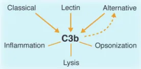
opsonic fragments derived from comple-ment components 3 and 4 (C3 and C4, respectively) on a target, and second, to ini-tiate the inflammatory response through release of the C3a anaphylatoxin. These 2 events occur in parallel and within min-utes, making complement activation an early (if not the earliest) part of the host defense response to a microbe. Three acti-vation pathways — classical (Figure 1), lectin (Figure 2), and alternative (Figure 3) — have been elucidated. They join at the step of C3 cleavage. Common to each path-way is the terminal half of the system, the membrane attack complex. This lytic sys-tem is initiated with the cleavage of C5 to C5a, another potent anaphylatoxin, and
C5b, which combines with C6, C7, C8, and C9 to form a pore or channel in the patho- gen’s cell membrane. Deficiencies in com- plement components predispose to infec-tion and autoimmunity. While it is rather straightforward to account for the former, it has not been so easy for immunologists to explain the latter.
The complement system is activated by lectins, representing a preantibody means of recognizing non-self by targeting the distinct sugar profiles of microorgan-isms (Figure 2). Not surprisingly, parts of the lectin complement cascade were then “borrowed” by the adaptive immune sys-
tem to form the antibody-triggered classi-Nonstandard abbreviations used: MASP, MBL-asso-ciated serine protease; MBL, mannan-binding lectin. Conflict of interest: The authors have declared that no conflict of interest exists.
[image:2.585.394.532.604.672.2]Citation for this article: J. Clin. Invest. 116:1215–1218 (2006). doi:10.1172/JCI28622.
Figure 1
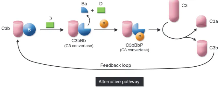
cal pathway. Complement also undergoes a continuous turnover of its key compo-nent, C3, which is the initiating event for the alternative pathway (Figure 3). Sponta- neous turnover of C3 continuously gener-ates C3b, allowing the alternative pathway to function independently of the classical or lectin pathways. This initial C3b can also be generated via the classical or lectin pathways. On cells lacking complement inhibitors, the deposited C3 fragment C3b is the nidus for amplification via the alter-native pathway.
Evolution of the complement system
The complement system is an ancient host defense pathway. As organisms became complex, the system evolved to be more efficient. The most primitive comple-ment system probably initially consisted
primarily of C3. C3 (and C4) possesses a biochemical mechanism whereby a fluid-phase protein may transfer to a surface. Specifically, cleavage of C3 exposes a highly reactive thioester bond that mediates cova-lent attachment to hydroxyl groups on cell surfaces. Once C3b is covalently bound to a pathogen, the alternative pathway’s feed-back loop becomes engaged (Figure 3). The end result of this amplification process is deposition of large amounts of C3b, which opsonize the pathogen, and the concomi-tant liberation of the proinflammatory C3a anaphylatoxin.
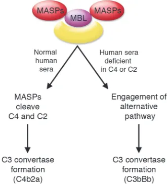
However, this early complement system lacks a more specific means to identify appropriate targets. This void was appar- ently first filled with lectins such as man- nan-binding lectin (MBL) or mannan-bind-ing protein. Lectins bind repeating sugar
moieties on pathogens and then activate complement by engaging MBL-associ-ated serine proteases (MASPs) (Figure 2). Initially, these serine proteases may have directly cleaved and thereby activated C3. To make the lectin pathway of complement activation more efficient, an intermedi-ate step employing C4 and C2 developed. Together they form an enzyme complex — C4b2a — that efficiently cleaves C3 to C3b (Figure 2). The void was next filled with the highly successful humoral immune system in which the complement activating agent antibody defines the target.
Discovery of bypass pathways
[image:3.585.88.319.84.264.2]Many fits and starts and reformulations undoubtedly occurred during the evolu-tion of the complement system. These less evolved cascades, though, are difficult to
Figure 2
[image:3.585.97.476.537.693.2]Lectin pathway. This pathway, also called the MBL pathway, is activated by the binding of MBL and other lectins to sugar moieties on the surface of pathogens, which results in cleav-age of C4 and C2 to produce the C3 conver-tase C4b2a. The converconver-tase that is formed is the same as that in the classical pathway of complement activation.
Figure 3
recognize. An obvious approach to uncover these earlier renditions of the complement system is to characterize the system in lower species. This is not so easy to achieve experimentally, however, especially in non- vertebrates, because there are over 30 dis-tinct interacting components, receptors, and regulators to consider. A second win-dow to evolutionary history is provided by the assessment of individuals with comple-ment component deficiencies. Thus the first such bypass system to come to light was identified by researchers at the NIH employing C4-deficient guinea pig serum (1–3). Although they require higher quanti- ties of sensitizing IgG or IgM than the tra-ditional classical pathway, sheep red blood indicator cells were lysed in this serum. This C4 bypass pathway also required a C1-like protein and the alternative pathway. At this juncture the lectin pathway had not yet been discovered, so the C1-like protein complex could have been a MASP. The NIH group further demonstrated that C2-defi-cient human serum could lyse cells under similar conditions (1–3). In other words, C4 and C2 were not necessary to lyse cells by the classical pathway.
These bypass pathways were next ana-lyzed by Matsushita and Okada, who showed that C4b deposited on cells could facilitate alternative pathway activation in human serum lacking C2 (4). Further work on these pathways also came about because of a serendipitous observation: a patient with complete C2 deficiency and systemic lupus erythematosus was reported by the clinical laboratory to have a total hemo-lytic complement titer approximately 50% of normal (5). As C2 is required for classical pathway activation, on multiple occasions
the titer in this patient had been measured as zero. Upon further analysis, it turned out that during this recent assay, approxi- mately 10 times the usual amount of sensi-tizing antibody had been employed (5, 6). While the total hemolytic complement titer of control (normal) serum was unchanged in the setting of these more heavily sensi- tized cells, serum of a C2-deficient indi-vidual was able to activate the cascade. The biochemical process that permitted this bypass pathway to be engaged was not through a hybrid classical pathway/alter-native pathway convertase (4, 6). Instead, the formation of the alternative pathway was facilitated, probably by providing a site relatively protected from regulators.
Improved assay systems to monitor complement activation
Traditionally it has been thought that lec-tins activate complement through a C4- and C2-dependent pathway analogous to the classical pathway. In this issue of the
JCI, Selander et al. examined the require- ments for mannan-binding protein activa-tion of the lectin pathway (7). Of note, they employed sensitive ELISA methodology in which the lectins in the test serum bound to the O oligosaccharides of several Salmo-nella species. The readout was the quantita- tion of C3 deposition on the plate. Impor-tant features of their assay system as well as similar ones developed by others (8) are specificity for a given pathway, greater sen- sitivity, and use of actual pathogen materi-al as substrates. Also, the concentrations of serum employed more closely match those in body secretions and blood. These types of assays will eventually become standard in the clinical immunology laboratory.
They provide a functional assessment, are easier to perform, and delineate more pre-cisely and in a more physiologic manner the complement pathway(s) being activated. They will replace the current total hemo-lytic complement test. The widely available total hemolytic complement assay mea-sures only the classical pathway and uses highly diluted serum and a sheep red blood cell target — none of which closely approxi-mate the complement system interacting with a microbe in vivo.
Using an improved assay system, Seland-er et al. (7) demonstrate that even in the absence of C2, C4, or a MASP, mannan-binding protein induces substantial C3 deposition. Analogous to the results noted above relative to bypass systems in the clas-sical cascade, these results point to a new pathway by which lectins may activate the complement pathway (Figure 4). Although this likely is not a major pathway of com-plement activation, it may be operative in the context of either acquired or naturally occurring complement deficiencies.
Implications
[image:4.585.50.215.84.266.2]Why is this the study by Selander et al. (7) important? The primary finding is the dem-onstration that individuals deficient in C4 or C2 possess a “backup” or bypass comple-ment activation system. Second, the study reaffirms the likely evolutionary history of the complement system. Third, these bypass pathways can mediate immunopathology. For example, the lysis of Giardia lamblia tro-phozoites (9), tissue damage in Forssman shock (10), binding of immune complexes to complement receptors (11, 12), and endothelial cell damage in hemolytic ure-mic syndrome (13) can all be shown to be
Figure 4
mediated by complement bypass systems. Fourth, and probably most important, the type of assay represents the wave of the future in detecting the activity of the com-plement system in human disease. As these more informative detection systems (like the one used in this report) come into routine clinical use, other examples of these bypass type pathways will likely be uncovered. For human diseases, these more specific and quantitative assay systems will establish which pathway of complement activation is playing a role in disease and elucidate which one to modulate with therapeutic agents. Finally, a word of caution is in order. These bypass pathways are often not considered by investigators attempting to define the role of the complement system in disease states. For example, C4-deficient animals are widely used to rule out a contribution of the classical pathway and/or lectin pathway in mouse models of human disease. One must be wary of such interpretations in view of bypass cascades that become opera-tive in “deficient” states. Thus the natural maturation of an antibody response to an infectious organism (i.e., to go rapidly into antibody excess) is all that is necessary to trigger these more ancient bypass pathways. Using our current methods, such pathways are not analyzed in clinical medicine or in animal models of human disease. Address correspondence to: John P. Atkin- son, Washington University School of Med-icine, 660 South Euclid Avenue, Box 8045, St. Louis, Missouri 63110, USA. Phone: (314) 362-8391; Fax: (314) 362-1366; E-mail: [email protected].
1. May, J.E., and Frank, M.M. 1973. A new comple-ment-mediated cytolytic mechanism-the C1-bypass activation pathway. Proc. Natl. Acad. Sci. U. S. A.
70:649–652.
2. May, J.E., and Frank, M.M. 1973. Hemolysis of sheep erythrocytes in guinea pig serum deficient in the fourth component of complement. I. Antibody and serum requirements. J. Immunol. 111:1661–1667. 3. May, J.E., and Frank, M.M. 1973. Hemolysis of sheep
erythrocytes in guinea pig serum deficient in the fourth component of complement. II. Evidence for involvement of C1 and components of the alternate complement pathway. J. Immunol. 111:1668–1676. 4. Matsushita, M., and Okada, H. 1986. Alternative
complement pathway activation by C4b deposited during classical pathway activation. J. Immunol.
136:2994–2998.
5. Farries, T.C., Knutzen Steuer, K.L., and Atkinson, J.P. 1990. The mechanism of activation of the alter-native pathway of complement by cell-bound C4b.
Mol. Immunol. 27:1155–1161.
6. Knutzen-Steuer, K.L., et al. 1989. Lysis of sensitized sheep erythrocytes in human sera deficient in the second component of complement. J. Immunol.
143:2256–2261.
7. Selander, B., et al. 2006. Mannan-binding lectin activates C3 and the alternative complement pathway without involvement of C2. J. Clin. Invest.
116:1425–1434. doi:10.1172/JCI25982.
8. Seelen, M.A., et al. 2005. Functional analysis of the classical, alternative, and MBL pathways of the com-plement system: standardization and validation of a simple ELISA. J. Immunol. Methods. 296:187–198.
9. Deguchi, M., Gillin, F.D., and Gigli, I. 1987. Mecha-nism of killing of Giardia lamblia trophozoites by complement. J. Clin. Invest. 79:1296–1302. 10. Wagner, E., Platt, J.L., Howell, D.N., Marsh, H.C.,
Jr., and Frank, M.M. 1999. IgG and complement- mediated tissue damage in the absence of C2: evi-dence of a functionally active C2-bypass pathway in a guinea pig model. J. Immunol. 163:3549–3558. 11. Traustadottir, K.H., Rafnar, B.O., Steinsson, K., Valdimarsson, H., and Erlendsson, K. 1998. Par-ticipation of factor B in residual immune complex red cell binding activity observed in serum from a C2-deficient systemic lupus erythematosus patient may delay the appearance of clinical symptoms.
Arthritis Rheum. 41:427–434.
12. Klint, C., Gullstrand, B., Sturfelt, G., and Truedsson, L. 2000. Binding of immune complexes to erythro- cyte CR1 (CD35): difference in requirement of clas- sical pathway components and indication of alter-native pathway-mediated binding in C2-deficiency.
Scand. J. Immunol. 52:103–108.
13. Nolin, L., et al. 1979. Possible C1q bypass loop acti-vation in the haemolytic uraemic syndrome. Clin. Exp. Immunol. 35:107–111.
The IL-23/IL-17 axis in inflammation
Yoichiro Iwakura and Harumichi Ishigame
Center for Experimental Medicine, Institute of Medical Science, University of Tokyo, Tokyo, Japan.
IL-23 induces the differentiation of naive CD4
+T cells into highly
pathogen-ic helper T cells (Th17/Th
IL-17) that produce IL-17, IL-17F, IL-6, and TNF-
α
,
but not IFN-
γ
and IL-4. Two studies in this issue of the
JCI
demonstrate that
blocking IL-23 or its downstream factors IL-17 and IL-6, but not the IL-12/
IFN-
γ
pathways, can significantly suppress disease development in animal
models of inflammatory bowel disease and MS (see the related articles
begin-ning on pages 1310 and 1317). These studies suggest that the IL-23/IL-17
pathway may be a novel therapeutic target for the treatment of chronic
inflammatory diseases.
Th17/ThIL-17 is a new CD4+ helper
T cell subset that produces IL-17
Upon antigenic stimulation, naive CD4+
T cells differentiate into 2 subsets, Th1 and Th2 cells, characterized by different cytokine production profiles and effector
functions (Figure 1). Th1 cells produce large quantities of IFN-γ and mediate cel-lular immunity while Th2 cells, which are involved in humoral immunity, primar-ily produce IL-4, IL-5, and IL-13. IL-12, a heterodimer of the p40 and p35 subunits, induces the differentiation of naive CD4+
T cells into IFN-γ–producing Th1 cells through activation of STAT4. IFN-γ signals are transduced by STAT1, which activates a downstream transcription factor, T-bet, that enhances the expression of genes spe-cific to Th1 cells. In contrast, IL-4 induces STAT6 activation, promoting the expres-sion of GATA-3, a transcriptional factor essential for both IL-4 production and Th2 cell differentiation. Recently, it was report-ed that CD4+ T cells isolated from the
inflamed joints of patients with Lyme dis-ease contain a subset of IL-17–producing CD4+ T cells that are distinct from those
producing either IL-4 or IFN-γ (Figure 1) (1). These IL-17–producing CD4+ T cells
were dubbed Th17 or ThIL-17 cells (2–4).
IL-17, a proinflammatory cytokine pre-dominantly produced by activated T cells, enhances T cell priming and stimulates fibroblasts, endothelial cells, macrophages, and epithelial cells to produce multiple pro-inflammatory mediators, including IL-1, IL-6, TNF-α, NOS-2, metalloprote-ases, and chemokines, resulting in the induction of inflammation (5, 6). IL-17 expression is increased in patients with a variety of allergic and autoimmune diseases, such as RA, MS,
inflamma-Nonstandard abbreviations used: CIA, collagen-induced arthritis; IBD, inflammatory bowel disease; IL-1Ra, IL-1 receptor antagonist; R, receptor. Conflict of interest: The authors have declared that no conflict of interest exists.