heart of the matter
Michael E. Mendelsohn
J Clin Invest.
2005;
115(4)
:840-844.
https://doi.org/10.1172/JCI24806
.
Blood pressure abnormalities are thought to originate from intrinsic changes in the kidney, a
concept that has been largely unchallenged for more than 4 decades. However, recent
molecular, cellular, and transgenic mouse studies support an alternative hypothesis:
primary abnormalities in vascular cell function can also directly cause abnormalities of
blood pressure. In this issue of the
JCI
, Crowley and coworkers describe the application of
an elegant cross-renal transplant model to type 1A angiotensin (AT
1A) receptor–deficient
mice and their wild-type littermates to explore the relative contributions of renal and
extrarenal tissues to the low blood pressure seen in the AT
1Areceptor–deficient animals.
Their studies further support the emerging paradigm that primary abnormalities of the
vasculature can make unique, nonredundant contributions to blood pressure regulation; the
findings have potentially important implications for the ways we diagnose and treat blood
pressure diseases in humans.
Commentary
Find the latest version:
sion in peripheral blood lymphocytes from patients with systemic lupus erythematosus. Autoimmunity.
29:111–120.
23. Kammer, G.M., Khan, I.U., and Malemud, C.J. 1994. Deficient type I protein kinase A isozyme activity in systemic lupus erythematosus T lymphocytes.
J. Clin. Invest.94:422–430.
24. Tada, Y., Nagasawa, K., Yamauchi, Y., Tsukamoto, H., and Niho, Y. 1991. A defect in the protein kinase C system in T cells from patients with systemic lupus erythematosus. Clin. Immunol. Immunopathol.
60:220–231.
25. Gergely, P., Jr., et al. 2002. Persistent mitochondrial hyperpolarization, increased reactive oxygen inter-mediate production, and cytoplasmic alkaliniza-tion characterize altered IL-10 signaling in patients with systemic lupus erythematosus. J. Immunol.
169:1092–1101.
26. Gergely, P., Jr., et al. 2002. Mitochondrial hyper-polarization and ATP depletion in patients with systemic lupus erythematosus. Arthritis Rheum.
46:175–190.
27. Yi, Y., McNerney, M., and Datta, S.K. 2000. Regula-tory defects in Cbl and mitogen-activated protein kinase (extracellular signal-related kinase) pathways cause persistent hyperexpression of CD40 ligand in human lupus T cells. J. Immunol.165:6627–6634. 28. Juang, Y.-T., Tenbrock, K., Nambiar, M.P., Gourley,
M.F., and Tsokos, G.C. 2002. Defective produc-tion of funcproduc-tional 98-kDa form of Elf-1 is respon-sible for the decreased expression of TCR z-chain in patients with systemic lupus erythematosus.
J. Immunol.169:6048–6055.
29. Kyttaris, V.C., Juang, Y.-T., Tenbrock, K., Wein-stein, A., and Tsokos, G.C. 2004. Cyclic adenosine 5′-monophosphate response element
modula-tor is responsible for the decreased expression of c-fos and activator protein-1 binding in T cells from patients with systemic lupus erythematosus.
J. Immunol.173:3557–3563.
30. Wong, H.K., Kammer, G.M., Dennis, G., and Tso-kos, G.C. 1999. Abnormal NF-kB activity in T lym-phocytes from patients with systemic lupus ery-thematosus is associated with decreased p65-RelA protein expression. J. Immunol.163:1682–1689. 31. Deng, C., et al. 2001. Decreased
ras-mitogen-acti-vated protein kinase signaling may cause DNA hypomethylation in T lymphocytes from lupus patients. Arthritis Rheum.44:397–407.
32. Grolleau, A., Kaplan, M.J., Hanash, S.M., Beretta, L., and Richardson, B.C. 2000. Impaired translational response and increased protein kinase PKR expres-sion in T cells from lupus patients. J. Clin. Invest.
106:1561–1568.
In hypertension, the kidney is not always
the heart of the matter
Michael E. Mendelsohn
Molecular Cardiology Research Institute, Tufts–New England Medical Center, Boston, Massachusetts, USA.
Blood pressure abnormalities are thought to originate from intrinsic
changes in the kidney, a concept that has been largely unchallenged for
more than 4 decades. However, recent molecular, cellular, and transgenic
mouse studies support an alternative hypothesis: primary abnormalities in
vascular cell function can also directly cause abnormalities of blood
pres-sure. In this issue of the
JCI
, Crowley and coworkers describe the application
of an elegant cross-renal transplant model to type 1A angiotensin (AT
1A)
receptor–deficient mice and their wild-type littermates to explore the
rela-tive contributions of renal and extrarenal tissues to the low blood pressure
seen in the AT
1Areceptor–deficient animals (see the related article beginning
on page 1092). Their studies further support the emerging paradigm that
primary abnormalities of the vasculature can make unique, nonredundant
contributions to blood pressure regulation; the findings have potentially
important implications for the ways we diagnose and treat blood pressure
diseases in humans.
Nonstandard abbreviations used: AT1A, type 1A
angiotensin; BKCa2+ large conductance Ca2+-activated K+
channel; CO, cardiac output; D+R+, donor+ recipient+;
IP3, inositol triphosphate; MLC, myosin light chain;
MLCK, MLC kinase; PKGI, cGMP-dependent protein kinase type I; PP1M, myosin phosphatase; RAS, renin-angiotensin system; RGS2, regulator of G protein signaling 2; SVR, systemic vascular resistance.
Conflict of interest: The author has declared that no conflict of interest exists.
Citation for this article:J. Clin. Invest.115:840–844 (2005). doi:10.1172/JCI200524806.
Introduction
Abnormalities of blood pressure, especially high blood pressure or hypertension, are widespread, cause extensive morbidity and mortality, and are insidious, because they rarely generate symptoms. But what causes hypertension? This simple question is remarkably difficult to answer, in large
part because blood pressure, which is mea-surable only in intact animals, is an inte-grated value determined by contributions from a complex mix of rheologic, renal, neural, vascular, and hormonal variables. It is therefore difficult to distinguish primary or causal factors for abnormal blood pres-sure from those responses that are second-ary to the changes in pressure.
Blood pressure is proportional to 2 funda-mental hemodynamic factors: cardiac out-put (CO) and systemic vascular resistance (SVR), and the equation BP = CO × SVR is taught to every first-year physiology stu-dent. The pioneering studies of Guyton and colleagues provide abundant support for the central, causal role of abnormal renal sodium handling in the etiology of hypertension (1, 2). This work
emphasiz-es the primacy of the kidney’s “premphasiz-essure control system,” which is recruited when systemic pressure is altered and causes a renally mediated increase or decrease in blood volume to establish long-term blood pressure control. Elegant genetic studies published over the past decade have provided further strong support for the hypothesis that alterations of net salt balance underlie abnormalities of blood pressure in humans (3). But are the alterations in renal salt balance that cause abnormal blood pressure always direct, as has been argued for many years, or could renal alterations in some cases be indirect or secondary to primary abnormalities of nonrenal tissues? Does the kidney always cause abnormal blood pressure by altering blood volume, which in turn alters CO and SVR? Or do intrinsic abnormalities of the vasculature in some cases give rise to a pri-mary abnormality of SVR, causing hyper-tension? Surprisingly, we do not yet know the answer to this simple dilemma.
In this issue of the JCI, Crowley and col-leagues (4) describe cross-renal transplan-tation studies with type 1A angiotensin (AT1A) receptor–deficient (Agtr1a–/–) mice
deter-mining blood pressure. AT1A
receptor–defi-cient mice are hypotensive, and their lower blood pressures have been attributed to renal changes in the function of the renin-angiotensin system (RAS). As the authors explain, their experiments were designed to explore the relative contributions of individual tissue compartments to the low blood pressure seen in AT1A-deficient mice,
since there is at present a surprising lack of direct proof of the primacy of the kidney in the regulation of blood pressure by RAS.
In the current study, 4 experimental groups of mice were created, and mice in all 4 groups received a new kidney by transplantation (4). The 4 groups studied included wild-type animals that received a wild-type kidney (group I or donor+
recipient+ [D+R+]); wild-type animals
that received an AT1A receptor–knockout
mouse kidney (group II or D–R+); AT1A
receptor–knockout mice that received a wild-type kidney (group III or D+R–); and
AT1A receptor–knockout mice that received
an AT1A receptor–knockout mouse kidney
(group IV or D–R–). Mice in all 4 groups
also underwent implantation of radiote-lemetry transmitters 6–8 days after kidney transplant procedure to monitor blood pressure. Untransplanted wild-type and AT1A receptor–knockout mice also received
blood pressure transmitters and served as additional controls.
The results of these experiments are both highly informative and provocative. Blood pressures in D+R+ mice were no different
from those in nontransplanted, wild-type mice, which confirmed that the transplant
procedure itself does not significantly alter systemic pressure (4). D–R+ mice had
sig-nificant reductions in blood pressure, as expected, which supports the importance of the kidney in determining systemic pres-sure. However, the D+R– mice had similar,
significant reductions in blood pressure. This is the “punch line” of the article: nonrenal tissues have an equal and inde-pendent role in determining blood sure in these mice. Furthermore, the pres-ence of intact AT1A receptor signaling in
the kidneys of D+R– mice was insufficient
to compensate for the absence of AT1A
receptors in their nonrenal tissues. The authors performed additional studies to determine whether lower aldosterone pro-duction in the D+R– mice might account
for their lower blood pressures, using an aldosterone clamping approach in which mice received steady infusions of this hor-mone. However, despite supraphysiological and equivalent aldosterone levels, blood pressures remained significantly lower in the D+R– mice compared with D+R+ mice,
which demonstrates the critical impor-tance of nonrenal tissues in the determina-tion of blood pressure.
These cross-transplantation studies in AT1A receptor–deficient mice (4) do not
precisely localize the nonrenal tissues responsible for determining blood pres-sure, but they provide strong hints that one such tissue is the vasculature. Sympathetic nervous system activation is thought to be important to the initiation and mainte-nance of high blood pressure and to con-tribute to hypertension (reviewed in refs. 5, 6). However, renal norepinephrine levels (an accepted measure of renal sympathetic nerve activity) were reduced equally in the D+R+ and D+R– mice, which indicates that
altered sympathetic activity is an unlikely explanation for the reduced blood pres-sures observed in D+R– mice. The chronic
reduction in renal norepinephrine levels confirms the persistent absence of sym-pathetic innervation in the transplanted kidneys and supports the notion that the reduced blood pressure observed in the D+R– mice results from direct vascular
effects of AT1A receptor deficiency.
Primary vascular abnormalities as a cause of abnormal blood pressure
[image:3.585.43.371.79.389.2]Though blood vessel tone is a major deter-minant of blood pressure, and abnormali-ties in vascular tone are common in hyper-tensive and ischemic disorders, the notion that abnormalities of vascular tone may in
Figure 1
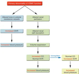
some cases actually cause high or low blood pressure remains unproven. Work over the past 5 years, including that of Crowley et al. discussed here (4), supports the hypoth-esis that VSMC abnormalities that alter the intrinsic contractile state of the cell can directly cause abnormal vascular tone and disorders of blood pressure regulation, including hypertension. Put differently, according to this hypothesis, primary vaso-dilatation or vasoconstriction is alone suf-ficient to cause hypotension or hyperten-sion, respectively. Vasoconstriction could produce hypertension in at least 2 different ways (Figure 1). Hypertension could arise de novo from alterations in the set point of resting vascular tone in the resistance vessels that regulate blood pressure. Alternatively, intrinsic abnormalities in the VSMCs of renal vessels could alter renal vacular tone and perfusion, thus secondarily creating the recognized changes in salt handling by the kidney that promote and/or sustain blood pressure elevations. In either case, however, the fundamental abnormality resides in the
critical VSMC proteins that regulate the contractile state of the cell, which focuses our attention on different diagnostic and therapeutic targets.
Regulation of smooth muscle cell tone
VSMCs are the principal cell of the blood vessel wall, and VSMC contractile state is dynamically and rapidly regulated by
[image:4.585.53.527.86.333.2]hor-monal and neural inputs. However, the basal state of VSMC tone is determined by a small number of proteins that are part of a final common pathway regulating force generation by the cell’s actinomyosin-based contractile apparatus. VSMC con-traction is initiated by a rise in intracellular calcium, which can occur as a result of either: entry of extracellular calcium into the cell via calcium channels following
Figure 2
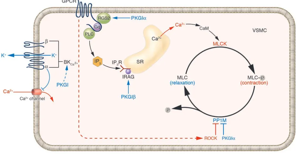
Vascular tone is dynamically regulated by MLC phosphorylation and dephosphorylation in VSMCs. Increases in VSMC intracellular calcium level via receptor-activated (pharmacomechanical) or ion channel–activated (electromechanical) pathways lead to MLCK activation. MLCK phosphor-ylates MLCs (MLC-P), activating the myosin ATPase, actinomyosin cross-bridging, and an increase in tension. PP1M dephosphorphosphor-ylates MLC-P, decreasing cell tension. PP1M is activated by the NO and nitrovasodilator effector PKGI, which has 2 isoforms (PKGIα and PKGIβ). RGS2, which is essential for normal blood pressure, causes VSMC relaxation by attenuating Gq protein–coupled receptor activation and associated rises in intracellular calcium concentration; it too is activated by PKGIα. PP1M is inhibited by Rho/Rho kinase (ROCK). The calcium-activated potassium channel, BKCa2+, is activated by PKG and by local calcium sparks, hyperpolarizing the cell, decreasing calcium entry, and decreasing
MLCK activity. This shifts the equilibrium between MLC and MLC-P, causing relaxation. Blue, relaxant pathway; red, contractile pathway. GPCR, G protein–coupled receptor; IP3R, IP3 receptor; SR, sarcoplasmic reticulum; PLC, phospholipase C; CaM, calmodulin; IRAG, IP3R-associated
cGMP kinase substrate.
Table 1
Mouse models of vascular contractile dysfunction and hypertension
Mouse model Phenotype Reference
BKCa2+ channel β1 subunit KO Vascular contractile dysfunction, HTN (12)
Estrogen receptor β KO Loss of K+ channels, vascular contractile (13)
dysfunction, HTN
VSMC Sur2 K(ATP) channel KO Vasospasm, HTN (14)
eNOS KO Vascular contractile dysfunction, HTN (21)
RGS2 KO Vascular contractile dysfunction, HTN (23)
PKGI KO Vascular contractile dysfunction, HTN (27)
PKGIα leucine zipper mutant knockin Vascular contractile dysfunction, HTN (28)
[image:4.585.222.533.625.730.2]membrane depolarization; or mobilization of intracellular calcium stores following activation of membrane receptors by con-tractile agonists (Figure 2) (reviewed in ref. 7). When intracellular calcium rises, myo-sin light chain kinase (MLCK) is activated (Figure 2) and phosphorylates the regula-tory myosin light chains (MLCs), which causes contraction (7). Conversely, when MLCs are dephosphorylated by the criti-cal regulatory protein myosin phosphatase (PP1M), relaxation occurs (reviewed in refs. 7, 8). Thus, smooth muscle contrac-tile state is determined by the state of MLC phosphorylation, which is directly deter-mined by the balance of MLCK and PP1M activities. It is not surprising that MLCK and PP1M are themselves highly regulated and that alterations in their activities can shift resting VSMC tone. Proteins regulat-ing VSMC intracellular calcium levels and MLC phosphorylation state are therefore good candidates for primary regulators of blood pressure.
Abnormalities in contractile pathway pro-teins can cause primary changes in VSMC tone and hypertension. Voltage-gated cal-cium channels, which allow calcal-cium ions to enter VSMCs, are sensitive to the resting potential of the cell and therefore regu-lated by potassium channels, which make major contributions to determining cellu-lar potential. In VSMCs, the cellu-large conduc-tance Ca2+-activated K+ channel (BK
Ca2+) is
an important primary regulator of vascular relaxation (9). BKCa2+ is activated by local
calcium sparks (reviewed in ref. 10) and by cGMP-dependent protein kinase (PKG) (11); this results in hyperpolarization of the cell, which decreases calcium entry and causes relaxation. VSMCs from mice with several potassium channel abnormalities display abnormal vascular contraction and hypertension (12–14) (Table 1). Many contractile agonists work by activating G protein–coupled receptors, which trigger mobilization of intracellular calcium and activation of MLCK (15). These receptors also activate Rho/Rho kinase, which direct-ly inhibits PP1M, augmenting contraction (8) (Figure 2). A recently recognized class of Rho kinase inhibitors lowers blood pres-sure (16), and one such compound, fasudil, shows promise in clinical trials as a new antihypertensive and vasospastic therapy (17, 18). It is also worth reconsidering the mechanisms of action of molecules we have assumed work solely through renal mecha-nisms to cause hypertension, such as the mineralocorticoid receptor (19, 20).
VSMC relaxation is also tightly regulated and the NO-cGMP-cGMP–dependent pro-tein kinase type I (NO-cGMP-PKGI–depen-dent) pathway contains molecules that also are good candidates for primary vascular regulators of blood pressure. Mice lacking eNOS have hypertension (21), and though the mechanism of abnormal blood pres-sure regulation in these animals is not yet understood, it likely involves PKGI (Figure
2).The endogenous vasodilator NO and
related compounds cause vascular relax-ation by activating PKGIα, which binds directly to and activates PP1M, reducing MLC phosphorylation and causing VSMC relaxation (22) (Figure 2). PKGIα also can relax smooth muscle cells by direct inter-action with the regulator of G protein sig-naling 2 (RGS2), a second PKGI substrate. RGS2 terminates signaling by Gq-coupled contractile agonist receptors, which medi-ate the actions of many physiologically important vascular contractile agonists (23). RGS2-deficient mice have abnor-mal vascular contraction and relaxation and are hypertensive (23, 24). The PKGIβ
isoform regulates release of calcium from intracellular stores, and mice lacking the PKGIβ target protein inositol triphos-phate receptor–associated cGMP kinase substrate (IRAG) have abnormal vascular relaxation (25, 26). Mice fully lacking PKGI (27) or with PKGI mutations that prevent the PKGI-PP1M interaction (28) also are hypertensive (Table 1).
The hypotensive AT1A receptor–deficient
mice studied by Crowley et al. (4) represent a loss-of-function model. It will also be important to apply the cross-renal trans-plantation approach to mice with single gene abnormalities that cause hyperten-sion (Table 1). More studies of mice har-boring VSMC-specific changes in candidate blood pressure–determining genes are also needed. Ultimately, however, nonrenal can-didate genes for blood pressure determina-tion will need to be explored in humans. Careful scrutiny of genomic associations and polymorphisms in candidate genes will be required. Several hypertension-associated loci that have been identified in genome-wide linkage studies contain candidate genes that we and others are cur-rently exploring, including the Rho kinases (ROCK1, located at chromosome18q11.2; and ROCK2, at 2p24) (29, 30) and the BK channel β subunit (on 5q34) (31, 32). Iden-tification of new, nonrenal genes capable of determining blood pressure in humans would have important implications for
diagnosis and therapy of a wide spectrum of cardiovascular disorders.
Address correspondence to: Michael E. Men-delsohn, Molecular Cardiology Research Institute, Tufts-New England Medical Cen-ter, 750 Washington St., Box 080, Boston, Massachusetts 02111, USA. Phone: (617) 636-9370; Fax: (617) 636-1444; E-mail: [email protected].
1. Guyton, A.C., et al. 1972. Arterial pressure regula-tion. Overriding dominance of the kidneys in long-term regulation and in hypertension. Am. J. Med.
52:584–594.
2. Guyton, A.C. 1991. Blood pressure control — spe-cial role of the kidneys and body fluids. Science.
252:1813–1816.
3. Lifton, R.P., Gharavi, A.G., and Geller, D.S. 2001. Molecular mechanisms of human hypertension [review]. Cell. 104:545–556.
4. Crowley, S.D., et al. 2005. Distinct roles for the kid-ney and systemic tissues in blood pressure regula-tion by the renin-angiotensin system. J. Clin. Invest.
115:1092–1099. doi:10.1172/JCI200523378. 5. Ferrario, C.M., and Averill, D.B. 1991. Do primary
dysfunctions in neural control of arterial pres-sure contribute to hypertension? Hypertension.
18(Suppl. 3):I38–I51.
6. Sved, A.F., Ito, S., and Sved, J.C. 2003. Brainstem mechanisms of hypertension: role of the rostral ven-trolateral medulla. Curr. Hypertens. Rep. 5:262–268. 7. Somlyo, A.P., and Somlyo, A.V. 1994. Signal
transduction and regulation in smooth muscle.
Nature. 372:231–236.
8. Hartshorne, D.J., Ito, M., and Erdodi, F. 2004. Role of protein phosphatase type 1 in contrac-tile functions: myosin phosphatase. J. Biol. Chem.
279:37211–37214.
9. Brayden, J.E., and Nelson, M.T. 1992. Regulation of arterial tone by activation of calcium-dependent potassium channels. Science. 256:532–535. 10. Jaggar, J.H., and Nelson, M.T. 2000. Calcium
sparks in smooth muscle. Am. J. Physiol., Cell Physiol.
278:C235–C256.
11. Robertson, B.E., Schubert, R., Hescheler, J., and Nel-son, M.T. 1993. cGMP-dependent protein kinase activates Ca-activated K channels in cerebral artery smooth muscle cells. Am. J. Physiol., Cell Physiol.
265:C299–C303.
12. Brenner, R., et al. 2000. Vasoregulation by the B1 subunit of the calcium-activated potassium chan-nel. Nature. 407:870–876.
13. Zhu, Y., et al. 2002. Abnormal vascular function and hypertension in mice deficient in estrogen receptor β. Science.295:505–508.
14. Chutkow, W.A., et al. 2002. Episodic coronary artery vasospasm and hypertension develop in the absence of Sur2 K(ATP) channels. J. Clin. Invest.
110:203–208. doi:10.1172/JCI200215672. 15. Demoliou-Mason, C.D. 1998. G-protein-coupled
receptors in vascular smooth muscle cells. Biol. Sig-nals. 7:90–97.
16. Uehata, M.,et al. 1997. Calcium sensitization of smooth muscle mediated by a Rho-associated pro-tein kinase in hypertension. Nature. 389:990–994. 17. Masumoto, A., et al. 2001. Possible involvement of
rho-kinase in the pathogenesis of hypertension in humans. Hypertension. 38:1307–1310.
18. Hirooka, Y., Shimokawa, H., and Takeshita, A. 2004. Rho-kinase, a potential therapeutic target for the treatment of hypertension. Drug News Perspect.
17:523–527.
20. Jaffe, I.Z., and Mendelsohn, M.E. 2005. Angiotensin II and aldosterone regulate gene transcription via functional mineralocortocoid receptors in human coronary artery smooth muscle cells. Circ. Res. doi:10.1161/01.RES.0000159937.05502.d1. 21. Huang, P.L., et al. 1995. Hypertension in mice
lack-ing the gene for endothelial nitric oxide synthase.
Nature. 377:239–242.
22. Surks, H.K., et al. 1999. Regulation of myosin phosphatase by a specific interaction with cGMP-dependent protein kinase Iα. Science. 286:1583–1587. 23. Tang, M., et al. 2003. Regulator of G-protein signal-ing 2 mediates vascular smooth muscle relaxation and blood pressure. Nat. Med. 9:1506–1512. 24. Le, T.H., and Coffman, T.M. 2003. RGS2: a
“turn-off” in hypertension. J. Clin. Invest. 111:441–443.
doi:10.1172/JCI200317836.
25. Schlossmann, J., et al. 2000. Regulation of intracellular calcium by a signalling complex of IRAG, IP3 receptor and cGMP kinase Iβ. Nature.
404:197–201.
26. Geiselhoringer, A., et al. 2004. IRAG is essential for relaxation of receptor-triggered smooth muscle con-traction by cGMP kinase. EMBO J. 23:4222–4231. 27. Pfeifer, A., et al. 1998. Defective smooth muscle
reg-ulation in cGMP kinase I-deficient mice. EMBO J.
17:3045–3051.
28. Mendelsohn, M.E. 2005. Molecular mechanisms reg-ulating vascular smooth muscle cell tone and blood pressure [abstract]. Keystone Symposia: The Cellular Biol-ogy of Atherosclerosis. Keystone, Colorado, USA. 29. Levy, D., et al. 2000. Evidence for a gene
influenc-ing blood pressure on chromosome 17: genome scan linkage results for longitudinal blood pres-sure phenotypes in subjects from the Framingham Heart Study. Hypertension. 36:477–483.
30. Angius, A., et al. 2002. A new essential hypertension susceptibility locus on chromosome 2p24-p25, detected by genomewide search. Am. J. Hum. Genet.
71:893–905.
31. Krushkal, J., et al. 1999. Genome-wide linkage analyses of systolic blood pressure using highly discordant siblings. Circulation. 99:1407–1410. 32. Fernandez-Fernandez, J.M., et al. 2004.
Gain-of-function mutation in the KCNMB1 potassium channel subunit is associated with low prevalence of diastolic hypertension. J. Clin. Invest. 113:1032–1039. doi:10.1172/JCI200420347.
Ikaros transcription factors:
flying between stress and inflammation
George P. Chrousos1,2 and Tomoshige Kino2
1First Department of Pediatrics, University of Athens, Athens, Greece. 2Reproductive Biology and Medicine Branch,
National Institute of Child Health and Human Development, NIH, Bethesda, Maryland, USA.
The hypothalamic-pituitary-adrenal axis (a major component of the stress
system) and the immune system contribute to the maintenance of
homeo-stasis at rest and during stress. Because of their essential roles for the
sur-vival of self and species, the activities of these systems have evolutionarily
developed in parallel and are intertwined at many levels. In this issue of the
JCI
, Ezzat et al. demonstrate that Ikaros, a differentiation factor of leukocyte
lineage, also influences the maturation of the fetal pituitary corticotroph
and, hence, the secretion of adrenocorticotropic hormone before and after
birth (see the related article beginning on page 1021). These results indicate
that Ikaros is an ontogenetic and phylogenetic integrator of the stress and
immune systems and that abnormalities in its function may produce
endo-crine and/or immune pathologies.
The stress and immune systems influence each other’s activity
The stress and immune systems play essen-tial roles in the maintenance of resting and stress-related homeostasis (1–3). The for-mer regulates behavioral, cardiovascular, metabolic, and immune homeostasis, so that individuals can survive under various stressful conditions. The latter distinguish-es between self and non-self and between injurious and noninjurious agents and protects the individual from external and
internal pathogens by eliminating them via several discrete mechanisms. As these 2 sys-tems have been very important for survival of the self and species, they have evolved in parallel, sharing regulatory and mediatory molecules and mutually influencing each other’s activities in both a cell- and a tissue- and time-specific manner.
When the stress and immune systems are activated during inflammation, their inter-action is particularly evident in the relation-ship between the HPA axis and the inflam-matory/immune reaction (Figure 1 and Table 1). The HPA axis, together with the arousal and autonomic systems, form the stress system. The central components of this system are located in the hypothalamus and brain stem. These centers, the para-ventricular nucleus (PVN) and the locus caeruleus/norepinephrine system, receive signals of internal and external changes, respectively, from both the central nervous
Nonstandard abbreviations used: ACTH, adreno-corticotrophic hormone; AVP, arginine vasopressin; CRH, corticotropin-releasing hormone; GR, glucocor-ticoid receptor; HPA, hypothalamic-pituitary-adrenal;
α-MSH, α–melanocyte-stimulating hormone; POMC, proopiomelanocortin.
Conflict of interest: The authors have declared that no conflict of interest exists.
Citation for this article:J. Clin. Invest.115:844–848 (2005). doi:10.1172/JCI200524886.
system and distant organs. The PVN trans-duces these signals to the pituitary gland by secreting corticotropin-releasing hormone (CRH) and arginine vasopressin (AVP) into the hypophyseal portal system. The pitu-itary gland, in particular its component corticotroph cells, is synergistically acti-vated by these neuropeptides and secretes adrenocorticotropic hormone (ACTH) into the systemic circulation, stimulating the cortices of the adrenal glands to produce glucocorticoids. These systemic end-effec-tor hormones interact with the ubiquitous glucocorticoid receptors (GRs) in target tis-sues and alter the biologic activities of the brain and most organs and tissues, includ-ing the immune system.