Syntaxin 1A is expressed in airway epithelial
cells, where it modulates CFTR Cl
–
currents
Anjaparavanda P. Naren, … , Deborah J. Nelson, Kevin L.
Kirk
J Clin Invest.
2000;
105(3)
:377-386.
https://doi.org/10.1172/JCI8631
.
The CFTR Cl
–channel controls salt and water transport across epithelial tissues.
Previously, we showed that CFTR-mediated Cl
–currents in the
Xenopus
oocyte expression
system are inhibited by syntaxin 1A, a component of the membrane trafficking machinery.
This negative modulation of CFTR function can be reversed by soluble syntaxin 1A
peptides and by the syntaxin 1A binding protein, Munc-18. In the present study, we
determined whether syntaxin 1A is expressed in native epithelial tissues that normally
express CFTR and whether it modulates CFTR currents in these tissues. Using
immunoblotting and immunofluorescence, we observed syntaxin 1A in native gut and
airway epithelial tissues and showed that epithelial cells from these tissues express
syntaxin 1A at >10-fold molar excess over CFTR. Syntaxin 1A is seen near the apical cell
surfaces of human bronchial airway epithelium. Reagents that disrupt the CFTR-syntaxin
1A interaction, including soluble syntaxin 1A cytosolic domain and recombinant Munc-18,
augmented cAMP-dependent CFTR Cl
–currents by more than 2- to 4-fold in mouse
tracheal epithelial cells and cells derived from human nasal polyps, but these reagents did
not affect CaMK II–activated Cl
–currents in these cells.
Article
Introduction
The cystic fibrosis transmembrane conductance regu-lator (CFTR) is a cAMP-activated Cl–channel that is
localized to the apical membranes of epithelial cells lin-ing the airway, gut, and exocrine glands (1). CFTR is causal in 2 major human diseases: cystic fibrosis (CF) and secretory diarrhea. CF is caused by mutations in the CFTR gene that reduce the synthesis or the func-tional activity of the CFTR Cl–channel. This autosomal
recessive disorder affects approximately 1 in 2,500 Cau-casians in the United States (1). The severest forms of the disease are associated with early onset of
Pseudomonas aeruginosainfection in airway epithelia and premature death (1). Currently, lung transplantation is the only effective therapy in CF. Other symptoms include pancreatic insufficiency, meconium ileus, and infertility. Whereas reduced CFTR activity causes CF, excessive CFTR activity is implicated in cases of toxin-induced secretory diarrhea (e.g., by cholera toxin and heat stable Escherichia colienterotoxin) that stimulate
cAMP or cGMP production in the gut. It is estimated that approximately 12,600 children from Asia, Africa, and Latin America die each day as a result of diarrhea and that the majority of these cases are caused by enterotoxigenic E. coli(2).
Recently, we demonstrated that CFTR-mediated Cl–
currents in Xenopusoocytes can be inhibited by recom-binant syntaxin 1A (3, 4). Syntaxin 1A is highly expressed in the brain, where it regulates synaptic vesi-cle fusion (5) possibly in concert with Munc-18, a syn-taxin 1A–binding protein (6, 7). Synsyn-taxin 1A is also expressed in certain CFTR-expressing colonic carcino-ma cell lines (3), although the expression of syntaxin 1A protein in native gut or airway epithelia has not been reported. In addition to regulating vesicle fusion at the synapse, syntaxin 1A has been observed to bind direct-ly to presynaptic Ca2+channels and to modulate the
gating of these channels (8). It has been proposed that the binding of syntaxin 1A to Ca2+channels may
spa-tially and temporally couple the exocytotic machinery
Syntaxin 1A is expressed in airway epithelial cells,
where it modulates CFTR Cl
–currents
Anjaparavanda P. Naren,
1Anke Di,
2Estelle Cormet-Boyaka,
1Prosper N. Boyaka,
3Jerry R. McGhee,
3Weihong Zhou,
4Kimio Akagawa,
5Tomonori Fujiwara,
5Ulrich Thome,
6John F. Engelhardt,
4Deborah J. Nelson,
2and Kevin L. Kirk
11Gregory Fleming James Cystic Fibrosis Center and Department of Physiology and Biophysics,
University of Alabama at Birmingham, Birmingham, Alabama 35294, USA 2Department of Neurology, University of Chicago, Chicago, Illinois 60637, USA
3The Immunobiology Vaccine Center and Department of Microbiology, University of Alabama at Birmingham,
Birmingham, Alabama 35294, USA
4Department of Anatomy and Cell Biology, University of Iowa, Iowa City, Iowa 52242, USA 5Department of Physiology, Kyorin University School of Medicine, Tokyo 181-8611, Japan
6Department of Pediatrics, University of Alabama at Birmingham, Birmingham, Alabama 35294, USA
Address correspondence to: Kevin L. Kirk, Department of Physiology and Biophysics, University of Alabama at Birmingham, 1918 University Boulevard, Birmingham, Alabama 35294, USA.
Phone: (205) 934-3122; Fax: (205) 934-1445; E-mail: [email protected].
Anjaparavanda P. Naren and Anke Di contributed equally to this work.
Received for publication October 6, 1999, and accepted in revised form December 15, 1999.
The CFTR Cl–channel controls salt and water transport across epithelial tissues. Previously, we showed
that CFTR-mediated Cl–currents in the Xenopusoocyte expression system are inhibited by syntaxin 1A,
a component of the membrane trafficking machinery. This negative modulation of CFTR function can be reversed by soluble syntaxin 1A peptides and by the syntaxin 1A binding protein, Munc-18. In the present study, we determined whether syntaxin 1A is expressed in native epithelial tissues that nor-mally express CFTR and whether it modulates CFTR currents in these tissues. Using immunoblotting and immunofluorescence, we observed syntaxin 1A in native gut and airway epithelial tissues and showed that epithelial cells from these tissues express syntaxin 1A at >10-fold molar excess over CFTR. Syntaxin 1A is seen near the apical cell surfaces of human bronchial airway epithelium. Reagents that disrupt the CFTR-syntaxin 1A interaction, including soluble syntaxin 1A cytosolic domain and recom-binant Munc-18, augmented cAMP-dependent CFTR Cl–currents by more than 2- to 4-fold in mouse
tracheal epithelial cells and cells derived from human nasal polyps, but these reagents did not affect CaMK II–activated Cl–currents in these cells.
to Ca2+influx at the synapse (9). Recent evidence that
syntaxins may also regulate the activities of epithelial Na+channels (ENaC) and plant K+and Cl–channels
implies that syntaxins may be relatively general regula-tors of ion channel activity (10, 11).
The regulation of CFTR Cl–channels by recombinant
syntaxin 1A in Xenopusoocytes appears to be mediated in part by a direct protein-protein interaction. The inhi-bition of CFTR activity and modulation of Ca2+
chan-nel gating each requires the membrane anchor of syn-taxin 1A (4, 8). Soluble synsyn-taxin 1A peptides and recombinant Munc-18 can reverse the inhibition of CFTR activity by syntaxin 1A in oocytes (3), apparent-ly by blocking the physical interaction between CFTR and membrane-anchored syntaxin 1A. It has been reported recently that syntaxin 1A inhibits CFTR Cl–
channel activity in Xenopusoocytes by altering CFTR trafficking to the cell surface (12). However, syntaxin 1A directly binds to the NH2-terminal tail of CFTR,
which is a region that modulates CFTR gating (13).
Thus, it is conceivable that this interaction can influ-ence CFTR function at multiple levels, i.e., channel gat-ing and intracellular traffic.
The broad goal of the present study was to determine whether syntaxin 1A is expressed in native epithelial tis-sues and whether syntaxin 1A regulates CFTR activity in these tissues. Syntaxin 1A has been argued to be a neural-specific protein (14, 15); whether it is expressed in normal gastrointestinal or airway epithelial cells is unknown. Moreover, nearly all the functional data regarding the reg-ulation of CFTR activity by syntaxin 1A are derived from studies in heterologous expression systems such as Xeno-pusoocytes. In this study, we establish that syntaxin 1A is expressed in native epithelial tissues, where it appears to limit the functional activity of the CFTR Cl–channel.
Methods
[image:3.612.97.499.56.385.2]Isolation of epithelial cells from mouse trachea and intestine. Mouse intestinal epithelium (MIE) and mouse tra-cheal epithelium (MTE) cells were isolated from C57
Figure 1
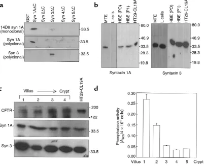
BL/6 mice aged 8–12 weeks. Intestinal epithelial cells from villi tips and crypt portions of mouse intestine were isolated from fragments of small intestine as described previously (16). The villi tip or crypt origin of epithelial cells in each fraction was determined by analysis of alkaline phosphatase activity, which is enriched in villus (Figure 1d). For this purpose, 1.9 mM p-nitrophenylphosphate (Sigma Chemical Co., Saint Louis, Missouri, USA) in 50 mM carbonate buffer was added to 5 ×10⁴cells, and the absorbance
was measured for 10 minutes at 405 nm (16).
For the isolation of tracheal cells for Western blot-ting, MTE were longitudinally opened and subjected to 5 sequential incubations (10 minutes at 37°C) in F12 media. Supernatants were pooled and layered onto dis-continuous Percoll gradients made of 75%, 55%, 40%,
and 20% Percoll layers. The epithelial cells were collect-ed at the 20-40% Percoll layer interface (16).
Seeding MTE cells and human nasal polyps for patch clamp studies. Primary cells from MTE were prepared and cul-tured for 2–3 days as described previously (17). Cells were split into 35-mm dishes and cultured 4–6 days before whole-cell patch clamp studies. Nasal polyp specimens were obtained from 2 subjects according to an Institu-tional Review Board–approved protocol. The methods for obtaining and enzymatically dispersing human air-way epithelial cells were adapted from Coleman et al. (18). Dissociated nasal polyp cells were cultured for 6–7 days on 35-mm dishes before patch clamp analysis.
Bronchial airway epithelial cell culture. Primary human bronchial epithelial (HBE) cell cultures were generated as described previously (19) from human bronchial
tis-Figure 2
[image:4.612.58.531.262.594.2]sue acquired at the time of lung transplantation. Briefly, surface airway epithelial cells were isolated from bronchial samples by protease XIV digestion and cul-tured in hormonally defined BEGM media for less than 1 week (Clonetics Inc., San Diego, California, USA). Freshly isolated (P0) and primary cultures (P1) of bronchial surface airway epithelial cells were analyzed for syntaxin 1A and 3 protein by Western blotting.
Antibodies. Syntaxin 1A– and syntaxin 3–specific anti-bodies were generated by immunizing rabbits with puri-fied GST-syntaxin fusion proteins containing the respec-tive cytoplasmic domains (amino acids 1–266). The antibodies were first subjected to Protein A affinity chro-matography and further purified by passing them through a column containing the respective syntaxin fusion protein lacking GST that was immobilized on CNBr-activated Sepharose. The monoclonal syntaxin 1A antibody (14D8) was developed as described earlier (20). Syntaxin 1A is highly conserved across species and shares approximately 93% amino acid identity between mouse
and human; therefore, the syntaxin 1A antibodies (both 14D8 and affinity-puri-fied polyclonal) recognize mouse as well as human syntaxin 1A. mAb’s to the COOH-terminal tail of CFTR (C-CFTR) were pro-cured from Genzyme Pharmaceuticals (Cambridge, Massachusetts, USA). This antibody recognizes the last 4 amino acids of human CFTR (i.e., DTRL). Murine CFTR varies slightly at the COOH-termi-nal tail (ETRL), but it is still efficiently immunoprecipitated by this antibody (Fig-ure 1c). Rabbit polyclonal C-CFTR anti-body was raised against a synthetic peptide mimicking the last 14 amino acids of human CFTR (1467–1480). Munc-18 anti-bodies were procured from Transduction Laboratories (Lexington, Kentucky, USA). Horseradish peroxidase–conjugated (HRP-conjugated) secondary antibodies (anti-rabbit and anti-mouse) were from Pierce Chemical Co. (Rockford, Illinois, USA).
Immunoprecipitation and Western blotting. CFTR immunoprecipitations (IPs) were performed in cell lysates (PBS containing 1% Triton X-100 and protease inhibitors: 1 mM PMSF, 1 µg/mL leupeptin, 1
µg/mL pepstatin, and 1 µg/mL aprotinin) using 0.5 µg C-CFTR mAb. Nonimmune mouse IgG (Santa Cruz Biotechnology Inc., Santa Cruz, California, USA) was used as a control. The IgG was cross-linked to protein A/G agarose using 10 mM dimethylpimelimi-date (DMP; Pierce Chemical Co.) for 30 minutes at 22°C. Western blotting was performed on proteins transferred to PVDF membrane (BioRad Laboratories, Inc., Hercules, California, USA) and blocked with 5% dry milk. The primary antibody dilution depended on the antibody being used (e.g., anti–C-CFTR monoclonal: 0.25 µg/mL; anti–syntaxin 1A monoclonal: 0.1 µg/mL), and the secondary antibody HRP conjugate was used at 0.02 µg/mL. An enhanced chemiluminescence detec-tion system was used (NEN Life Science Products Inc., Boston, Massachusetts, USA). Signals were quantified by densitometry using recombinant fusion proteins as standards (see later here).
[image:5.612.61.348.49.496.2]Quantitation of syntaxin, Munc-18, and CFTR protein lev-els. Syntaxin 1A and 3 protein levels in HT29-CL19A cells (3) were quantitated by immunoblotting 50 µg of
Figure 3
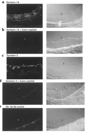
Immunofluorescence localization of syntaxin 1A (a) and 3 (c) in human bronchus. Nonimmune control lacking primary antibody (e) and inhibition of staining by fusion peptide competition (band d) are also shown. Arrows represent the apical pole of the bronchial epithelium. Affinity-purified syntaxin 1A and 3 polyclonal antibodies were used at 5
lysate (PBS-1% Triton X-100) and comparing signals to the respective syntaxin standards; i.e., GST-fusion pro-teins. Purified fusion proteins lacking the COOH-ter-minal membrane anchor (syn 1A∆C and syn 3∆C: where ∆C refers to deletion of the 21–23 amino acid membrane anchor) were used over a wide range of con-centrations to quantitate the particular syntaxin pres-ent in the lysate. To quantitate Munc-18, 10 µg of GST-syn 1A∆C was added to cell lysates containing 200 µg total protein. The complex (GST-syn 1A∆C-Munc-18) was then pulled down using excess glutathione agarose, eluted in SDS-sample buffer, and immunoblotted after SDS-PAGE. We were able to deplete the lysates of Munc 18 by more than 90% by syntaxin 1A pulldown (data not shown), as expected given the high affinity of this interaction (21). Recombinant GST-Munc-18a was used as a standard for quantifying Munc-18 protein levels. CFTR was quantitated by immunoprecipitation from 1 mg of cell lysate (PBS-1% Triton-X-100) using the monoclonal C-CFTR antibody. Under the condi-tions used, this antibody immunodepletes more than 95% of CFTR (data not shown) from cell lysates. Mal-tose binding protein-C-CFTR (MBP-C-CFTR; residues 1387–1480) was used as standard. The CFTR protein was run on a 10% gel so that the mature CFTR band could be detected as a sharp band, which could be quantitated by densitometry.
Immunofluorescence localization of syntaxin 1A and 3 in native human bronchus. Immunofluorescence localization of syntaxin 1A and 3 was performed on fresh frozen sec-tions (5 µm) of native human bronchus. Bronchial sam-ples were harvested at the time of lung transplantation and frozen directly in OCT media using liquid nitrogen. Three independent samples were evaluated from non-smokers undergoing transplantation for chronic obstructive pulmonary disease (COPD). Sections were fixed in –20°C methanol for 30 minutes and post-fixed in 3% paraformaldehyde/PBS for 15 minutes followed by quenching in 50 mM NH4Cl/PBS for 10 minutes. Slides
were blocked in 3% BSA/PBS for 45 minutes at 22°C and incubated with 5 µg/mL of affinity-purified rabbit syn-taxin 1A and synsyn-taxin 3 antibodies in 1% BSA/PBS overnight at 4°C. Slides were then blocked in 5% donkey serum/PBS for 45 minutes at 22°C before incubation with FITC-conjugated donkey anti-rabbit IgG (Jackson ImmunoResearch Laboratories Inc., West Grove, Penn-sylvania, USA) at a concentration of 25 µg/mL for 45 minutes at 22°C. Competition experiments were per-formed by preabsorption of each syntaxin antibody with 5 µg/mL of the relevant syntaxin fusion protein before application to tissue section.
Transient expression of membrane-anchored syntaxin 1A in L cells. cRNA coding for full-length, membrane-anchored syntaxin 1A was microinjected into individual attached, nonconfluent L cells expressing CFTR (22) using an Eppendorf 5242 microinjection system (Eppendorf, Hamburg, Germany). Commercially avail-able sterile, RNAse free Femtotips (Eppendorf) were filled with the cRNA solution and manipulated under
microscopic observation using an Eppendorf 5171 micromanipulator under remote control. Cells were injected with 0.5% Texas Red diluted with DEPC-treat-ed water for later identification or with dye plus 1 ng/µL syntaxin 1A cRNA approximately 24 hours before use in the electrophysiological studies. Injected cells were read-ily observable under fluorescent magnification.
Electrophysiology. Whole-cell patch clamping and data acquisition were carried out according to methods described previously (3, 4, 23). CFTR currents were blocked using a site-directed antibody (0.1 µg/mL) against a synthetic peptide, corresponding to amino acids 505–511 of CFTR, as described previously (24).
Results
Syntaxin 1A is expressed in native airway and intestinal epithe-lial cells. Multiple isoforms of syntaxin have been described, 2 of which are reportedly brain specific
(syn-Figure 4
[image:6.612.307.537.53.348.2]taxin 1A and 1B; ref. 14). In the present study, we observed by immunoblotting that syntaxin 1A is also expressed in native airway and gut epithelial cells. Two syntaxin 1A–specific antibodies were used; an mAb (14D8) and an affinity-purified rabbit polyclonal body (see Methods for details). These 2 syntaxin 1A anti-bodies do not cross react with other syntaxins that we have tested, as shown in Figure 1a (14D8 mAb has been shown not to cross react with syntaxin 1B as well; ref. 20). A syntaxin 3–specific polyclonal antibody (Figure 1a) was also used to verify the expression of this broad-ly distributed syntaxin isoform in epithelial cells. Syn-taxin 1A protein was detected in freshly isolated MTE cells, native HBE cells, and HT29-CL19A colonic carci-noma cells using 14D8 mAb (Figure 1b). The expression of syntaxin 1A protein in these tissues and cells was also detected using the affinity-purified polyclonal antibody (data not shown). Conversely, syntaxin 1A protein was not detected in lysates of mouse fibroblasts (L cells). However, these cells and all others we have tested do express syntaxin 3. Intestinal epithelial cells freshly iso-lated from mouse small intestine (MIE cells) were also observed to express both syntaxin 1A and 3 protein. Nei-ther syntaxin isoform exhibited a detectable gradient of expression from villus to crypt (Figure 1c) when the iso-lated intestinal cells were fractionated by Percoll gradi-ent cgradi-entrifugation and assayed for alkaline phosphatase activity, which is enriched in villus (Figure 1d; ref. 16). Thus, by using 2 different isoform specific antibodies, we were able to detect expression of syntaxin 1A in native airway and intestinal epithelial cells.
Syntaxin 1A is expressed in epithelial cells at much lower lev-els than in brain but is in molar excess over CFTR. At first glance, these results seem inconsistent with initial reports that syntaxin 1A is highly neural specific (14, 15). To resolve this apparent discrepancy, we quantitat-ed the expression of syntaxin 1A in epithelial cells rela-tive to brain, and then directly compared the expression
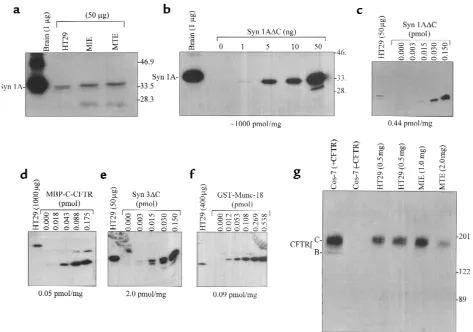
of syntaxin 1A to that of CFTR in the same epithelial cell type. Syntaxin 1A is expressed abundantly in the brain when compared with MIE or MTE cells (Figure 2a). In rat brain lysate (SDS-detergent extract), syntax-in 1A constitutes approximately 1% (wt/wt) of the total detergent extractable protein (Figure 2b), as determined by quantitative immunoblotting using rat syntaxin 1A fusion protein as a standard. On the other hand, syn-taxin 1A comprises only about 0.002% of total deter-gent-extractable cell protein (wt/wt) in HT29-CL19A colonic epithelial cells (Figure 2c), MIE cells, and MTE cells (data not shown). The MTE lysates tested negative for synaptophysin, a synaptic marker, which indicates that these cells were not contaminated with nerve end-ings in the process of isolation (data not shown).
[image:7.612.60.404.52.278.2]Whereas syntaxin 1A is expressed at low levels in epithelial cells relative to brain, it is nonetheless at con-siderable excess over CFTR in epithelial cells (Figure 2, c–f). With the use of quantitative immunoblotting, we determined that the amounts of syntaxin 1A and CFTR in HT29-CL19A cells correspond to approximately 0.44 pmol/mg and 0.05 pmol/mg of total protein, respec-tively (Figure 2, c and d). Syntaxin 3 and Munc-18 pro-tein levels in HT 29-CL19A cells were similarly quanti-tated (Figure 2, d and e). Syntaxin 3 is considerably more abundant than syntaxin 1A in HT29-CL19A cells as well as in T84 colonic epithelial cells and native MIE cells (data not shown). We also attempted to estimate the relative amounts of syntaxin 1A and CFTR in MTE cells. We were able to detect syntaxin 1A protein in freshly isolated MTE cells, where the protein amount was comparable to that found in MIE cells (Figure 2a). However, the CFTR protein amount was considerably lower in the MTE cells compared with the MIE cells (Figure 2g). This is consistent with previous reports that CFTR is more abundant in gut than in airway epithelial cells (25) and indicates that syntaxin 1A is at an even greater excess over CFTR in MTE cells.
Figure 5
Syntaxin 1A resides at the apical poles of airway epithelial cells. To analyze the distribution of syntaxin 1A and 3 in epithelial cells, we immunolocalized these isoforms in sections of native human bronchus using affinity-puri-fied syntaxin 1A and syntaxin 3 antibodies. Specific syn-taxin 1A staining was observed at or near the apical cell surface as well as in numerous intracellular puncta (Fig-ure 3). Syntaxin 3 staining was more intense, and the dis-tribution was almost exclusively at or near the apical cell surface. Nonimmune controls were negative for staining. Importantly, syntaxin 1A staining at the apical pole could be blocked by competition with excess GST-syn 1A∆C fusion protein (Figure 3), but not by GST-syn 3∆C fusion protein (data not shown). Conversely, syntaxin 3 staining could be blocked by excess GST-syn 3∆C, but not by GST-syn 1A∆C. We also observed a punctate pat-tern of staining for syntaxin 1A at the apical surfaces of HT29-CL19A cells (results not shown). These results confirm that syntaxins 1A and 3 are expressed in HBE cells and that they have partially overlapping distribu-tions in the vicinity of the apical plasma membrane.
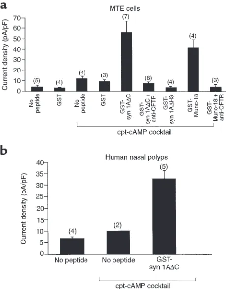
Syntaxin 1A modulates CFTR function in airway epithelial cells. Given that native epithelial cells express syntaxin 1A in addition to CFTR, we determined whether CFTR activity is modulated by syntaxin 1A in airway epithe-lial cells. Toward this end, we used 2 reagents that block the physical interaction between CFTR and syn-taxin 1A and were previously documented to rescue CFTR from syntaxin 1A inhibition in Xenopusoocytes (3, 4); namely, the cytosolic domain of syntaxin 1A as a GST fusion protein (GST-syn 1A∆C) and the syntax-in 1A bsyntax-indsyntax-ing protesyntax-in, Munc-18 (GST-Munc 18a). Both reagents markedly potentiated cAMP-dependent chloride currents in primary cultures of MTE cells when introduced into the cells through whole-cell patch pipettes (Figure 4a), as expected if CFTR activi-ty is limited by its interaction with native syntaxin 1A. The addition of a cAMP cocktail increased the current amplitude at +110 mV by about 2-fold in the absence of peptide and by 6- to 10-fold in the presence of either GST-syn 1A∆C or GST-Munc-18. Basal currents in the absence of cAMP were not affected by either reagent. In addition, control peptides that do not bind to CFTR or affect CFTR currents in Xenopusoocytes (GST and GST-syn 1A∆H3, which lacks the CFTR binding domain [H3]; ref. 4) had no effect on cAMP-dependent current amplitude. The large cAMP-dependent cur-rents that were potentiated by GST-syn 1A∆C or GST-Munc-18 reached a maximum in 5–10 minutes after exposure of the cell to cAMP cocktail (see Figure 4 leg-end). The potentiation of Cl– currents by GST-syn
1A∆C and GST-Munc-18 could be completely inhibit-ed by a CFTR-neutralizing antibody (Figure 4a). In a limited number of experiments, we also observed that GST-syn 1A∆C could stimulate cAMP-dependent Cl–
currents in human nasal polyp cells (Figure 4b). These cAMP-mediated currents in nasal polyp cells could also be completely blocked by CFTR-neutralizing anti-body (data not shown).
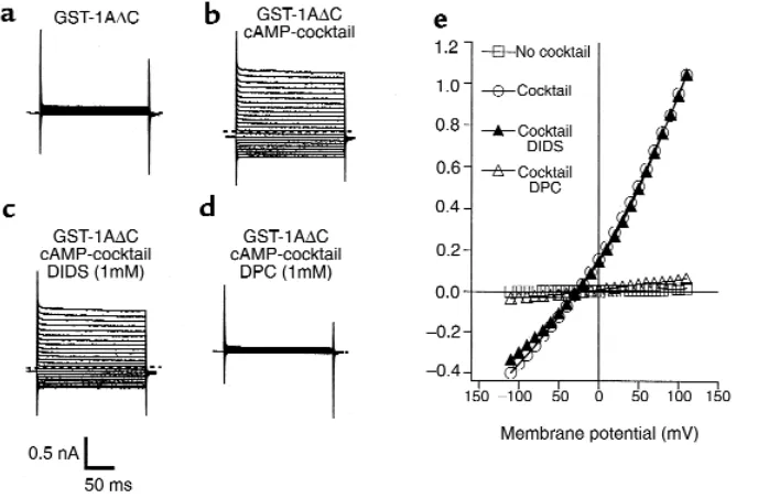
To verify further that the large currents in MTE cells that were induced by GST-syn 1A∆C were CFTR medi-ated, we characterized the I–V relationship and inhibitor sensitivity of these currents (Figure 5). The large Cl–
cur-rents in the presence of GST-syn 1A∆C were not affect-ed by DIDS, which blocks a number of Cl–channels
other than CFTR (26). Conversely, diphenylamine car-boxylate (DPC), which does block CFTR Cl–channels
(27), completely inhibited the currents that were poten-tiated by GST-syn 1A∆C (Figure 5). The I–V relationship in the presence of either reagent was also approximately linear, which is a hallmark of CFTR Cl–channels (28). In
combination with the blocking effect of the CFTR-neu-tralizing antibody on the syntaxin 1A–modulated cur-rents (Figure 4a), these results confirm that the large cAMP-dependent currents in MTE cells that are poten-tiated by GST-syn 1A∆C are indeed mediated by CFTR. We next determined whether the functional effects of the syntaxin 1A peptide on CFTR currents in MTE cells could be explained by the disruption of an
inter-Figure 6
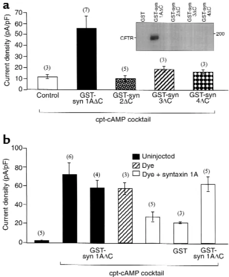
[image:8.612.305.540.309.593.2]action between CFTR and native syntaxin 1A. In this regard, the physical interaction between human CFTR and syntaxin 1A was previously determined to be syntaxin 1A–isoform specific (3), unlike most other syntaxin binding interactions that do not dis-criminate between isoforms (29). Figure 6a shows the results of a pulldown assay that indicate that mouse CFTR like human CFTR also binds to syntaxin 1A in a highly isoform-specific manner (Figure 6, inset). In addition, of the 4 syntaxin peptides that were tested for CFTR binding and for effects on CFTR currents, only the syntaxin 1A peptide substantially potentiat-ed CFTR activity in MTE. Moreover, Figure 6b shows that the syntaxin 1A peptide had no effect on the Cl–
currents mediated by recombinant CFTR in mouse L cells that do not express syntaxin 1A (see Figure 1b). However, this syntaxin 1A peptide did stimulate CFTR-mediated chloride currents in L cells that were microinjected with recombinant cRNA coding for full-length syntaxin 1A. Thus, the simplest interpre-tation of our peptide data is that GST-syn 1A∆C stimulates CFTR-mediated Cl–currents in MTE cells
by competing with native, membrane-anchored syn-taxin 1A for binding to CFTR.
Syntaxin 1A does not regulate CaM Kinase II–activated Cl–currents in airway epithelial cells. Epithelial cells also
exhibit a calcium-calmodulin–dependent protein kinase II–regulated (CaMK II–regulated) chloride conductance that is not mediated by CFTR. To deter-mine the specificity of the effects of the syntaxin 1A peptide on Cl–currents in airway epithelial cells, we
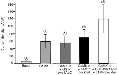
also tested the effects of this reagent on CaMK II–activated Cl–currents (Figure 7). CaMK II, when
introduced into the cell via the patch pipette, increased the Cl–current amplitude in MTE cells
sim-ilar to that previously reported for colonic epithelial cells (30). The CaMK II–activated currents were not
affected by inclusion of GST-syn1A∆C in the pipette. However, the currents did tend toward higher levels in the presence of GST-syn 1A∆C when the cells were activated with CaMK II plus the cAMP cocktail, as expected if CaMK II and cAMP activate parallel Cl–
conductances. These results indicate that the effects of GST-syn 1A∆C in airway epithelial cells are specif-ic for CFTR-mediated chloride currents.
Discussion
Previous studies have shown that syntaxin 1A regu-lates CFTR chloride channels and epithelial sodium channels (ENaCs) when these proteins are expressed in
Xenopusoocytes (3, 4, 10, 12). Although those studies have provided evidence that syntaxin 1A has the capac-ity to regulate CFTR and ENaC under certain condi-tions, they do not speak to the issue of whether syn-taxin 1A modulates ion channel function in native epithelial tissues that are targets for diseases such as cystic fibrosis (CFTR), secretory diarrhea (CFTR), and Liddle’s syndrome (ENaC). This issue is particularly significant given that syntaxin 1A was originally reported to be neural specific (14, 15). The present results indicate that syntaxin 1A protein is expressed in cells that normally express CFTR; namely, native MTE cells, MIE cells, and HBE cells. The amount of syntaxin 1A protein expressed in these native epithe-lial cells is much lower than in brain, which probably explains why some groups have difficulty in detecting this protein by immunoblotting or by immunofluo-rescence in colonic epithelial cells lines such as CaCo-2 (ref. 31; although we have detected syntaxin 1A expression in CaCo-2 cells as well [data not shown]). We have confidence in these data as we have used at least 6 different antibodies to detect syntaxin 1A pro-tein in epithelial cells, of which the 2 described in this study are highly isoform specific (Figure 1a). Impor-tantly, although syntaxin 1A is present in relatively low abundance in the gut and trachea compared with in brain, it is nonetheless considerably more abundant than CFTR in epithelial cells. In particular, gut and air-way epithelial cells express syntaxin 1A protein at a greater than 10-fold molar excess over CFTR.
[image:9.612.58.292.56.207.2]Our immunofluorescence results place syntaxin 1A at the apical poles of HBE cells, although this protein was also detected in multiple compartments within the cell. Syntaxin 1A is also rather broadly distributed in neu-rons, where it has been observed along the axon and in endosomes and synaptic vesicles (32). CFTR protein has also been localized to multiple intracellular compart-ments including endosomes as well as the apical mem-brane (33). The intracellular localization of syntaxin 1A and CFTR raises the possibility that these proteins could interact in intracellular compartments as well as at the apical surface. In contrast to syntaxin 1A, syntax-in 3 protesyntax-in was more precisely localized to the apical cell surface. A very similar pattern has been reported for syntaxin 3 in other epithelial cells such as MDCK cells (34) and CaCo2 colonic epithelial cells (35), although
Figure 7
there is 1 report that this syntaxin isoform is localized to the basolateral surfaces of renal collecting duct cells (36). Interestingly, syntaxin 3 does not appear to direct-ly interact with CFTR or to regulate CFTR Cl–currents
(Figure 6a), in spite of the fact that it is more abundant than syntaxin 1A and is more precisely localized to the apical membranes of airway and gut epithelial cells.
The results of our functional studies indicate that syntaxin 1A downregulates CFTR activity in airway epithelial cells. CFTR-mediated chloride currents were stimulated by 2 reagents that rescue CFTR from inhi-bition by membrane anchored syntaxin 1A in Xenopus
oocytes (3, 4) and mouse L fibroblasts (Figure 6b). These reagents perturb the interaction between CFTR and membrane-anchored syntaxin 1A by different mechanisms. Munc-18 blocks the regulation of CFTR currents by binding to native syntaxin 1A and inhibit-ing the physical interaction between these proteins. GST-syn 1A∆C appears to have a dominant negative effect on this interaction by competing with native syntaxin 1A for CFTR. The potentiation of CFTR-mediated chloride currents in mouse tracheal epithe-lial cells by the syntaxin 1A peptide was highly iso-form specific, as was the binding of this peptide to mouse CFTR (Figure 6a). This agrees with our earlier biochemical evidence for an isoform-specific binding interaction between syntaxin 1A and the NH2
-termi-nal cytoplasmic tail of human CFTR (ref. 3; the NH2
-terminal tails of mouse and human CFTR are 80% identical). In contrast, syntaxins generally interact with broad specificity with other components of the membrane traffic machinery (29); thus, it seems unlikely that the specific effect of the syntaxin 1A peptide on CFTR activity occurred through some generic effect on membrane trafficking. However, this does not exclude the possibility that the intracellular traffic of CFTR is more specifically influenced by its physical interaction with syntaxin 1A. For example, Peters et al. (12) have recently argued that syntaxin 1A (but not syntaxin 3) inhibits CFTR delivery to the plasma membranes of Xenopus oocytes. To what extent syntaxin 1A regulates CFTR activity in epithe-lial cells by this mechanism or by effects on channel gating is unknown.
In addition to regulating CFTR, syntaxin 1A also appears to regulate presynaptic calcium channels and the physical coupling of these channels to the exocy-totic machinery. A plant syntaxin has also been iden-tified on the basis of its ability to regulate K+ and Cl–
channels in guard cells (11). Moreover, syntaxin 1A reportedly inhibits ENaC currents in Xenopusoocytes, possibly by affecting the gating properties of this sodi-um channel (10). On the other hand, it is clear from the present results and from previous oocyte expres-sion studies that there are numerous other ion chan-nels that are not affected by syntaxin 1A (3, 10, 12). For example, we observed no effect of GST-syn1A∆C on the CaMK II–activated Cl–currents in mouse
tracheo-cytes. Conceivably, there exists a subset of ion channels
that physically and functionally interact with syntax-ins as a means to coordinate the regulation of ion transport and membrane traffic in certain tissues.
The present results set the stage for determining whether syntaxin 1A regulates the activity of wild-type CFTR or disease-associated mutants in intact ani-mals. Cystic fibrosis can be caused by 1 of more than 800 different CFTR mutations, many of which lead to partial-loss-of-function phenotypes. Conceivably, the functional activity of certain disease-associated mutants could be augmented by reagents that block their interaction with syntaxin 1A in epithelial cells. The results of the present study indicate that the development of cell permeant reagents that could block the CFTR-syntaxin 1A interaction in vivo is worth considering as an alternative approach to stim-ulating mutant CFTR activity.
Acknowledgments
The authors thank G. Li and S. Reddy for excellent technical assistance. Our work is primarily supported by a Research Grant from the Cystic Fibrosis Founda-tion (NAREN 99GO for APN) and grants from the National Institutes of Health (DK 51868 and DK 50830 for K.L. Kirk).
1. Welsh, M.J., Tsui, L.-C., Boat, T.F., and Beaudet, A.L. 1995. Cystic fibro-sis. In The metabolic and molecular basis of inherited diseases: membrane trans-port systems. C.R. Scriver, A.L. Beaudet, W.S. Sly, and D. Valle, editors. Vol-ume 3. McGraw-Hill. New York, NY. 3799–3876.
2. Guerrant, R.L., Hughes, J.M., Lima, N.L., and Crane, J. 1990. Diarrhea in developed and developing countries: magnitude, special settings, and etiologies. Rev. Infect. Dis.12(Suppl.):41–50.
3. Naren, A.P., et al. 1997. Regulation of CFTR chloride channels by syn-taxin and munc-18 isoforms. Nature.390:302–305.
4. Naren, A.P., Quick, M.W., Collawn, J.F., Nelson, D.J., and Kirk, K.L. 1998. Syntaxin 1A inhibits CFTR chloride channels by means of domain-specif-ic protein-protein interactions. Proc. Natl. Acad. Sci. USA.95:10972–10977. 5. Bennett, M.K. 1995. SNARES and the specificity of transport vesicle
tar-geting. Curr. Opin. Cell Biol.7:581–586.
6. Hata, Y., Slaughter, L.A., and Sudhof, T.C. 1993. Synaptic vesicle fusion complex contains unc-18 homolog bound to syntaxin. Nature.
366:347–351.
7. Pevsner, J., Hsu, S., and Scheller, R.H. 1994. N-Sec1: a neuronal-specific syntaxin binding protein. Proc. Natl. Acad. Sci. USA.91:1445–1449. 8. Bezprozvanny, I., Scheller, R.H., and Tsien, R.W. 1995. Functional impact
of syntaxin on gating of N-type and Q-type calcium channels. Nature.
378:623–626.
9. Rettig, J., et al. 1997. Alteration of Ca2+ dependence of neurotransmit-ter release by disruption of Ca2+ channel/syntaxin inneurotransmit-teraction. J. Neu-rosci.17:6647–6656.
10. Saxena, S., Quick, M.W., Tousson, A., Oh, Y., and Warnock, D.G. 1999. Interaction of syntaxins with the amiloride-sensitive epithelial sodium channel. J. Biol. Chem.274:20812–20817.
11. Leyman, B., Geelen, D., Quintero, F.J., and Blatt, M.R. 1999. A tobacco syntaxin with a role in hormonal control of guard cell ion channels. Sci-ence.283:537–540.
12. Peters, K.W., Qi, J., Watkins, S.C., and Frizzell, R.A. 1999. Syntaxin 1A inhibits regulated CFTR trafficking in Xenopus oocytes. Am. J. Physiol.
277:C174–C180.
13. Naren, A.P., et al. 1999. CFTR chloride channel regulation by an inter-domain interaction. Science.286:544–548.
14. Bennett, M.K., et al. 1993. The syntaxin family of vesicular transport receptors. Cell. 74:863–873.
15. Inoue, A., and Akagawa, K. 1993. Neuron specific expression of a mem-brane protein, HPC-1: tissue distribution, and cellular and subcellular localization of immunoreactivity and mRNA. Brain Res. Mol. Brain Res.
19:121–128.
17. Devor, D.C., Singh, A.K., Bridges, R.J., and Frizzell, R.A. 1997. Psoralens: novel modulators of Cl-secretion.Am. J. Physiol.272:C976–C988. 18. Coleman, D.L., Tuet, I.K., and Widdicombe, J.H. 1984. Electrical
prop-erties of dog tracheal epithelial cells grown in monolayer culture. Am. J. Physiol.246:C355–C359.
19. Duan, D., Zhang, Y., and Engelhardt, J.F. 1999. Gene delivery to the air-way. In Current protocols in human genetics. N.C. Dracopoli et al., editors. John Wiley & Sons Inc. New York, NY.
20. Kushima, Y., Fujiwara, T., Sanada, M., and Akagawa, K. 1997. Charac-terization of HPC-1 antigen, an isoform of syntaxin-1, with the isoform-specific monoclonal antibody, 14D8. J. Mol. Neurosci.8:19–27. 21. Pevsner, J., et al. 1994. Specificity and regulation of a synaptic vesicle
docking complex.Neuron.13:353–361.
22. Yang, Y., et al. 1993. Molecular basis of defective anion transport in L cells expressing recombinant forms of CFTR. Hum. Mol. Genet.2:1253–1261. 23. Chan, H.-C., Goldstein, J., and Nelson, D.J. 1992. Alternate pathways for chloride conductance activation in normal and cystic fibrosis airway epithelial cells. Am. J. Physiol.262:C1273–C1283.
24. Chan, H.C., Kaetzel, M.A., Nelson, D.J., Hazarika, P., and Dedman, J.R. 1992. Antibody against a cystic fibrosis transmembrane conductance regulator-derived synthetic peptide inhibits anion currents in human colonic cell line T84. J. Biol. Chem.267:8411–8416.
25. Tizzano, E.F., Chitayat, D., and Buchwald, M. 1993. Cell-specific local-ization of CFTR mRNA shows developmentally regulated expression in human fetal tissues. Hum. Mol. Genet.2:219–224.
26. Singh, A.K., Afink, G.B., Venglarik, C.J., Wang, R.P. and Bridges, R.J. 1991. Colonic Cl channel blockade by three classes of compounds. Am. J. Physiol.261:C51–C63.
27. McCarty, N.A., et al. 1993. Voltage-dependent block of the cystic
fibro-sis transmembrane conductance regulator Cl– channel by two closely related arylaminobenzoates. J. Gen. Physiol.102:1–23.
28. Kartner, N., et al. 1991. Expression of the cystic fibrosis gene in non-epithelial invertebrate cells produces a regulated anion conductance.
Cell.64:681–691.
29. Yang, B., et al. 1999. SNARE interactions are not selective. Implication for membrane fusion specificity. J. Biol. Chem.274:5649–5653. 30. Xie, W., et al. 1998. Regulation of Ca2+-dependent Cl- conductance in a
human colonic epithelial cell line (T84): cross-talk between Ins(3,4,5,6)P4 and protein phosphatases. J. Physiol. (Lond.).51:661–673. 31. Galli, T., et al. 1998. A novel tetanus neurotoxin-insensitive vesicle-asso-ciated membrane protein in SNARE complexes of the apical plasma membrane of epithelial cells. Mol. Biol. Cell.9:1437–1448.
32. Garcia, E.P., McPherson, P.S., Chilcote, T.J., Takei, K., and De Camilli, P. 1995. rbSec1A and B colocalize with syntaxin 1 and SNAP-25 through-out the axon, but are not in a stable complex with syntaxin. J. Cell Biol.
129:105–120.
33. Bradbury, N.A, Cohn, J.A, Venglarik, C.J., and Bridges, R.J. 1994. Bio-chemical and biophysical identification of cystic fibrosis transmembrane conductance regulator chloride channels as components of endocytic clathrin-coated vesicles. J. Biol. Chem.269:8296–8302.
34. Riento, K., et al. 1998. Interaction of Munc-18-2 with syntaxin 3 controls the association of apical SNAREs in epithelial cells. J. Cell Sci.
111:2681–2688.
35. Delgrossi, M.H., Breuza, L., Mirre, C., Chavrier, P., and Le Bivic, A. 1997 Human syntaxin 3 is localized apically in human intestinal cells. J. Cell Sci. 110:2207–2214.