Beich Frandsen, M. and Vecerek, B. and Konarev, P.V. and Sjoblom, B. and
Kloiber, K. and Hammerle, H. and Rajkowitsch, L. and Miles, Andrew J. and
Kontaxis, G. and Wallace, Bonnie A. and Svergun, D.I. and Konrat, R. and
Blasi, U. and Djinovic Carugo, K. (2011) Structural insights into the dynamics
and function of the C-terminus of the E. coli RNA chaperone Hfq. Nucleic
Acids Research 39 (11), pp. 4900-4915. ISSN 0305-1048.
Downloaded from:
Usage Guidelines:
Please refer to usage guidelines at
or alternatively
BIROn
- Birkbeck Institutional Research Online
Enabling open access to Birkbeck’s published research output
Structural insights into the dynamics and function of the
C-terminus of the E. coli RNA chaperone Hfq
Journal Article
http://eprints.bbk.ac.uk/3320
Version: Published (Refereed)
Citation:
© 2011
Birkbeck College
Publisher version
______________________________________________________________
All articles available through Birkbeck ePrints are protected by intellectual property law, including copyright law. Any use made of the contents should comply with the relevant law.
______________________________________________________________
Deposit Guide
Contact: [email protected]
Mads Beich-Frandsen, Branislav Večerek, Petr V. Konarev, Björn
Sjöblom, Karin Kloiber, Hermann Hämmerle, Lukas Rajkowitsch,
Andrew J. Miles, Georg Kontaxis, B. A. Wallace, Dimitri I. Svergun,
Robert Konrat, Udo Bläsi and Kristina Djinović-Carugo. (2011)
Structural insights into the dynamics and function of the C-terminus of
the E. coli RNA chaperone Hfq–
Structural insights into the dynamics and function
of the C-terminus of the
E. coli
RNA chaperone Hfq
Mads Beich-Frandsen
1, Branislav Vecˇerek
2, Petr V. Konarev
3, Bjo¨rn Sjo¨blom
1,
Karin Kloiber
1, Hermann Ha¨mmerle
2, Lukas Rajkowitsch
2, Andrew J. Miles
4,
Georg Kontaxis
1, B. A. Wallace
4, Dimitri I. Svergun
3, Robert Konrat
1,
Udo Bla¨si
2,* and Kristina Djinovic´-Carugo
1,5,*
1
Department of Structural and Computational Biology, Max F. Perutz Laboratories, University of Vienna, Campus Vienna Biocenter 5, A-1030 Vienna,2Department of Microbiology, Immunobiology and Genetics, Max F. Perutz Laboratories, University of Vienna, Dr. Bohrgasse 9, A-1030 Vienna, Austria,3EMBL-Hamburg c/o DESY, Notkestrasse 85, D-22603, Hamburg, Germany, 4Department of Crystallography, Institute of Structural and Molecular Biology, Birkbeck College, University of London, Malet Street, London WC1E 7HX, UK and 5
Department of Biochemistry, Faculty of Chemistry and Chemical Technology, University of Ljubljana, Asˇkercˇeva 5, 1000 Ljubljana, Slovenia
Received September 28, 2010; Revised December 18, 2010; Accepted December 22, 2010
ABSTRACT
The hexamericEscherichia coliRNA chaperone Hfq (HfqEc) is involved in riboregulation of target mRNAs
by small trans-encoded RNAs. Hfq proteins of dif-ferent bacteria comprise an evolutionarily conserved core, whereas the C-terminus is variable in length. Although the structure of the conserved core has been elucidated for several Hfq proteins, no structural information has yet been obtained for the C-terminus. Using bioinfor-matics, nuclear magnetic resonance spectroscopy, synchrotron radiation circular dichroism (SRCD) spectroscopy and small angle X-ray scattering we provide for the first time insights into the conform-ation and dynamic properties of the C-terminal ex-tension of HfqEc. These studies indicate that the
C-termini are flexible and extend laterally away from the hexameric core, displaying in this way features typical of intrinsically disordered proteins that facilitate intermolecular interactions. We identified a minimal, intrinsically disordered region of the C-terminus supporting the interactions with longer RNA fragments. This minimal region together with rest of the C-terminal extension provides a flexible moiety capable of tethering long and struc-turally diverse RNA molecules. Furthermore, SRCD
spectroscopy supported the hypothesis that RNA fragments exceeding a certain length interact with the C-termini of HfqEc.
INTRODUCTION
In bacteria small trans-encoded regulatory RNAs (sRNAs) can modulate target gene expression in response to various stress conditions, a mechanism known as riboregulation. The majority of functionally characterizedEscherichia colisRNAs act as negative regu-lators by preventing ribosome loading onto the target mRNA through base-pairing with or in the vicinity of the ribosome binding site. As a result, the respective mRNA is prone to rapid decay [for review see refs (1–3)]. Positive regulation by sRNAs has been reported less frequently. In this case the sRNAs can act by an ‘anti-antisense’ mechanism and melt intramolecular sec-ondary structures, which impede ribosome binding (4–6). Alternatively, an mRNA can act as a decoy for a sRNA leading to translation of another mRNA that is negatively controlled by this sRNA (7). Similarly, a sRNA can protect a homologous sRNA from degradation, which in turn results in activation of a target mRNA by the latter (8). A number of bacterial sRNAs associate with the global regulator Hfq (9) and often require this protein for function (10,11). Part of the Hfq requirement may be ascribed to Hfq-mediated stabilization of sRNAs (12–15).
*To whom correspondence should be addressed. Tel: +43 1 4277 52203; Fax: +43 1 4277 9522; Email: [email protected] Correspondence may also be addressed to Udo Bla¨si. Tel: +43 1 4277 54609; Fax: +43 1 4277 9546; Email: [email protected] Present address:
Lukas Rajkowitsch, Lexogen GmbH, Brunnerstrasse 69/Obj. 3, A-1230 Vienna, Austria.
ßThe Author(s) 2011. Published by Oxford University Press.
However, as many sRNAs display imperfect and non-contiguous target complementarity, the requirement for Hfq in riboregulation has mainly been attributed to its RNA chaperone function, which is believed to facilitate the interaction between the sRNA and the cognate mRNA (13,16–19).
Hfq belongs to the eukaryotic and archaeal family of Sm- and Sm-like proteins (13,19,20). Hfq orthologues have been found in a number of prokaryotes. The Hfq proteins of different organisms display an evolutionarily conserved core consisting of amino acid residues 7–66, whereas there is considerable variation at the C-terminal extension (20). The first X-ray structure of the
Staphylococcus aureus Hfq protein (HfqSa) in complex with a short RNA oligonucleotide, was reported by Schumacher et al. (21). Since then 13 unique structures of Hfq-proteins from seven different organisms have been deposited in the Protein Data Bank [S. aureus
(HfqSa; amino acids 1–77) 1KQ1, 1KQ2 (21); E. coli
(HfqEc; amino acids 1–72) 1HK9 (22), 3GIB (amino
acids 2–69) (23);Pseudomonas aeruginosa(HfqPae; amino
acids 1–82) 1U1T, 1U1S (24), 3M4G, 3INZ (25);
Methanococcus jannaschii (HfqMj; amino acids 1–71)
2QTX (26); Synechocystis sp. (HfqCSyn; amino acids 1–
70) 3HFO; Anabaena sp. (HfqCAna; amino acids 1–72)
3HFN (27); Bacillus subtilis (HfqBs; amino acids 1–78)
3HSB (28), and 3HSA (to be released)].
The study by Schumacheret al. (21) showed that HfqSa forms homo-hexamers with a central pore and that a short poly(U) oligoribonucleotide was bound in a circular con-formation along the inner basic rim. A mutational analysis performed with E. coli Hfq (HfqEc) has later provided evidence that sRNAs bind to the same positively charged proximal face of HfqEc (29). Unlike uridine-containing sequences, a recent structural study revealed binding of a poly(A) tract to the distal face of HfqEc using six tripartite binding motifs (23). Interestingly, a poly(A) tract in the 50UTR of rpoS mRNA was shown to be relevant for Hfq-mediated riboregulation by the sRNA DsrA (30), and a mutational analysis by Mickulecky et al. (29), suggested that rpoS
mRNA contacts the distal site. Nevertheless, a truncated HfqEc protein containing the conserved common core (amino acids 1–65) was deficient in binding to the full-length 50-untranslated region (50-UTR) of rpoS, sug-gesting that mRNA contacts to the distal site are not suf-ficient for stable binding of this ligand (18). Further studies provided genetic and biochemical evidence that the C-terminus of HfqEc is additionally required for binding of rpoS and other mRNAs, i.e. it constitutes a RNA interaction surface with specificity for longer RNAs (18).
Only the Hfq proteins ofg- andb-proteobacteria have an extended C-terminus, whereas some Gram-positive bacteria including S. aureus have Hfq proteins with short C-terminal extensions (20). Interestingly, Hfq-mediated riboregulation of mRNAs by sRNAs has been mainly demonstrated in bacterial species bearing Hfq proteins with C-terminal extensions (31). As yet, structural data on the C-terminus of HfqEc are lacking. Previous analyses predicted that the C-terminus of HfqEc
is structurally disordered (18,32), which is a hallmark of RNA chaperones (33).
Here we have used an integrated approach employing bioinformatics, biophysical and structural studies to analyze the dynamic properties, and to determine the con-formation and shape of the C-terminal extension of HfqEc. Collectively, these studies revealed that the C-terminus is intrinsically disordered and flexible, which appears to con-tribute to the interactions of Hfq with RNA substrates.
MATERIALS AND METHODS
Bacterial strains and growth conditions
The E. coli strains BL21 DE3 (Novagen), AM111 (MC4100 hfq1::) (34) and the corresponding F0 (lacIq
) variant (35) have been described. They were grown at 37C in Luria–Bertani (LB) medium or in M9 medium supplemented with 0.2% glucose, 2 mM MgSO4, 0.1 mM CaCl2and 10mg/ml thiamine. For RpoS synthesis the cells were grown in LB medium at 22C. Where appropriate, ampicillin (100mg/ml), kanamycin (25mg/ml), tetracycline (30mg/ml) or chloramphenicol (15mg/ml) was added to the medium.
Construction of plasmids and expression ofhfqvariants
The plasmids pUH5Hfq and pUH65 used for synthesis of full-length HfqEcand HfqEc65, respectively, have previous-ly been described (18,36). In these plasmids, the corres-ponding hfq gene was placed under transcriptional control of the lac promoter in plasmid pUC19 (New England Biolabs). To avoid negative translational autoregulation (37), the 50-UTR of the hfq gene was replaced by the ribosome binding site of phage T7 gene
10using a PCR approach.
The plasmids pUH85 and pUH75 used for synthesis of HfqEc85 and HfqEc75 were constructed as follows. The forward primer (50-GCTCTAGAAATATAATAGTTTA
ACTTTAAGAAGGAGATATACATATGGCTAAGGG
GCAATCTTTACAAGATCCGTTCCT-30) contained the sequence derived from plasmid pET22b (underlined; Novagen) including the XbaI site and the Shine and Dalgarno sequence of gene 10 abutted with the first 35 nucleotides of the hfq gene (given in bold). The reverse primers (50-TTGAATTCATTAATGATGGTAGTTACT GCTGGTACCGCCACC-30) and (50-TTGGATCCCTG
CAGTTACTAGGCGTTGTTACTGTGATGAG-30)
contain two stop codons after the triplet encoding the Hfq-specific amino acids His85 and Ala75 as well as an
EcoRI and BamHI site, respectively. The resulting PCR products were cleaved with XbaI andEcoRI/BamHI and then cloned into the same sites of plasmid pUC19 yielding plasmids pUH85 and pUH75, respectively.
Full-length HfqEc was produced in E. coli BL21 DE3 (Novagen) and the S. aureus, B. subtilis and truncated
E. coli Hfq proteins were produced in the hfq deficient strain AM111F0 (lacIq
). One liter of Luria–Bertani medium was supplemented with 100mg/ml of ampicillin, 50mg/ml kanamycin and 15mg/ml tetracycline where ap-propriate. Protein for NMR-studies was obtained from cells grown in M9-medium, supplemented with 15NH4Cl (Isotec) and13C-glucose (Isotec). The cells were grown to an OD600of 0.6–0.8 and the expression of the respective
hfqvariants was induced by addition of 0.5 mM Isopropyl b-D-1-thiogalactopyranoside (IPTG) (Sigma). After 4 h
the cells were harvested by centrifugation (4,000g/15 min).
In vitrosynthesis of RNA
For hfq126 mRNA synthesis plasmid pUhfqwt (37) cleaved with AflIII was used as a template for in vitro
transcription with the T7-MEGAshortscript kit (Ambion). The run-off transcripts were purified on 6% polyacrylamide-8M urea gels following standard proced-ures. The mRNA concentration was determined spectro-photometrically measuring absorption at 260 nm. The DsrA34 RNA, comprising the nucleotides 26–59 of the DsrA-sequence was purchased from Dharmacon, USA.
Purification of Hfq proteins
The Hfq proteins are heat-stable, and the purification therefore involved an initial fractionation by heating and subsequent processing by FPLC (A¨kta, GE-Healtcare). The final purification-scheme for E. coli Hfq and variants thereof comprised four steps: (i) An initial washing step by Ni2+-affinity (HfqEc and HfqEc85) or hydrophobic interaction chromatography (HfqEc75, HfqEc65); (ii) a filtration over an anion-exchange column to remove nucleic acids; (iii) a concentrating step by re-spectively Ni2+-affinity or HIC; and (iv) a final step of size-exclusion chromatography. The detailed procedure is described in Supplementary Data. Purification of HfqBs and HfqSa was performed as previously described (18).
Western blot analysis
The RpoS, Hfq and L14 (loading control) protein levels were determined in strain AM111 harboring plasmids pACYC184 (control), pAHfq, pAH65, pAH75 and pAH85, respectively. The strains were grown at 22C in LB medium to OD600of 0.5, at which time 2 ml aliquots were withdrawn, pelleted and boiled in protein sample buffer. Equal amounts of total protein were separated on 12% SDS–polyacryamide gels and blotted to a nitro-cellulose membrane. The blots were blocked with 5% dry milk in TBS buffer, and then probed with anti-RpoS (NeoClone, Madison), anti-Hfq (Pineda, Berlin) and anti-L 14 (provided by Dr I. Moll) antibodies to detect the RpoS, Hfq and L14 proteins, respectively. The antibody–antigen complex was visualized as described (38).
FRET assays
This method is described in more detail in refs (32,39). Two complementary, fluorophore-tagged RNA 21mers [Cy5-50-AUGUGGAAAAUCUCUAGCAGU-30
(Cy5-21R+) and Cy3-50-ACUGCUAGAGAUUUUCCA CAU-30 (Cy3-21R
)] were used in the annealing experi-ment. The tagged RNA oligonucleotides were purchased from VBC-Biotech (Vienna, Austria). Using a Tecan GENios Pro microplate reader, the first oligoribo-nucleotide was injected into wells with or without Hfq protein (100 nM final Hfq-hexamer concentration), and the measurement was started with the injection of the second oligoribonucleotide. The reaction was performed in annealing buffer (50 mM Tris–HCl pH 7.5, 3 mM MgCl2and 1 mM DTT) at 37C. The final concentration of the RNAs was 5 nM, in a sample volume of 40ml. The reaction was allowed to proceed for 180 s, and with Cy3 excited, donor and acceptor dye fluorescence emissions were measured once every second. The time-resolved ratio of the fluorescence emissions (FRET index FCy5/
FCy3) was normalized to 1 at t180s and least-square
fitted with Prism 4.03 (GraphPad Software Inc., San Diego, CA, USA) with the second-order reaction equation for equimolar initial reactant concentrations:
y=A [1(kobs t+1)
1
], where y= fraction annealed,
kobs= observed annealing reaction constant,
A= maximum reaction amplitude. The observed reaction constantkobsshown in Figure 1A was calculated as the average of three individually fitted reactions.
SRCD Spectroscopy
SRCD spectra were collected on the CD1 beamline at the ISA facility, University of Aarhus, Denmark. For each protein, the extinction coefficient at 280 nm {HfqEc 0.34, HfqEc85 0.41, HfqEc75 0.30, HfqEc65 0.35 [(mg/ml)1cm1]} was calculated from its amino acid sequence (http://www.scripps.edu/cdputnam/protcalc .html) and the concentration derived from the A280 measured on a Nanodrop 1000 spectrophotometer imme-diately prior to the SRCD spectrum being acquired. For the complexes with RNA, the protein concentration was assessed prior to RNA addition to allow for a 1:1 molar ratio of Hfq hexamer to RNA. The protein and RNA was mixed at elevated temperature (60C), and incubated for 5 h at 65C, and then allowed to slowly cool to room tem-perature. Subsequently the complexes were centrifuged for 1 h at 10 000g, at room temperature.
values [calculated as molecular mass/number of residue1
] of 110.6, 114.3, 113.7 and 112.5 for the HfqEc, HfqEc85, HfqEc75, and HfqEc65 constructs, respectively, based on molecular weights of 11166.3, 7317.5, 8417.7, 9415.7, and numbers of residues of 102, 64, 75, 85. The low wavelength cutoff of the data was determined as previously described (42). Data were analysed with the DichroWeb analysis server (43) and are reported as the averaged results (±1 SD) from the CONTINLL (44,45), SELCON3 (46) and CDDSTR (47) algorithms, using the SP175 reference dataset (48). The NRMSD is a goodness-of-fit parameter (49) calculated for the CONTINLL-calculated structure and the experi-mental data.
NMR
All NMR experiments were performed at 310 K on a Varian Inova 600 MHz spectrometer equipped with 5 mm triple resonance cryo-probe and pulsed field gradi-ents, and a Varian Inova 800 MHz spectrometer equipped with 5 mm conventional triple resonance probe equipped with pulsed field gradients. NMR spectra were processed with NMRPipe (50) and analyzed with Sparky software. The protein samples for NMR-experiments were all prepared by size-exclusion chromatography in 50 mM Na-PO4 pH 7.2, 200 mM NaCl and concentrated to 1 mM HfqEc65 (with respect to the monomer). Samples were supplemented with10% (v/v)2H2O to provide the deuterium signal for the field-frequency lock, as well as 0.1–0.2% (w/v) NaN3 to inhibit bacterial growth. Backbone signal assignment for the C-terminally truncated mutant HfqEc65 was obtained by a suite of standard (sensitivity enhanced) three-dimensional (3D) triple resonance experiments, acquired pairwise: HNCA (51)/HN(CO)CA (52), HNCACB (53), HNCO (54) were recorded for sequential backbone chemical shift assign-ment.15N relaxation times (T1,T2) were measured using well-known gradient sensitivity-enhanced 2D methods with1H detection (55,56).
SAXS
Small angle X-ray scattering experiments were acquired at beamline X33 (57,58) at the DORIS III synchrotron storage-ring (DESY, Hamburg, Germany), using a MAR345 image-plate detector and X-ray wavelength of
= 1.5 A˚, with a sample to detector distance of 2.7 m. This setup covers a range of momentum transfer 0.12<s<0.45 nm1(s = 4p siny/, where 2y is the scat-tering angle). The data were processed in the ATSAS program-package (59). Data were acquired at 37C from four concentrations of the full-length HfqEc(18.5, 9.3, 4.6, 2.3 mg/ml), and similar concentration of the C-terminally truncated core-construct HfqEc65 (19.0, 9.5, 4.8, 2.4 mg/ml). Protein concentration was measured with 2ml samples on a Nanodrop 1000 UV/vis spectrophotom-eter at 280 nm, immediately prior to data-acquisition. The data were averaged and normalized to the intensity of the incident beam prior to subtraction of buffer
scattering. The difference data curves were scaled, merged and initial analysis performed using PRIMUS (60). Invariants were derived by standard approaches for comparison (61). The intrinsic scattering (I0) and radius of gyration (Rg) was derived by Guinier analysis for the data range s<1.3/Rg, approximating I(s) =I0 exp[(s Rg)2/3]. The reverse transform program GNOM (62) was employed for calculation of the distance-distribution function, under the assumption of correct estimation of the particle maximum dimension (Dmax). The program also outputs a value for I0 and Rg. The particle excluded volume was calculated from the Porod Equation (63):
V¼22I0=
Z1
s2IexpðsÞds, ð1Þ
whereIexp(s) is the experimental data.
In addition, the program Dammif (64) run without symmetry restraints outputs the particle excluded volume. The solute molecular mass was further evaluated by the standard approach of comparison of the intrinsic forward scattering (I0) with that recorded from a reference solution of known molecular mass and concentration. In the present study a single sample of 3.68 mg/ml bovine serum albumin (Mr = 66.4 kDa) was used.
Low-resolution shape-models of the SAXS data were generated ab initiousing the program Dammif (64). The program represents the protein as a collection of densely packed dummy-residues inside a sphere with the diameter
DMAX. Each dummy-residue belongs either to the particle
or the solvent, and the shape is described by a binary string of length M. Starting from a random string, simulated annealing is employed to search for a compact model that fits the shape scattering curveI(s) to minimize discrepancy2:
2¼ 1
N1
X
j
IðsjÞ cIcalcðsjÞ
ðsjÞ
2
, ð2Þ
where N is the number of experimental points, c is a scaling factor and Icalc(sj) and s(sj) are the calculated
in-tensity and the experimental error at the momentum transfer sj, respectively.
Equation (2). The program allows for specifying restraints for the conformation of the flexible chains. In present study, the disordered nature of the C-terminus was repre-sented by lowering of the restraint on the dihedral angle between adjacent dummy residues.
In order to test the packing of the C-terminus into the groove formed by amino acids 35–55 of one subunit and the b-sheet of the adjacent subunit as observed in the crystal structure of HfqEc65 (amino acids 6–65), we calculated rigid body models using the program BUNCH applying distance restraints on His70. When placing His70 in the groove, to force the C-termini into the groove we observe a decrease in the quality of the fit. When placing His70 on the outside of the N-terminal helix, to constrain the C-terminus in the vicinity of the conserved patch of charged residues present in Hfq (22), no significant change to the quality of the fit was observed. All graphical representations were made using MacPymol (67).
RESULTS
Annealing deficiency of Hfq proteins naturally devoid of a C-terminus and of C-terminally truncatedE. coli
Hfq variants
We have recently shown using fluorescence resonance energy transfer assays (FRET) that full-length HfqEc stimulated annealing of two complementary RNA-oligonucleotides with a rate constant that was 3-fold higher than that observed for HfqEc65, comprising only the evolutionarily conserved core region of Hfq (amino acids 1–65) (18). When two non-complementary RNA oligonucleotides were used, no significant increase in the FRET signal was discernable in the presence of HfqEc65, whereas an increase in FRET was observed for HfqEc. In contrast to HfqEc, the dual rate binding constant kdbfor
HfqEc65 was 10-fold lower, suggesting that HfqEc65 is severely impaired in annealing of two RNA oligonucleo-tides (18).
To test whether Hfq proteins naturally lacking a C-terminal extension are deficient or impaired in anneal-ing activity, FRET assays were performed with the Hfq proteins of B. subtilis (HfqBs; 73 amino acids) and
S. aureus (HfqSa; 77 amino acids). When compared to the absence of protein and similarly to HfqEc65, neither HfqBsnor HfqSastimulated annealing of the complemen-tary oligonucleotides (Figure 1A), again suggesting that the evolutionarily conserved core is not sufficient to support duplex formation of two RNAs.
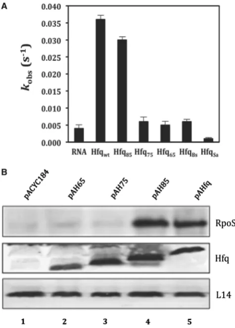
With the aim to identify sub-sequences in the C-terminus of HfqEc that support RNA binding, we tested the ability of two C-terminally truncated HfqEc variants, HfqEc75(amino acids 1–75) and HfqEc85(amino acids 1–85) for their capacity to mediate low temperature (20C) translational activation of rpoS mRNA by the sRNA DsrA during exponential growth (68). Under these conditions, HfqEc65, as shown before (18), and HfqEc75 were defective in stimulating DsrA-mediated RpoS synthesis, whereas the stimulation by HfqEc85was indistinguishable from that of full-length HfqEc
[image:7.612.313.551.188.520.2](Figure 1B). To verify these experiments the two C-terminally truncated HfqEc variants, HfqEc75 (amino acids residues 1–75) and HfqEc85 (amino acids residues 1–85) were analyzed in FRET assays with regard to their annealing activity. Interestingly, HfqEc85was profi-cient in stimulating annealing of the two RNA ligands, whereas HfqEc75 exhibited the same low annealing activity as HfqEc65(Figure 1A). As HfqEc75was defective
Figure 1. The region between amino acids 75 and 85 is instrumental for RNA binding andin vivo function of HfqEc. (A) RNA annealing
activities of HfqBc, HfqSa, full-length HfqEcand C-terminally truncated
variants thereof. Five nanomolar single-stranded, complementary 21-nt-long oligoribonucleotides with fluorophores at their 50-end were
annealed at 37C in the absence or presence of 100 nM protein.
Relative fluorescence resonance energy transfer was calculated as the ratio of acceptor to donor fluorescence (FCy5/FCy3) as described in ‘Materials and Methods’ section. The time-resolved curves were least-square fitted with the second-order reaction equation for equimolar initial reactant concentrations: y=A[1(kobs t+1)1];
y = fraction annealed, kobs= observed annealing reaction constant, A= maximum reaction amplitude. The reaction rate kobs was
calculated from the average of three independent experiments. (B) RpoS protein synthesis in theE. coli hfq-strain AM111 harboring plasmids pACYC184 (control, lane 1), pAH65 (encoding HfqEc65;
lane 2), pAH75 (encoding HfqEc75; lane 3), pAH85 (encoding
HfqEc85; lane 4) and pAHfq (encoding full-length HfqEc; lane 5),
in both, the FRET and in vivo assay, these studies implicated amino acids residues 75–85 in RNA binding.
Bioinformatics analyses predict an intrinsically disordered, solvent exposed C-terminus of HfqEc
The C-terminus of HfqEc(amino acids 66–102) contains a low portion (17%) of ‘order promoting’ residues (Asn, Cys, Ile, Leu, Phe, Trp, Tyr and Val), and a high fraction (64%) of ‘disorder promoting’ residues (Ala, Arg, Gln, Glu, Gly, Lys, Pro and Ser). As observed in several bioinformatic studies (69,70) this is a fingerprint of intrinsically disordered proteins. For comparison, in folded globular proteins, these values are 36% for ‘order’ and 47% for ‘disorder promoting’ residues, which gives a ratio of 0.76. Interestingly, in the first half of the HfqEc C-terminus (amino acids 68–85) this ratio is 0.63, whereas for the last half we calculate 0.08. Overall this indicated that the C-terminal extension, which was indispensable for function in the assays used in this study (Figure 1), is predicted to be disordered.
The metaPrDOS algorithm, which generates disorder predictions based on a consensus principle between the individual predictors PrDOS, DISOPRED2, DisEMBL, DISPROT-VSL2P, DISpro and IUPred (http://prdos .hgc.jp/meta) (71), predicts the C-terminal moiety comprising residues 69–102, to be intrinsically disordered, with high signals (above 0.70) for amino acids 73–102 (Supplementary Figure S1). Secondary-structure predic-tion servers Jpred (72) and PSIPRED (73) do not detect secondary-structure elements in the C-terminal segment (66–102) of HfqEc (data not shown).
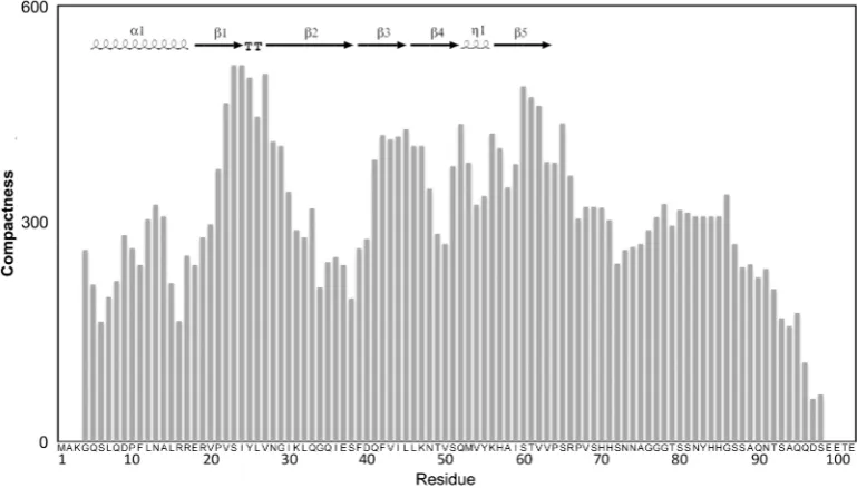
In addition, a protein meta-structural analysis was per-formed, which extracts two parameters for each residue on the basis of the protein primary sequence, one related to
compactness and the other to secondary structure (74). The compactness parameter (Ci) provides a quantitative
measure of the residue embedded in the protein ter-tiary structure. High values (>300) are found for residues in the buried core of globular domains, whereas low values correspond to surface exposed or conformationally flexible residues. The overall Ci values
for the C-terminal segment (amino acids 70–102) are lower than for the core of Hfq, suggesting that this region is conformationally flexible and surface exposed. In addition, the compactness of the Hfq-core region and that of the C-terminus were calculated separately. The average compactness value of the Hfq core region increased (from 292 of the full-length protein) to about 337, whereas the corresponding value for the C-terminal extension alone decreased to 170, indicating that the C-terminus is less compactly folded when compared to the Hfq core. The conclusion that the C-terminus is largely disordered is further supported by the fact that a similar average compactness value (Cav) were found for a set of experimentally characterized in-trinsically unstructured proteins [Myc C-terminal domain (75): Cav= 131; osteopontin (76):Cav= 181;a-synuclein (77): Cav= 255; TAU (78): Cav= 224; Sic1 (79):
Cav= 229].
[image:8.612.120.505.479.698.2]This bioinformatic analysis (Figure 2; Supplementary Figure S1) together with the 3D structure of Hfq (22), in which residues 65–71 are found in an extended conform-ation leaning on the body Hfq core, suggest that the segment encompassing amino acid residues 65 up to 75 displays propensity to be structured, while this tendency is much less pronounced for the rest of the C-terminal extension with high indications for being intrinsically disordered. These features of HfqEc resemble intrinsic-ally disordered protein sequences, which show a
Figure 2. Meta-structure analysis of HfqEc. Compactness values (Ci) are plotted with respect to residue position. A high value indicates a buried
residue (the averageCi of all residues in the Protein Data Bank is 300). Inserted in upper-left is the known secondary-structure content of Hfq
(amino acids 6–65; PDBid: 1HK9) generated by ESPript (http://espript.ibcp.fr/ESPript/ESPript/), TT denotesb-turn andZ1 the small 310-helix.
propensity to form local elements and/or contain pre-formed elements of secondary structure or hydrophobic clusters (80,81).
Nuclear magnetic resonance and synchrotron radiation circular dichroism spectroscopy reveal the unstructured nature of the C-terminal extension of HfqEc
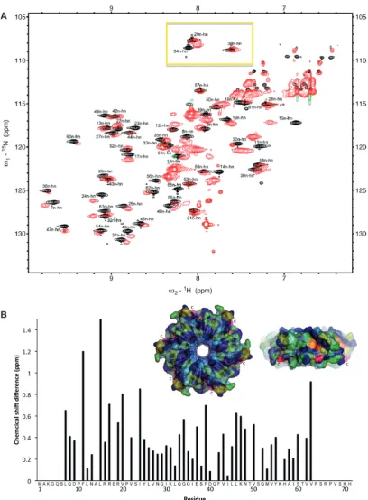
To obtain more information on the structural and dynamic properties of the C-terminus of HfqEc in solution we employed nuclear magnetic resonance spec-troscopy (NMR). A series of NMR-spectra were recorded for full-length HfqEc and for HfqEc65. All NMR experiments for sequential signal assignments were conducted with HfqEc65, which—due to its lower mo-lecular weight—was more amenable for NMR studies. Figure 3A shows the fingerprint region of the 15
N-HSQC spectra of HfqEc65 superimposed on the 15
N-HSQC spectra of full-length HfqEc. The spectrum of HfqEc65 is well resolved and sequential backbone signal assignment was possible for residues 6–65. Residues 1–5 were not observed possibly due to exchange broadening, suggesting some conformational flexibility of these residues. The spectra of HfqEc65 and full-length HfqEc overlapped largely, which allowed for assignment of the HfqEc spectra by simple peak comparison. However, many of the observed peaks corresponding to well-structured residues in the core of the protein show severe line broadening in the HfqEc spectrum due to fast relaxation. A plot of the 15N–1HN chemical shift differ-ences between HfqEc65and full-length HfqEc is shown in Figure 3B. Significant chemical shift differences were observed for residues in the region preceding the C-terminal segment (amino acids 45–55) and at the end of the N-terminala-helix (residues Phe11, Leu15, Arg17, Val20), indicating that the presence of the C-terminus affects residues in these regions. In the 3D structure from X-ray studies performed by Sauter et al. (22), the segment encompassing amino acids 65–70 was found to pack in an extended conformation against the core of the Hfq hexamer, in a groove formed by the N-terminal a-helix of one domain and b-sheets formed by amino acids 34–55 of the adjacent subunit. A conserved patch of charged residues comprised by Arg16, Arg17, Glu18 and Arg19 are present in Hfq. Interestingly, amino acids substitutions of Arg17 in HfqEc compromised phage Qß replication (82), suggesting that this region is of functional importance.
The spectrum of full-length HfqEc shows additional intense peaks, mainly in the region of 7.5–8.5 ppm (in the 1H dimension), corresponding to the partially dis-ordered C-terminus. Interestingly, in the region of the 15
N-HSQC-spectra expected for Gly residues in a random-coil conformation, we did not observe any of the four extra glycine residues present in the C-terminus, indicating that they are indeed not fully unstructured. One possible explanation for their disappearance is that they are subjected to an additional dynamic process, leading to further line broadening, which could result from conform-ational exchange between a partially structured and a fully unstructured state. Another cause for disappearance of
correlation peaks is rapid intermolecular exchange of amide protons with bulk water. This exchange process is particularly facilitated in disordered and conformationally flexible parts of the protein lacking stabilizing backbone hydrogen bonds.
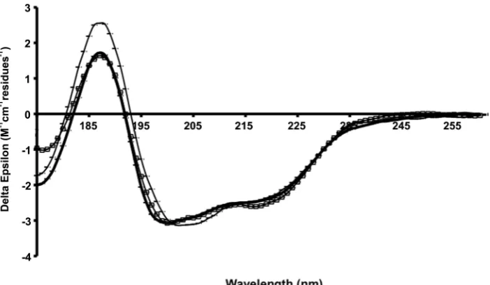
In order to study the secondary structure composition of the C-terminal extension of HfqEcin solution, we used synchrotron radiation circular dichroism (SRCD) spec-troscopy. The additional data in the low-wavelength region accessible by this technique relative to that attain-able in a conventional CD instrument is important for detecting natively-disordered types of secondary structure. The spectra of the protein constructs HfqEc65(amino acids 1–65), HfqEc75 (amino acids 1–75), HfqEc85 (amino acids 1–85) and HfqEc(full-length, amino acids 1–102) collected under the same experimental conditions are shown in Figure 4.
The spectrum of the HfqEc65 is typical for a mixed a/b protein and the calculated secondary structure based on these data produced values close to those calculated from the crystal structure (Table 1). When the HfqEc65 protein is extended by 10 residues (HfqEc75) there is a slight increase in spectral magnitude at both 222 and 190 nm, generally indicative of an increase in helix content, which is reflected in the analysis (Table 1) as a slight increase (2%) in the calculated helical content. This may be explained by stabilization of the N-terminal a-helix by the segment 65–75 through a similar packing arrangement observed in the crystal structure of E. coli
Hfq (amino acid residues 1–72 PDBid: 1HK9) (22), giving rise to increased ellipticity. This is in good agree-ment with the NMR observations showing that this C-terminal stretch of residues interacts with the N-terminala-helix.
In general, with each additional extension to the C-terminus (i.e. HfqEc85and full-length HfqEc) there is a reduction in magnitude of the spectrum combined with a blue shift of the peak located at208 nm. This is indica-tive of an increase in the contribution of naindica-tively disordered-type structures to the spectrum, consistent with the assumption in the ‘theoretical’ values that the additional C-terminal residues are almost entirely unstruc-tured; the percentage of ‘other’ increases as the number of residues are added.
Figure 3. Comparative NMR analysis of HfqEc65and full-length HfqEc. (A) Overlay of 15
N-HSQC spectra of HfqEc65[black (positive) and blue
(negative) contours] and full-length HfqEc[red (positive) and green (negative) contours] demonstrating the unstructured nature of the C-terminal
extension, with multiple intense peaks occurring in the 7.5–8.5 ppm region of the1H-dimension. The glycine region in the upper part of the HfqEc
spectra (yellow box) is lacking the expected four peaks for the four glycine residues of the C-terminus. (B) Chemical 1H–15N shift differences
calculated for assigned peaks plotted against residue positions. Chemical shift differences>0.5 ppm are considered significant. The insert shows a surface representation of the proximal face and side ofE. coliHfq (amino acids 6–65) hexamer. Chemical1H–15N shift differences between Hfq
Ec65
and full-length HfqEcare color-coded from blue (zero) to red (largest shift). Residues which could not be unambiguously assigned in the full-length
Solution structure, hydrodynamic properties and shape of the C-terminus of full-length HfqEc
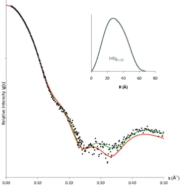
To further study the behavior of Hfq in solution, we employed a series of solution small angle X-ray scattering (SAXS) experiments in combination with NMR spectros-copy measurements of the transverse relaxation rate of Hfq. Figure 5 shows the fit between experimental X-ray scattering data and the calculated scattering curve (66) for residues 6–65 of Hfq (PDBid: 1HK9) (22), confirming the close agreement between crystal and solution structure of the hexameric Hfq core. By modeling the five missing N-terminal residues, we observe only a marginal improve-ment of the fit (21.55!1.40), indicating a modest con-tribution to the overall scattering of these 65 = 30 unstructured residues in HfqEc65.
For HfqEc65 we estimated a molecular mass of 44 ± 5 kDa and for full-length HfqEc58 ± 7 kDa by nor-malization of the intrinsic scattering (I0) to a standard
sample of bovine serum albumin (BSA) (84), which agrees with the theoretical values for hexamers of 43.2
and 67.2 kDa, respectively. We estimate the solute excluded volume (Porod’s volume) for HfqEc65 (70 ± 5)103 A˚3 and for full-length HfqEc (110 ± 8)103A˚3, further indicating that both proteins adopt their native hexameric quaternary structure. In addition, Guinier analysis established for HfqEc65an Rg of (24.2 ± 0.5) A˚, in good agreement with Rg of 23.5 A˚ calculated for the crystal structure of HfqEc65. Comparatively, for full-length HfqEc Rgwas (32.3 ± 0.5) A˚ (60). The estimated largest dimension (DMAX) of the particle was for HfqEc65 (68 ± 5) A˚ and for HfqEc (112 ± 8) A˚ (62).
By NMR 15N-relaxation measurements we measured for HfqEc65 (43.2 kDa) a T2= 40.0 ± 2.1 ms (R2= 25.0 ± 1.3 s1). These homogeneous 15N-relaxation data indicate that the core domain of Hfq is uniformly rigid and does not undergo large amplitude fluctuations on timescales relevant for NMR.
[image:11.612.129.470.71.264.2]For full-length HfqEc (67.2 kDa), we measured an ex-tremely fast15N relaxation ofT2= 5–10 ms for the signals of the core-region. Compared to HfqEc65, HfqEc exhibits
Figure 4. SRCD spectra of Hfq constructs. HfqEc65(thin dotted line), HfqEc75(thick dotted line), HfqEc85(thin solid line), full-length HfqEc(thick
solid line). The error bars represent one standard deviation between replicate scans.
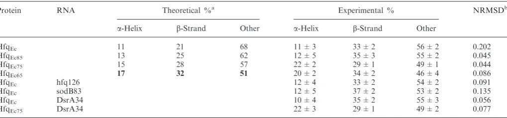
Table 1. Secondary structure content ofE. coliHfq constructs and their complexes with RNA, evaluated from SRCD spectra using the DichroWeb analysis server (43)
Protein RNA Theoretical %a Experimental % NRMSDb
a-Helix b-Strand Other a-Helix b-Strand Other
HfqEc 11 21 68 11 ± 3 33 ± 2 56 ± 2 0.202
HfqEc85 13 25 62 12 ± 5 35 ± 3 55 ± 2 0.045
HfqEc75 15 28 57 22 ± 2 29 ± 1 49 ± 1 0.044
HfqEc65 17 32 51 20 ± 2 34 ± 2 46 ± 4 0.086
HfqEc hfq126 12 ± 4 33 ± 2 54 ± 2 0.091
HfqEc sodB83 12 ± 5 37 ± 2 53 ± 2 0.135
HfqEc DsrA34 10 ± 4 35 ± 2 55 ± 3 0.056
HfqEc75 DsrA34 22 ± 3 29 ± 1 49 ± 2 0.077
a
Bold values listed under theoretical correspond to the crystal structure of HfqEc65; other ‘theoretical’ values are calculated based on the crystal
structure and assuming the extension residues are disordered.
[image:11.612.46.556.335.454.2]considerably reduced 15N-transverse relaxation times (T2= 5–10 ms). Consequently the precision of the T2 measurement is much lower due to the resulting lowerS/ N-ratio. Interestingly, there is a larger residue by residue variation of 15N T2 relaxation times in HfqEc than in HfqEc65, which is an indication of a more anisotropic behavior due to the extended C-termini. The observed re-laxation rate is much faster than what could be expected for the increase in molecular mass, indicating an unusual increase in the hydrodynamic radius. For a perfect solid sphere, the radius of gyration (Rg) is only slightly smaller than the hydrodynamic radius (Rh), whereas increased dis-crepancy between the two reflects an anisometric structure of the particle. With a relation of 1.33 between theRh3of HfqEc65 and HfqEc from SAXS measurements, reflecting the expected increase in molecular weight, the relation of 4–8 between the relaxation rates measured by NMR (which are directly proportional to Rh3) indicates a pronounced anisometric structure of HfqEc. A possible explanation for these observations is that the partially un-structured C-termini are laterally extended into the
surrounding solution. Thus the overall molecular shape is ‘puffed up’, causing the molecule to appear much larger than a comparable well-folded and tightly packed globular protein.
To validate this hypothesis, the observed15N T2 relax-ation properties were modeled through simulrelax-ation of the hydrodynamic properties of HfqEc65and HfqEcusing their molecular coordinates as input. For HfqEc65the coordin-ates from the X-ray crystal structure of Hfq of E. coli
(amino acid residues 6–65, PDBid: 1HK9) (22) were used, onto which the N-termini had been added as un-structured. For full-length HfqEc a representative all-atom ensemble was created by restrained simulated an-nealing using the X-plor/CNS software system (85) con-straining the backbone dihedral angles of the C-terminus to the b-region of the Ramachandran plot and applying the observed radius of gyration as overall restraint.
[image:12.612.131.497.73.447.2]Using the HYDRONMR software (86) for simulation of relaxation times both protein systems were very well reproduced in the simulation and good agreement was obtained between experimental and simulated relaxation
Figure 5. Overlay of experimental X-ray scattering data from HfqEc65(black dots), on the calculated scattering curves for Hfq crystal structure
(PDB: 1HK9, aa 6–65, red curve) and the complete model of HfqEc65(aa 1-65, green curve) where N-terminal residues were restored using the Bunch
behavior. For HfqEc6515N transverse relaxation times of
T2= 35.0 ± 2.0 ms (R2= 28.6 ± 1.6 s1) were predicted; for the ensemble of full-length HfqEc models an average value of 15N T2= 9.7 ± 2.0 ms (R2= 103.1 ± 21.3 s
1 ) was found in the simulation.
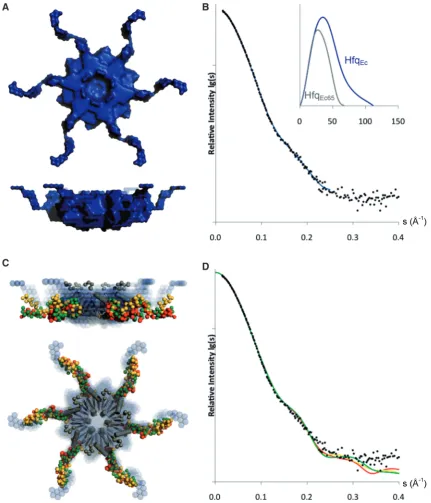
Implying 6-fold symmetry the low-resolution shape of HfqEc was constructed ab initio in the program Dammif (64). As shown in Figure 6A, the C-termini extend away
[image:13.612.86.518.67.571.2]from the core of the protein in a propeller like shape. The dimensions of the hexameric core of the ab initio model (diameter 72 A˚, thickness 35 A˚) are larger than those derived from the crystal structure (diameter65 A˚, thick-ness28 A˚) (22), which can be ascribed to the presence of the termini. The outputRg32.6 A˚ by Dammif is in agree-ment with the Rg derived from the Guinier-analysis (32.3 A˚), and the partial specific volume of the protein
Figure 6. Shape models of HfqEc. (A) Surface representation of ab initiomodel. (B) Calculated scattering curve ofab initiomodel (blue) overlaid
with experimental X-ray scattering data, inserted is the pair-distribution function for HfqEc65(grey) and full-length HfqEc(blue). (C) Superimposition
of the three best rigid-body HfqEcmodels from three iterations of modeling (red, gold and green). In the center, the crystal structure of the HfqEc
(amino acids 6–65) hexameric core is represented as grey ribbons and the N-termini as grey spheres. Theab initiomodel is represented as blue spheres in the background. (D) Calculated scattering curves to a maximum range of momentum transfer (s) of 0.4 A˚1for the three best rigid-body models
(HfqEc65 74103 A˚3 and HfqEc 116103 A˚3; calculated by Dammif without implying symmetry) is within range of the estimated invariant values. Figure 6B shows the calculated scattering curve of the ab initio
model, which agrees with the experimental data on full-length HfqEc, with a2of 1.31, and the distance dis-tribution function for HfqEc65 and for full-length HfqEc, with the difference clearly indicating the extended nature of the C-terminal domain of full-length HfqEc.
Next, we took an approach of rigid-body modeling with the program Bunch (65). We first built the N-terminal residues onto the crystal structure of HfqEc65(Figure 5), and proceeded to utilize the outcome model as a rigid-body in reconstruction of the C-terminal domains of full-length HfqEc, again implying 6-fold symmetry in the particle. Figure 6C shows the superposition of the model of HfqEc from three iterations of rigid-body modeling. Clear agreement was observed for the conserved hexameric core between the ab initio and rigid-body models, whereas some variability was observed for the C-termini. Iterative runs of Bunch (1000) generated models with low variability in the shape of the C-termini. By adjusting restraints in the Bunch-program we could fit the experimental data to
21.65–1.75. We observed bad fits (2>2.3) for models with the C-termini completely extended towards either the proximal or distal face of the hexamer. The calculated Rg 31.8 A˚ (average of three best fits) for the models is in good agreement though slightly smaller than the corresponding value of 32.3 A˚ derived from the Guinier plot. Figure 6D shows the fit with the experi-mental data for the three best rigid-body models, as determined by 2-values generated by the program Bunch. These models fit the experimental data to a range of momentum transfer of 0.35 A˚1, and indicate that the C-termini extend laterally away from the hexameric core. The overall trend appears to be a wave-like shape, with a characteristic loop in the begin-ning of the C-termini and some variability in the tip. Interestingly, this wave-like behavior is also seen in the meta-structure analysis, where a b-turn like structural element is found in the disordered C-terminus (Figure 2).
SRCD spectroscopy reveals an interaction of the C-terminus with RNA
In order to monitor changes in the structure of the C-terminal extension of HfqEc in the presence of RNA, we next used SRCD spectroscopy. Both HfqEc65 and full-length HfqEc bind the sRNA DsrA (82) and also the Hfq binding fragment of DsrA (DsrA34; data not shown) with comparable affinity, indicating that the C-terminus is not required for binding of the sRNA. As the SRCD spectra of HfqEc alone and in the presence of DsrA34 were indistinguishable (Figure 7), the known binding mode of DsrA to the inner core of the HfqEc hexamer (29), apparently does not affect the structure of the protein. In contrast to sRNAs, longer (m)RNA fragments did not bind to the HfqEc65but depended on the presence of the C-terminus (18). Interestingly a 126-nt long fragment ofhfqmRNA, which failed to bind to HfqEc65
but bound to full-length HfqEc (18),produced significant increases in the 185–200 nm region only upon binding to full-length HfqEc, reflecting an increase in the amount of ordered (b-strand ora-helical) conformations for this part of Hfq. The increase is consistent with a combination of decrease of disordered and increase in ordered residues. Altogether this suggests that RNA binding leads to an ordering of the C-terminal extension of Hfq. Many intrin-sically disordered proteins undergo transitions to more ordered states or fold into stable secondary or tertiary structures upon binding to their targets. They undergo coupled folding and binding, forming complexes with high specificity and relatively low affinity, which is critical for processes in which not only specific association but also subsequent dissociation of binding partners is required (87).
DISCUSSION
In this integrative study on the dynamics and structure of the C-terminus of the Hfq protein from E. coli, we used bioinformatics and biophysical methods combined with molecular and structural biology. In brief, the different methods collectively revealed that the C-termini of HfqEc are intrinsically disordered and that they extend laterally away from the doughnut shaped core of the hexamer. Such flexible, intrinsically disordered regions are believed to provide conformational fluctuations, which can facili-tate intermolecular interactions (33). Intrinsically dis-ordered protein sequences are known to contain preformed, structured recognition motifs with functional implications in a hierarchical model of binding (88). Conversely, the unstructured nature of a binding motif is also thought to have a functional implication in compensating the gain in enthalpy upon binding by the loss of conformational entropy (89). This effect, termed isothermal enthalpy/entropy compensation, effectively un-couples binding-specificity from binding-strength, which enables specific binding partners to associate and dissoci-ate reversibly. Recently this effect has been demonstrdissoci-ated in polyuridine-tract recognition of the splicing factor U2AF (90), and is known be valid for DNA-binding proteins (91). Interestingly, a recent study (92) revealed that Hfq65 and Hfq75 are defective in DNA binding, implicating the C-terminus as well as the distal surface in DNA interactions.
translational repression of sodB mRNA (16). When compared to self-annealing HfqSa reduced the annealing rate of the two oligonucleotides. Although we have not further studied this behaviour of HfqSa it appears possible that this protein binds one or the other RNA too tightly, thereby reducing self-annealing. In contrast, HfqEc85was functional in the annealing assay (Figure 1A) and stimulatedrpoSactivation by DsrA at low tempera-ture. Thus, this study implicates the region between amino acids 75 and 85 as a minimal portion supporting the inter-actions with longer RNA fragments, whereas the core region, i.e. HfqEc65 is fully proficient in sRNA binding. Thus, the structurally flexible and disordered C-termini appear to provide a moiety involved in tethering of longer and structurally diverse RNA molecules, which could be followed by stable accommodation of the sub-strate at the distal site, i.e. at the polyA binding motifs as suggested for rpoS mRNA (23). Such a rearrangement would be in line with the concept of isothermal enthalpy/entropy compensation, as discussed above. While we see a certain spread in the conformations of the C-termini from our SAXS analysis, with the best fitting conformations extending laterally away from the hexameric core—the crystallization process apparently selects conformers with the C-termini on the proximal face, indicating this conformation to be one of the low energy states of the molecule. Using NMR, SAXS and small angle neutron scattering, we have obtained evidence that the DsrA34 fragment binds to the proximal region and extends into the solvent (M. B.-F., unpublished data). Thus, annealing of the sRNA with the mRNA may occur either during the accommodation of mRNA at the distal site or after the mRNA is bound at the distal site.
Although these biophysical studies predestine the C-termini of HfqEc for interactions with RNA, they are seemingly at variance with the observations that the Hfq orthologues ofM. jannaschii(HfqMj; 71 amino acids) and
Listeria monocytogenes (HfqLm; 77 amino acids) comple-mented HfqEc in rpoS translation (at least at 37C), and RyhB-mediated decay of sodB mRNA (26,93). While HfqEc has relatively short predicted intrinsically dis-ordered segments in the N-terminus (the first 5–6 amino acids), in HfqMj the first 13 N-terminal residues are pre-dicted to be disordered, while the C-terminus is not (Supplementary Figure S2), which could indicate a role of the N-terminus in RNA binding. Nonetheless, HfqSa, being with 77 amino acids as long as HfqLm with also similar pattern of predicted unstructured regions, was in our hands defective in RyhB-mediated translational re-pression of sodBmRNA (18). Thus, the possibility exists that Hfq proteins of different origin interact differently with distinct substrates. In this regard it is notable that HfqEc65,although defective in the annealing assay and in supporting translation ofrpoS (Figure 1) was fully func-tional in phage Qbreplication.
SUPPLEMENTARY DATA
Supplementary Data are available at NAR Online.
ACKNOWLEDGEMENTS
[image:15.612.125.476.68.272.2]The group of D. Svergun, particularly Weifeng Shang, Adam Round, Alexei Kikhney and Manfred Roessle, at the X33 beamline, DESY, EMBL-Hamburg, is gratefully acknowledged for help with SAXS data-collection. Greg J. R. Ewing is acknowledged for help with shell scripting, and Euripedes de Almeida Ribeiro for helpful discussions. Access to the CD1 beamline at ISA (the Institute for Storage Ring Facilities, Aarhus University, Denmark) is acknowledged under the EU Integrated Infrastructure Initiative, Integrated Activity on Synchrotron and Free Electron Laser Science (IA-SFS), contract number RII3-CT-2004-506008.
Figure 7. SRCD spectra of HfqEc(thick line), in complex with DsrA34 (open squares), and hfq126 RNA (thin line). The error bars shown represent
FUNDING
The work in the K.Dj.-C., U.B. and R.K laboratories was supported by grants F1722 (K.Dj.-C.), F1720 (to U.B) and F1719 (to R.K.) in the framework of the Special Research Program (SFB17) on ‘Modulators of RNA fate and function’ by the Austrian Science Fund. U.K. Biotechnology and Biological Research Council (BBSRC) project grant (to B.A.W.); SRCD beamtime grants (to B.A.W.). Funding for open access charge: Austrian National Science fund.
Conflict of interest statement. None declared.
REFERENCES
1. Gottesman,S. (2004) The small RNA regulators ofEscherichia coli: roles and mechanisms.Annu. Rev. Microbiol.,58, 303–328. 2. Kaberdin,V.R. and Bla¨si,U. (2006) Translation initiation and the
fate of bacterial mRNAs.FEMS Microbiol. Rev.,30, 967–979. 3. Papenfort,K. and Vogel,J. (2009) Multiple target regulation by
small noncoding RNAs rewires gene expression at the post-transcriptional level.Res. Microbiol.,160, 278–287. 4. Morfeldt,E., Taylor,D., von Gabain,A. and Arvidson,S. (1995)
Activation of alpha-toxin translation inStaphylococcus aureusby the trans-encoded antisense RNA, RNAIII.EMBO J.,14, 4569–4577.
5. Repoila,F., Majdalani,N. and Gottesman,S. (2003) Small non-coding RNAs, co-ordinators of adaptation processes in
Escherichia coli: the RpoS paradigm.Mol. Microbiol.,48, 855–861.
6. Urban,J.H. and Vogel,J. (2008) Two seemingly homologous noncoding RNAs act hierarchically to activateglmSmRNA translation.PLoS Biol.,6, e64.
7. Mandin,P. and Gottesman,S. (2009) Regulating the regulator: an RNA decoy acts as an OFF switch for the regulation of an sRNA.Genes Dev.,23, 1981–1985.
8. Gorke,B. and Vogel,J. (2008) Noncoding RNA control of the making and breaking of sugars.Genes Dev.,22, 2914–2925. 9. Gottesman,S., McCullen,C.A., Guillier,M., Vanderpool,C.K.,
Majdalani,N., Benhammou,J., Thompson,K.M., FitzGerald,P.C., Sowa,N.A. and FitzGerald,D.J. (2006) Small RNA regulators and the bacterial response to stress.Cold Spring Harb. Symp. Quant. Biol.,71, 1–11.
10. Gottesman,S. (2005) Micros for microbes: non-coding regulatory RNAs in bacteria.Trends Genet.,21, 399–404.
11. Masse,E., Majdalani,N. and Gottesman,S. (2003) Regulatory roles for small RNAs in bacteria.Curr. Opin. Microbiol.,6, 120–124. 12. Moll,I., Leitsch,D., Steinhauser,T. and Bla¨si,U. (2003) RNA
chaperone activity of the Sm-like Hfq protein.EMBO Rep.,4, 284–289.
13. Moller,T., Franch,T., Hojrup,P., Keene,D.R., Bachinger,H.P., Brennan,R.G. and Valentin-Hansen,P. (2002) Hfq: a
bacterial Sm-like protein that mediates RNA-RNA interaction.
Mol. Cell,9, 23–30.
14. Sledjeski,D.D., Whitman,C. and Zhang,A. (2001) Hfq is necessary for regulation by the untranslated RNA DsrA.J. Bacteriol.,183, 1997–2005.
15. Sonnleitner,E., Schuster,M., Sorger-Domenigg,T., Greenberg,E.P. and Bla¨si,U. (2006) Hfq-dependent alterations of the
transcriptome profile and effects on quorum sensing in
Pseudomonas aeruginosa.Mol. Microbiol.,59, 1542–1558. 16. Kawamoto,H., Koide,Y., Morita,T. and Aiba,H. (2006)
Base-pairing requirement for RNA silencing by a bacterial small RNA and acceleration of duplex formation by Hfq.
Mol. Microbiol.,61, 1013–1022.
17. Moll,I., Afonyushkin,T., Vytvytska,O., Kaberdin,V.R. and Bla¨si,U. (2003) Coincident Hfq binding and RNase E cleavage sites on mRNA and small regulatory RNAs.RNA,9, 1308–1314.
18. Vecerek,B., Rajkowitsch,L., Sonnleitner,E., Schroeder,R. and Bla¨si,U. (2008) The C-terminal domain ofEscherichia coliHfq is required for regulation.Nucleic Acids Res.,36, 133–143. 19. Zhang,A., Wassarman,K.M., Ortega,J., Steven,A.C. and Storz,G.
(2002) The Sm-like Hfq protein increases OxyS RNA interaction with target mRNAs.Mol. Cell, 9, 11–22.
20. Sun,X., Zhulin,I. and Wartell,R.M. (2002) Predicted structure and phyletic distribution of the RNA-binding protein Hfq.Nucleic Acids Res.,30, 3662–3671.
21. Schumacher,M.A., Pearson,R.F., Moller,T., Valentin-Hansen,P. and Brennan,R.G. (2002) Structures of the pleiotropic translational regulator Hfq and an Hfq-RNA complex: a bacterial Sm-like protein.EMBO J.,21, 3546–3556. 22. Sauter,C., Basquin,J. and Suck,D. (2003) Sm-like proteins in
Eubacteria: the crystal structure of the Hfq protein from
Escherichia coli.Nucleic Acids Res.,31, 4091–4098. 23. Link,T.M., Valentin-Hansen,P. and Brennan,R.G. (2009)
Structure ofEscherichia coliHfq bound to polyriboadenylate RNA.Proc. Natl Acad. Sci. USA,106, 19292–19297. 24. Nikulin,A., Stolboushkina,E., Perederina,A., Vassilieva,I.,
Bla¨si,U., Moll,I., Kachalova,G., Yokoyama,S., Vassylyev,D., Garber,M.et al. (2005) Structure ofPseudomonas aeruginosaHfq protein.Acta Crystallogr. D Biol. Crystallogr.,61, 141–146. 25. Moskaleva,O., Melnik,B., Gabdulkhakov,A., Garber,M.,
Nikonov,S., Stolboushkina,E. and Nikulin,A. (2010) The structures of mutant forms of Hfq fromPseudomonas aeruginosa
reveal the importance of the conserved His57 for the protein hexamer organization.Acta Crystallogr. Sect. F Struct. Biol. Cryst. Commun.,66, 760–764.
26. Nielsen,J.S., Boggild,A., Andersen,C.B., Nielsen,G., Boysen,A., Brodersen,D.E. and Valentin-Hansen,P. (2007) An Hfq-like protein in archaea: crystal structure and functional
characterization of the Sm protein fromMethanococcus jannaschii.
RNA,13, 2213–2223.
27. Boggild,A., Overgaard,M., Valentin-Hansen,P. and Brodersen,D.E. (2009) Cyanobacteria contain a structural homologue of the Hfq protein with altered RNA-binding properties.FEBS J.,276, 3904–3915.
28. Baba,S., Someya,T., Kawai,G., Nakamura,K. and Kumasaka,T. (2010) Expression, crystallization and preliminary crystallographic analysis of RNA-binding protein Hfq (YmaH) fromBacillus subtilisin complex with an RNA aptamer.Acta Crystallogr. Sect. F Struct. Biol. Cryst. Commun.,66, 563–566.
29. Mikulecky,P.J., Kaw,M.K., Brescia,C.C., Takach,J.C., Sledjeski,D.D. and Feig,A.L. (2004)Escherichia coliHfq has distinct interaction surfaces for DsrA, rpoS and poly(A) RNAs.
Nat. Struct. Mol. Biol.,11, 1206–1214.
30. Soper,T.J. and Woodson,S.A. (2008) The rpoS mRNA leader recruits Hfq to facilitate annealing with DsrA sRNA.RNA,14, 1907–1917.
31. Jousselin,A., Metzinger,L. and Felden,B. (2009) On the facultative requirement of the bacterial RNA chaperone, Hfq.Trends Microbiol.,17, 399–405.
32. Rajkowitsch,L. and Schroeder,R. (2007) Coupling RNA annealing and strand displacement: a FRET-based microplate reader assay for RNA chaperone activity.Biotechniques,43, 304–310. 33. Tompa,P. and Csermely,P. (2004) The role of structural disorder
in the function of RNA and protein chaperones.FASEB J.,18, 1169–1175.
34. Muffler,A., Fischer,D. and Hengge-Aronis,R. (1996) The RNA-binding protein HF-I, known as a host factor for phage Qbeta RNA replication, is essential for rpoS translation in
Escherichia coli.Genes Dev.,10, 1143–1151.
35. Sonnleitner,E., Moll,I. and Bla¨si,U. (2002) Functional replacement of the Escherichia coli hfq gene by the homologue of
Pseudomonas aeruginosa.Microbiology,148, 883–891. 36. Vecerek,B., Moll,I., Afonyushkin,T., Kaberdin,V. and Bla¨si,U.
(2003) Interaction of the RNA chaperone Hfq with mRNAs: direct and indirect roles of Hfq in iron metabolism ofEscherichia coli.Mol. Microbiol.,50, 897–909.
38. Vecerek,B., Beich-Frandsen,M., Resch,A. and Bla¨si,U. (2010) Translational activation of rpoS mRNA by the non-coding RNA DsrA and Hfq does not require ribosome binding.Nucleic Acids Res.,38, 1284–1293.
39. Mayer,O., Rajkowitsch,L., Lorenz,C., Konrat,R. and
Schroeder,R. (2007) RNA chaperone activity and RNA-binding properties of theE. coliprotein StpA.Nucleic Acids Res.,35, 1257–1269.
40. Lees,J.G., Smith,B.R., Wien,F., Miles,A.J. and Wallace,B.A. (2004) CDtool-an integrated software package for circular dichroism spectroscopic data processing, analysis, and archiving.
Anal. Biochem.,332, 285–289.
41. Miles,A.J., Wien,F., Lees,J.G., Rodger,A., Janes,R.W. and Wallace,B.A. (2003) Calibration and standardisation of
synchrotron radiation circular dichroism and conventional circular dichroism spectrophotometers.Spectroscopy,17, 653–661. 42. Miles,A.J. and Wallace,B.A. (2006) Synchrotron radiation circular
dichroism spectroscopy of proteins and applications in structural and functional genomics.Chem. Soc. Rev.,35, 39–51.
43. Whitmore,L. and Wallace,B.A. (2004) DICHROWEB, an online server for protein secondary structure analyses from circular dichroism spectroscopic data.Nucleic Acids Res.,32, W668–673. 44. Provencher,S.W. and Glockner,J. (1981) Estimation of globular
protein secondary structure from circular dichroism.Biochemistry,
20, 33–37.
45. van Stokkum,I.H., Spoelder,H.J., Bloemendal,M., van Grondelle,R. and Groen,F.C. (1990) Estimation of protein secondary structure and error analysis from circular dichroism spectra.Anal. Biochem.,191, 110–118.
46. Sreerama,N. and Woody,R.W. (2000) Estimation of protein secondary structure from circular dichroism spectra: comparison of CONTIN, SELCON, and CDSSTR methods with an expanded reference set.Anal. Biochem.,287, 252–260.
47. Johnson,W.C. (1999) Analyzing protein circular dichroism spectra for accurate secondary structures. Proteins:Structure Function and Genetics,35, 307–312.
48. Lees,J.G., Miles,A.J., Wien,F. and Wallace,B.A. (2006) A reference database for circular dichroism spectroscopy covering fold and secondary structure space.Bioinformatics,22, 1955–1962. 49. Mao,D., Wachter,E. and Wallace,B.A. (1982) Folding of the
mitochondrial proton adenosinetriphosphatase proteolipid channel in phospholipid vesicles.Biochemistry,21, 4960–4968.
50. Delaglio,F., Grzesiek,S., Vuister,G.W., Zhu,G., Pfeifer,J. and Bax,A. (1995) NMRPipe: a multidimensional spectral processing system based on UNIX pipes.J. Biomol. NMR,6, 277–293. 51. Kay,L.E., Ikura,M., Tschudin,R. and Bax,A. (1990)
Three-dimensional triple-resonance NMR spectroscopy of isotopically enriched proteins.J. Magn. Reson.,89, 496–514. 52. Grzesiek,S. and Bax,A. (1993) Amino acid type determination in
the sequential assignment procedure of uniformly 13 C/15 N-enriched proteins.J. Biomol. NMR,3, 185–204.
53. Wittekind,M. and Mueller,L. (1993) HNCACB, a high-sensitivity 3 D NMR experiment to correlate amide-proton and nitrogen resonances with the alpha- and beta-carbon resonances in proteins.J. Magn. Reson. B,101, 201–205.
54. Kay,L.E., Ikura,M. and Bax,A. (1990) Proton-proton correlation via carbon-carbon couplings: A three-dimensional NMR approach for the assignment of aliphatic resonances in proteins labeled with carbon-13.J. Am. Chem. Soc.,112, 888–889.
55. Farrow,N.A., Muhandiram,R., Singer,A.U., Pascal,S.M., Kay,C.M., Gish,G., Shoelson,S.E., Pawson,T., Forman-Kay,J.D. and Kay,L.E. (1994) Backbone dynamics of a free and
phosphopeptide-complexed Src homology 2 domain studied by 15N NMR relaxation.Biochemistry,33, 5984–6003.
56. Farrow,N.A., Zhang,O., Szabo,A., Torchia,D.A. and Kay,L.E. (1995) Spectral density function mapping using 15 N relaxation data exclusively.J. Biomol. NMR,6, 153–162.
57. Roessle,M.W., Klaering,R., Ristau,U., Robrahn,B., Jahn,D., Gehrmann,T., Konarev,P., Round,A., Fiedler,S. and Hermes,C. (2007) Upgrade of the small-angle X-ray scattering beamline X33 at the European Molecular Biology Laboratory, Hamburg.
J. Appl. Crystallogr.,40, s190–s194.
58. Round,A.R., Franke,D., Moritz,S., Huchler,R., Fritsche,M., Malthan,D., Klaering,R., Svergun,D.I. and Roessle,M. (2008)
Automated sample-changing robot for solution scattering experiments at the EMBL Hamburg SAXS station X33.
J Appl. Crystallogr.,41, 913–917.
59. Petoukhov,M.V., Konarev,P.V., Kikhney,A.G. and Svergun,D.I. (2007) ATSAS 2.1-towards automated and web-supported small-angle scattering data analysis.J. Appl. Crystallogr,40, S223–S228.
60. Konarev,P.V., Volkov,V.V., Sokolova,A.V., Koch,M.H.J. and Svergun,D.I. (2003) PRIMUS: a Windows PC-based system for small-angle scattering data analysis.J. Appl. Crystallogr.,36, 1277–1282.
61. Putnam,C.D., Hammel,M., Hura,G.L. and Tainer,J.A. (2007) X-ray solution scattering (SAXS) combined with crystallography and computation: defining accurate macromolecular structures, conformations and assemblies in solution.Q. Rev. Biophys.,40, 191–285.
62. Semenyuk,A.V. and Svergun,D.I. (1991) GNOM-a program package for small-angle scattering data processing.J. Appl. Crystallogr.,24, 537–540.
63. Porod,G. (1982) General theory. In Kratky,O.G.a.O. (ed.),
Small-angle X-ray Scattering, Vol. 17. Academic Press, London, p. 51.
64. Franke,D. and Svergun,D.I. (2009) DAMMIF, a program for rapid ab-initio shape determination in small-angle scattering.
J. Appl. Crystallogr.,42, 342–346.
65. Petoukhov,M.V. and Svergun,D.I. (2005) Global rigid body modeling of macromolecular complexes against small-angle scattering data.Biophys. J.,89, 1237–1250.
66. Svergun,D., Barberato,C. and Koch,M.H.J. (1995) CRYSOL-a program to evaluate X-ray solution scattering of biological macromolecules from atomic coordinates.J. Appl. Crystallogr.,
28, 768–773.
67. DeLano,W.L. (2007)MacPyMOL: A PyMOL-based Molecular Graphics Application for MacOS X. DeLano Scientific LLC, Palo Alto, CA.
68. Sledjeski,D.D., Gupta,A. and Gottesman,S. (1996) The small RNA, DsrA, is essential for the low temperature expression of RpoS during exponential growth inEscherichia coli.EMBO J.,
15, 3993–4000.
69. Dunker,A.K., Lawson,J.D., Brown,C.J., Williams,R.M., Romero,P., Oh,J.S., Oldfield,C.J., Campen,A.M., Ratliff,C.M., Hipps,K.W.et al. (2001) Intrinsically disordered protein.
J. Mol. Graph. Model,19, 26–59.
70. Haynes,C., Oldfield,C.J., Ji,F., Klitgord,N., Cusick,M.E., Radivojac,P., Uversky,V.N., Vidal,M. and Iakoucheva,L.M. (2006) Intrinsic disorder is a common feature of hub proteins from four eukaryotic interactomes.PLoS Comput. Biol.,2, e100. 71. Ishida,T. and Kinoshita,K. (2008) Prediction of disordered
regions in proteins based on the meta approach.Bioinformatics,
24, 1344–1348.
72. Cole,C., Barber,J.D. and Barton,G.J. (2008) The Jpred 3 secondary structure prediction server.Nucleic Acids Res.,36, W197–W201.
73. McGuffin,L.J., Bryson,K. and Jones,D.T. (2000) The PSIPRED protein structure prediction server.Bioinformatics,16, 404–405. 74. Konrat,R. (2009) The protein meta-structure: a novel concept for
chemical and molecular biology.Cell Mol. Life Sci.,66, 3625–3639.
75. Fieber,W., Schneider,M.L., Matt,T., Krautler,B., Konrat,R. and Bister,K. (2001) Structure, function, and dynamics of the dimerization and DNA-binding domain of oncogenic transcription factor v-Myc.J. Mol. Biol.,307, 1395–1410.
76. Schedlbauer,A., Ozdowy,P., Kontaxis,G., Hartl,M., Bister,K. and Konrat,R. (2008) Backbone assignment of osteopontin, a cytokine and cell attachment protein implicated in tumorigenesis.Biomol. NMR Assign.,2, 29–31.
77. Bertoncini,C.W., Jung,Y.S., Fernandez,C.O., Hoyer,W., Griesinger,C., Jovin,T.M. and Zweckstetter,M. (2005) Release of long-range tertiary interactions potentiates aggregation of natively unstructured alpha-synuclein.Proc. Natl Acad. Sci. USA,102, 1430–1435.
(2009) Structural polymorphism of 441-residue tau at single residue resolution.PLoS Biol.,7, e34.
79. Mittag,T., Marsh,J., Grishaev,A., Orlicky,S., Lin,H., Sicheri,F., Tyers,M. and Forman-Kay,J.D. (2010) Structure/function implications in a dynamic complex of the intrinsically disordered Sic1 with the Cdc4 subunit of an SCF ubiquitin ligase.Structure,
18, 494–506.
80. Demchenko,A.P. (2001) Recognition between flexible protein molecules: induced and assisted folding.J. Mol. Recognit.,14, 42–61.
81. Dyson,H.J. and Wright,P.E. (2002) Coupling of folding and binding for unstructured proteins.Curr. Opin. Struct. Biol.,12, 54–60.
82. Sonnleitner,E., Napetschnig,J., Afonyushkin,T., Ecker,K., Vecerek,B., Moll,I., Kaberdin,V.R. and Bla¨si,U. (2004) Functional effects of variants of the RNA chaperone Hfq.Biochem. Biophys. Res. Commun.,323, 1017–1023.
83. Arluison,V., Folichon,M., Marco,S., Derreumaux,P., Pellegrini,O., Seguin,J., Hajnsdorf,E. and Regnier,P. (2004) The C-terminal domain ofEscherichia coliHfq increases the stability of the hexamer.Eur. J. Biochem.,271, 1258–1265.
84. Akiyama,S. (2010) Quality control of protein standards for molecular mass determinations by small-angle X-ray scattering.
J. Appl. Crystallogr.,43, 237–243.
85. Brunger,A.T., Adams,P.D., Clore,G.M., DeLano,W.L., Gros,P., Grosse-Kunstleve,R.W., Jiang,J.S., Kuszewski,J., Nilges,M., Pannu,N.S.et al. (1998) Crystallography & NMR system: A new software suite for macromolecular structure determination.Acta Crystallogr. D Biol. Crystallogr.,54, 905–921.
86. Garcia de la Torre,J., Huertas,M.L. and Carrasco,B. (2000) HYDRONMR: prediction of NMR relaxation of globular
proteins from atomic-level structures and hydrodynamic calculations.J. Magn. Reson.,147, 138–146.
87. Dyson,H.J. and Wright,P.E. (2005) Intrinsically unstructured proteins and their functions.Nat. Rev. Mol. Cell Biol.,6, 197–208.
88. Rajamani,D., Thiel,S., Vajda,S. and Camacho,C.J. (2004) Anchor residues in protein-protein interactions.Proc. Natl Acad. Sci. USA,101, 11287–11292.
89. Fuxreiter,M., Simon,I., Friedrich,P. and Tompa,P. (2004) Preformed structural elements feature in partner recognition by intrinsically unstructured proteins.J. Mol. Biol.,338, 1015–1026.
90. Jenkins,J.L., Shen,H., Green,M.R. and Kielkopf,C.L. (2008) Solution conformation and thermodynamic characteristics of RNA binding by the splicing factor U2AF65.J. Biol. Chem.,283, 33641–33649.
91. Jen-Jacobson,L., Engler,L.E. and Jacobson,L.A. (2000) Structural and thermodynamic strategies for site-specific DNA binding proteins.Structure,8, 1015–1023.
92. Updegrove,T.B., Correia,J.J., Galletto,R., Bujalowski,W. and Wartell,R.M. (2010)E. coliDNA associated with isolated Hfq interacts with Hfq’s distal surface and C-terminal domain.
Biochim. Biophys. Acta,1799, 588–596.
93. Nielsen,J.S., Lei,L.K., Ebersbach,T., Olsen,A.S., Klitgaard,J.K., Valentin-Hansen,P. and Kallipolitis,B.H. (2010) Defining a role for Hfq in Gram-positive bacteria: evidence for Hfq-dependent antisense regulation inListeria monocytogenes.Nucleic Acids Res.,