Neuroscience Institute Dissertations Neuroscience Institute
5-13-2016
Cross-talk between the Oxytocin and Vasopressin
Systems in the Brian: Roles in Social Behavior
Zhimin Song
Follow this and additional works at:https://scholarworks.gsu.edu/neurosci_diss
This Dissertation is brought to you for free and open access by the Neuroscience Institute at ScholarWorks @ Georgia State University. It has been accepted for inclusion in Neuroscience Institute Dissertations by an authorized administrator of ScholarWorks @ Georgia State University. For more information, please [email protected].
Recommended Citation
Song, Zhimin, "Cross-talk between the Oxytocin and Vasopressin Systems in the Brian: Roles in Social Behavior." Dissertation, Georgia State University, 2016.
BRAIN: ROLES IN SOCIAL BEHAVIOR
by
ZHIMIN SONG
Under the Direction of H. Elliott Albers, PhD
ABSTRACT
dissertation is to examine cross-activation of OT receptors by AVP and of AVP1a receptors by OT, and the functional significance of this cross-talk in the regulation of social behavior. Three specific aims are addressed using Syrian hamsters as animal models: Aim 1 was to test the hypothesis that central OT enhances social communicative behavior by acting on V1aRs; Aim 2 was to test the hypothesis that central OT and AVP prolong social recognition via activation of the same receptor, OTRs or V1aRs. Aim 3 was to test the hypothesis that AVP in the ventral tegmental area (VTA) enhances social reward via activation of OTRs. Our data showed intraccerebroventricular (ICV)
injections of OT or AVP act on V1aRs to induce social communication; ICV injections of OT and AVP act on OTRs to prolong social recognition; OT and AVP in the VTA act on OTRs and not V1aRs to enhance social reward. These results demonstrate the ability of OT and AVP to facilitate three essential aspects of a social interaction: communication, recognition, and reward. Our findings also strongly suggest OT and AVP act on both OTRs and V1aRs to influence social behavior but that OTRs regulate some social behaviors while V1aRs regulate other social behaviors.
BRAIN: ROLES IN SOCIAL BEHAVIOR
by
ZHIMIN SONG
A Dissertation Submitted in Partial Fulfillment of the Requirements for the Degree of Doctor of Philosophy
in the College of Arts and Sciences Georgia State University
Copyright by Zhimin Song
BRAIN: ROLES IN SOCIAL BEHAVIOR
by
ZHIMIN SONG
Committee Chair: Elliott Albers
Committee: Kim Huhman Geert de Vries Walter Wilczynski Zuoxin Wang
Electronic Version Approved:
DEDICATION
ACKNOWLEDGEMENTS
I would like to take this opportunity to thank all of the people who contributed to this work and who made it possible for me to complete this dissertation.
First and foremost, I would like to thank Elliott Albers, my advisor and mentor. You have helped me so much over the years and have made me a better scientist and a better person. You taught me the importance of communicating well both in an oral presentation and in scientific writing. You taught me the importance of fully respecting people who have different opinions than ours. You also taught me to be positive during difficult times. I learned so much from you and for that I am truly grateful.
I would also like to thank my dissertation committee members Drs. Geert de Vries, Zuoxin Wang, Kim Huhman, and Walt Wilczynski. Thank you all for all your support and your guidance throughout my dissertation work. Kim, thank you for teaching me to be straightforward and simple in my experiments. Zuoxin, I am so grateful for you to be on my committee. I love the research that you do in the lab and the way you present your data. I know the odds are pretty small, but I hope one day I will be able to become a scientist like you. Geert and Walt, thank you so much for always being there for me whenever I have questions and thank you for helping me refine my dissertation proposal and see the broader context wherein my research lies.
was so worried and sad that our more than two-year long project would end up with nothing; then I was so excited and happy that we got alpha-MSH to work. Alisa, I won’t forget the day Atlanta was shut down after the ¾ inch snow a couple of years ago. We both managed to drive to the lab and ended up standing in the street for 1~2 hours during the middle of my CPP experiment, due to a door that wouldn’t shut on the
penthouse of PSC. Tony, you were the best tech in the world and I am so excited we will be on the same campus again. Also, thank you to all the undergraduates who have
worked with me and without your help I could not have completed my research projects. They are Abiel Berhane, Taylor Kahl, Nathan Hardcastle, Alex Ausborn, Maureen
O’Malley, Ansa Riaz, Ngoc-Thao Nguyen, and Shavindya Kaluarachchige.
I would like to thank Neuroscience Institute faculty and staff and fellow students who have helped me navigate throughout the years. I thank Dr. Donald Edwards, Dr. Bill Walthall, Dr. Paul Katz, Dr. Sarah Pallas, Dr. Aras Petrulis, Dr. Brad Cooke, Dr. Tim Bartness, Dr. Kim Huhman, Dr. Anne Murphy, and Dr. Laura Carruth for their inspiring instruction. I thank Mary Karom, Anwar Lopez and Ryan Sleeth for their technical assistance. I also thank Tenia Wright, Emily Hardy, and Reagan Koski for
administrative assistance over the years.
Finally, I would like to thank my wife Rebecca for her support and for taking care of our two children at home. I thank my parents for their love and support of me
TABLE OF CONTENTS
ACKNOWLEDGEMENTS ... v
LIST OF TABLES ... xi
LIST OF FIGURES ... xii
CHAPTER 1: INTRODUCTION ... 1
1.1 General Introduction ... 1
1.2 Roles of OT and AVP in social behavior ... 3
1.3 Cross-talk between OT and AVP: evolution, release patterns, and selectivity 6 1.4 Why Syrian hamsters? ... 9
1.5 Goal of Dissertation ... 9
CHAPTER 2: OXYTOCIN INDUCES SOCIAL COMMUNICATION BY ACTIVATING ARGININE-VASOPRESSIN V1A RECEPTORS AND NOT OXYTOCIN RECEPTORS ... 12
2.1 Abstract ... 12
2.2 Introduction ... 13
2.3 Materials and Methods ... 14
2.3.1 Animals ... 14
2.3.2 Surgery, microinjections, and histology ... 15
2.3.3 Drugs ... 15
2.3.5 Data Analysis and Statistics ... 18
2.4 Results ... 18
2.4.1 Dose-dependent effects of OT and AVP on Flank Marking ... 18
2.4.2 Effects of OTR and V1aR Agonists on Flank Marking ... 19
2.4.3 Effects of OTR and V1aR Antagonists on OT-induced Flank Marking 19 2.4.4 Effects of Alpha-MSH on Odor-stimulated Flank Marking ... 20
2.5 Discussion ... 20
2.6 Acknowledgements ... 23
2.7 Chapter 2 Figures ... 24
CHAPTER 3: OXYTOCIN (OT) AND ARGININE-VASOPRESSIN (AVP) ACT ON OT RECEPTORS AND NOT AVP V1A RECEPTORS TO ENHANCE SOCIAL RECOGNITION IN ADULT SYRIAN HAMSTERS (MESOCRICETUS AURATUS) ... 28
3.1 Abstract ... 28
3.2 Introduction ... 29
3.3 Materials and Methods ... 31
3.3.1 Animals ... 31
3.3.2 Surgery, microinjections, and histology ... 32
3.3.3 Drugs ... 32
3.3.5 Data analyses and statistics ... 36
3.4 Results ... 37
3.4.1 Recognition of social odors ... 37
3.4.2 Recognition of non-social odors... 40
3.5 Discussion ... 42
3.6 Conclusion ... 46
3.7 Acknowledgements ... 46
3.8 Chapter 3 Figures ... 47
CHAPTER 4 ACTIVATION OF OXYTOCIN RECEPTORS IN THE VENTRAL TEGMENTAL AREA IS ESSENTIAL FOR THE REWARDING PROPERTIES OF SOCIAL INTERACTIONS ... 53
4.1 Abstract ... 53
4.2 Introduction ... 54
4.3 Materials and Methods ... 56
4.3.1 Animals ... 56
4.3.2 Surgery, and microinjections ... 57
4.3.3 Drugs ... 58
4.3.4 Behavioral testing ... 58
4.3.5 Data analyses and statistics ... 60
4.5 Discussion ... 63
4.6 Conclusion: ... 65
4.7 Chapter 4 Figures ... 66
CHAPTER 5 GENERAL DISCUSSION AND CONCLUSION ... 72
5.1 Summary ... 72
5.2 Implications of this work ... 73
5.3 Cross-talk of OT and AVP in other social behaviors ... 75
5.3.1 Aggressive behavior ... 75
5.3.2 Pair bonding ... 76
5.3.3 Sexual behavior ... 77
5.3.4 Parental behavior ... 77
5.4 Future directions ... 78
5.4.1 Binding affinities of OT and AVP to V1aRs and OTRs in hamsters .. 78
5.4.2 Functional significance of cross-talk between OT and AVP systems 79 5.5 Conclusion ... 79
5.6 Figures for Chapter 5 ... 81
LIST OF TABLES
LIST OF FIGURES
Figure 2.1 Effects of OTR and V1aR agonists on flank marking. ... 24
Figure 2.2 Effects of OTR and V1aR agonists on flank marking. ... 25
Figure 2.3 Effects of OTR and V1aR antagonists on OT-induced flank marking. 26 Figure 2.4 Effects of alpha-MSH on odor-stimulated flank marking and involvement of V1aR. ... 27
Figure 3.1 Duration of social recognition in hamsters ... 47
Figure3.2 OT and AVP enhance social recognition ... 48
Figure 3.3 Effects of V1aR and OTR agonists on social recognition ... 49
Figure 3.4 Effects of V1aR and OTR antagonists on social recognition ... 50
Figure 3.5 Duration of recognition for non-social cues ... 51
Figure 3.6 Neither V1aR nor OTR antagonist blocked non-social recognition ... 52
Figure 4.1 Representatives of VTA cannulation placement in all studies. ... 66
Figure 4.2 Effects of OT and AVP in the VTA on CPP. ... 67
Figure 4.3 Effects of OT and AVP in the VTA on social behavior. ... 68
Figure 4.4 Line-crossing of all hamsters in the post-test. ... 69
Figure 4.5 Effects of OTR and V1aR agonists on CPP ... 70
Figure 4.6 Effects of OTR and V1aR antagonists on CPP ... 71
LIST OF ABBREVIATIONS
AH, anterior hypothalamus AVP, arginine vasopressin
α-MSH, alpha-melanocyte-stimulating hormone BNST, the bed nucleus of the stria terminalis CeA, central amygdaloid nucleus
Fos, protein product of the immediate early gene c-fos LS, lateral septum
ICV, intracerebroventricle
MeA, medial nucleus of the amygdala MPOA, medial preoptic area
MPOA--‐AH, medial preoptic area--‐anterior hypothalamus continuum NAcc, nucleus accumbens
OT, oxytocin
OTA, oxytocin receptor antagonist OTAG, oxytocin receptor agonist OTR, oxytocin receptor
PAG, periaqueductal gray
PVN, paraventricular nucleus of the hypothalamus SON, supraoptic nucleus of the hypothalamus T, Testosterone
V1aR, vasopressin 1a receptor V1bR, vasopressin 1b receptor V2R, vasopressin 2 receptor
CHAPTER 1: INTRODUCTION
1.1 General Introduction
Oxytocin (OT) and arginine vasopressin (AVP) are two evolutionarily conserved nonapeptides that serve as neurotransmitters and neurohormones influencing a variety of complex social behaviors in mammalian species. Both peripheral and central effects of OT and AVP on social behaviors have been well documented in a wide range of species including humans in the past few decades (Choleris et al., 2013; Albers, 2015). Less is known, however, about the neuronal responses produced by OT and AVP activation in the regulation of social behavior. OT and AVP are structurally similar neuropeptides differing by only two of nine amino acids sequences (Light & Du
discuss the functional significance of this cross-talk in the signaling of OT and AVP in the physiological level.
OT and AVP
The nonapeptides OT and AVP are primarily synthesized in the magnocellular neurons of the paraventricular (PVN) and supraoptic nuclei (SON) of the hypothalamus, where they are transported to the posterior pituitary and released into the blood (Stoop, 2012). OT and AVP released in this peripheral pathway exert classic functions such as uterine contraction and milk ejection for OT, vasoconstriction and antidiuretic functions for AVP. OT and AVP are also synthesized in the parvocellular neurons in the PVN, which project to various brain regions (Stoop, 2012). Furthermore, AVP neurons also exist in brain regions outside of the hypothalamus, including medial amygdala (MeA) and the bed nucleus of the stria terminalis (BNST) (Rood et al., 2013). OT and AVP act in multiple brain structures that form a decision-making neural network (O'Connell & Hofmann, 2011b) to influence many complex social behaviors (Lee et al., 2009; Albers, 2012). OT and AVP are released in the CNS in response to physical and emotional stress (Nishioka et al., 1998; Wotjak et al., 2001) and OT is also known to be released upon positive social interactions including mating and affectionate physical contact (Uvna¨s-Moberg, 1998; Ross et al., 2009).
Receptors for OT and AVP
expressed centrally (Barberis et al., 1998) and the expression of the V1b receptor is very restricted in the brain (Dhakar et al., 2013).
Although the behavioral and physiological effects of OT and AVP have been well documented, our knowledge on the intra-cellular signaling pathways after activation of OTRs and V1aRs is still limited. Both OTRs and V1aRs are coupled to a class of GTP-binding proteins that stimulate phospholipase C and subsequent hydrolysis of
phosphatidylinositol, which results in the formation of inositol-1,4,5-trisphosphate (IP3) and diacylglycerol (DAG) (Gimpl & Fahrenholz, 2001; Stoop, 2012). Little is known about the intra-cellular signaling mechanisms upon activation of OTRs and V1aRs that lead to their distinct functions in complex social behavior. Such distinct effects of OTRs and V1aRs are likely to be dependent on neuron phenotypes and brain regions involved.
1.2 Roles of OT and AVP in social behavior
maintaining any social relationship and where the functions of OT and/or AVP have been relatively well established.
Social communication
Perhaps the most robust effect of central AVP is its ability to induce flank
marking behavior in Syrian hamsters (Ferris et al., 1984), a stereotypic behavior where hamsters rub their sebaceous glands on the dorso-lateral flanks against objects in the environment (Johnston & Lee, 1976). This behavior, by dispersing olfactory signals in the environment, serves functions of social communication including claiming
territories, guarding mates, and establishing dominance status (Johnston & Lee, 1976). Microinjection of AVP in the anterior hypothalamus, the lateral septum, and the
periaqueductal gray, stimulates vigorous flank marking behavior (Albers et al., 1986; Irvin et al., 1990; Hennessey et al., 1992; Albers & Cooper, 1995). The flank-marking stimulating effect of AVP is mediated by V1aRs because selective V1aR agonists and antagonists block AVP-stimulated flank marking behavior (Albers et al., 1986). While this flank marking behavior is somewhat unique to Syrian hamsters, studies of this behavior can potentially provide an important window into the mechanisms underlying the effects of AVP in the regulation of social behavior, particularly social communicative behavior, as many species engage in other forms of scent marking behavior.
Social recognition
well-established tests in studying social recognition. Both paradigms utilize the natural tendency of animals to decrease investigation time on familiar conspecifics compared to novel conspecifics, the social discrimination paradigm, however, is a more direct way of assessing social recognition in that it allows simultaneous assessment of the animals’ ability to discriminate between a familiar stimulus and a novel stimulus in the same test (Choleris et al., 2009).
Both central OT and AVP have been implicated in playing essential roles in social recognition (Gabor et al., 2012). For instance, OT gene knockout induces social amnesia in mice (Ferguson et al., 2000). Microdialysis administration of AVP into the septum improves social recognition in Brattleboro rats, a mutant rat strain that naturally lacks AVP (Engelmann & Landgraf, 1994). A majority of these studies tested social
recognition after a short period time, ranging from 10min to 2hr; few studies, however, have examined social recognition after a relatively longer period of time. This is in part due to the fact that juvenile individuals or ovariectomized females were used as stimulus animals in most studies because they provide a relatively ‘neutral’ social stimulus. It is important, however, to use more ethologically relevant social stimuli such as adult conspecifics and test relatively long-term social recognition as animals in nature likely can recognize conspecifics for more than just a few hours. As a result, we developed a new model for studying social recognition employing odors from adult male hamsters. Social reward
Social interaction is essential for emotional well-being, establishment and maintenance of social structure, and reproductive success in humans and animals. It has been revealed that sexual behavior, parental behavior, and social play have
conditioned place preference (CPP), operant lever pressing, and T-maze tests (Trezza et al., 2011). There is also strong evidence that interactions with same-sex peers are
rewarding among juveniles as well as adult individuals (Trezza et al., 2011). Adult rats show preference for an environment paired with a socially active same sex partner (Van den Berg et al., 1999). Adult rats in a T-maze task spend more time interacting with a same sex conspecific than time spent alone in the opposite arm (Taylor, 1981).
A large body of evidence indicates that OT and AVP play important roles in motivated social behavior by acting in multiple ‘nodes’ of the social behavior neural network (SBNN) including hypothalamus, amygdala, BNST, and lateral septum (Ferris et al., 1984; Huhman & Albers, 1993; Bamshad & Albers, 1996; Martinez et al., 2010; Gil
et al., 2011; Bredewold et al., 2014), and there is growing evidence also shows OT and
AVP act in multiple structures of the midbrain reward circuitry to influence behavior. For instance, AVP in the ventral pallidum, a structure within the basal ganglia, is important for pair bonding in male prairie voles as V1aR antagonism in this region disrupts this behavior (Lim & Young, 2004). OT in the VTA influences maternal behavior in female rats by interacting with the midbrain dopamine system (Baskerville & Douglas, 2010). Interestingly, even though the VTA is a key component of the
midbrain reward circuitry, few studies have investigated the effects of OT or AVP in the VTA on social reward.
1.3 Cross-talk between OT and AVP: evolution, release patterns, and selectivity
OT and AVP are structurally very similar nonapeptides that only differ in two of the nine amino acids (Light & Du Vigneaud, 1958; Stoop, 2012). They both have a disulfide bridge between cysteine residues one and six, resulting a six-amino acid cyclic part and a three-residue tail (Light & Du Vigneaud, 1958). OT and AVP are believed to have evolved from one ancestral precursor via gene duplication and in all mammalian species OT and AVP genes are on the same chromosome locus but are transcribed in opposite directions (Gimpl & Fahrenholz, 2001). OT and AVP belong to a superfamily of OT- or AVP-like peptides found in a wide range of species including both vertebrates and invertebrates. Most invertebrates have only one OT- or AVP-like homolog system, such as annetocin in annelid worms, conopressin in snails, inotocin in insects
(Donaldson & Young, 2008). These peptides in the invertebrates serve functions related to both reproduction (Fujino et al., 1999) and learning and memory (Bardou et al., 2010), similar to the functions of the OT and AVP systems in mammals. Taken together, these data suggest the intertwined relationship between OT and AVP and support the possibility that they may alter behavior via cross-activating each other’s receptors. Synaptic and nonsynaptic transmissions of OT and AVP
OT and AVP can be released in a highly localized manner from synaptic terminals following the activation of voltage sensitive Ca2+ channels, a classic process termed as
synaptic transmission (Albers, 2015). The peptides are released by this manner from hypothalamic parvocellular neuron axons that project to a variety of brain regions. In addition, OT and AVP can also be released diffusely from all parts of a neuron including soma and dendrites (i.e., non-synaptically) as the result of mobilization of intracellular stores of Ca2+; a phenomenon called volume transmission (for review, see Trueta &
this manner also contribute to the central effects elicited by these peptides. Volume transmission results in the diffuse release of OT or AVP that has been estimated to spread as far as 4-5 mm from its site of release (Engelmann et al., 2000; Albers, 2015). Due to volume transmission, cross-talk seems highly likely because the release of OT or AVP can activate either OTRs or V1aRs or both within its relatively large diffuse range. Selectivity of OT and AVP in binding OTRs and V1aRs
OT and AVP can activate each other’s receptors due to the structural similarities between the peptides and their receptors. Species differences exist, however, in the selectivity of OT and AVP in binding their receptors (See Table 1.1). Most of the selectivity data of OT and AVP on OTRs and V1aRs are from rats, mice, and humans, and from studies on the peptide and non-peptide agonists and antagonists of OT and AVP as therapeutic agents or research tools (Manning et al., 2012). Bioassays and receptor binding assays were applied in these studies. For bioassays, oxytocic and vasopressor effects of OT and AVP were determined for their affinity and selectivity in binding OTRs and V1aRs, respectively. For receptor binding assays, cells lines that express OTRs and V1aRs were applied and radiolabelled ligands were then added to the cell membrane preparations. Radio-signaling for bound ligand-receptor complex was assessed to determine affinity of the ligands in binding to the receptors.
Examples of cross-talk between OT & AVP in behavior
analgesia in V1aR knock-out mice and OT-induced analgesia in wild-type mice can be blocked by pretreatment of V1aR but not OTR antagonists (Schorscher-Petcu et al., 2010). Another study also suggests that OT can produce behavioral effects by acting on V1aRs; OT rescues autistic-like deficits of OTR null mice in sociability, aggression, as well as cognitive flexibility by acting on V1aRs (Sala et al., 2011).
1.4 Why Syrian hamsters?
Rodent species such as mice, rats, voles, and hamsters have been extensively used in studying the neurobiology underlying a variety of social behavior. We used Syrian hamsters in our studies because the behavioral models to study social behavior
including social communication and social recognition are well established. In fact, one of the first studies that show the direct effects of AVP on social behavior was done in Syrian hamsters. AVP injection in the anterior hypothalamus of Syrian hamsters induces flank marking, a form of olfactory communication (Ferris et al., 1984). There are also numerous studies that have investigated social recognition using Syrian hamsters (Petrulis et al., 2004; Lai et al., 2005; Johnston & Peng, 2008). All these studies provide well-established behavioral models to examine the roles of OT and AVP and the possibility of cross-talk in the regulation of social behavior.
1.5 Goal of Dissertation
communication by acting on V1aRs in Syrian hamsters? b) Do central OT and AVP enhance social recognition by acting on either OTRs or V1aRs in Syrian hamsters? c) Do OT and AVP in the VTA influence social reward by acting on OTRs in Syrian hamsters? In asking these questions, I first examine the effects of OT and AVP in social
Table 1.1 Species differences in binding affinities (Ki) of OT and AVP to OTRs and V1aRs
Affinity values (in nM) were obtained on cells expressing the oxytocin receptor (OTR), V1a, V1b and V2 receptor subtypes. Ki = (Conc. Ligand) x (Conc. Receptor) / (Conc. of Ligand Receptor Complex); so a smaller number of Ki indicates a higher
affinity. Modified from (Manning et al., 2012). References: (Elands et al., 1988; Chini et al., 1995; Mouillac et al., 1995; Thibonnier et al., 1997; Derick et al., 2002; Terrillon et
al., 2002; Serradeil-Le Gal et al., 2007)
Human Rat Mouse
OTR V1aR V1b OTR V1aR V1b OTR V1aR V1b
OT 0.8 120 >1000 1.0 71 294 0.6 46.1 494
CHAPTER 2: OXYTOCIN INDUCES SOCIAL COMMUNICATION BY
ACTIVATING ARGININE-VASOPRESSIN V1A RECEPTORS AND NOT
OXYTOCIN RECEPTORS
Zhimin SONG, Katharine E. MCCANN, John K. MCNEILL, IV, Tony E. LARKIN, II, Kim L. HUHMAN, H. Elliott ALBERS
Neuroscience Institute, Center for Behavioral Neuroscience, Georgia State University, Atlanta, GA USA
Previously published in Psychoneuroendocrinology 50C:14-19
2.1 Abstract
marking. Lastly, injection of alpha-melanocyte-stimulating hormone (α-MSH), a peptide that stimulates OT but not AVP release, significantly increased odor-induced flank marking, and these effects were blocked by a V1a receptor antagonist. These data demonstrate that OT induces flank marking by activating AVP V1a and not OT
receptors, suggesting that the V1a receptor should be considered to be an OT receptor as well as an AVP receptor.
Keywords: flank marking, aggression, prosocial behavior, α-MSH, social behavior, social recognition
2.2 Introduction
Oxytocin (OT) and arginine-vasopressin (AVP) are evolutionarily conserved mammalian neuropeptides that play a major role in many complex social behaviors (Lee et al., 2009; Albers, 2012). OT and AVP are structurally very similar, differing only in
together, these data suggest the hypothesis that the central effects of both OT and AVP can be mediated by OT, V1a and/or V1b receptors.
Perhaps the most robust effect of AVP on behavior is its ability to induce high levels of flank marking in Syrian hamsters following its injection into the brain (Ferris et al., 1984; Bamshad & Albers, 1996). Flank marking is a form of social communication
in which hamsters deposit flank gland secretions by rubbing their flank glands against objects in the environment (Johnston & Lee, 1976; Johnston & Brenner, 1982). Studies of flank marking have not only provided an important window into the mechanisms underlying the control of social behavior by AVP (Ferris et al., 2013), they have also proven to be a powerful bioassay for the behavioral effects of AVP/OT agonists and antagonists in the brain (Albers et al., 1986; Ferris et al., 1988). In the following studies we tested the hypothesis that OT can influence flank marking by acting on V1a
receptors.
2.3 Materials and Methods
2.3.1 Animals
Adult male Syrian hamsters (Harlan Laboratories Inc., Prattville, AL, and Charles River Laboratories Inc., Wilmington, MA, USA), 8-10 weeks old, weighing between 110-130g were used for all experiments. Hamsters were individually housed in
2.3.2 Surgery, microinjections, and histology
Hamsters were deeply anesthetized with 5% isoflurane in an induction chamber and maintained under anesthesia with 3.75% isoflurane throughout all surgical
procedures. All animals were implanted with a 4mm, 26-gauge cannula guide aimed at the lateral ventricle. The stereotaxic coordinates were +0.8mm anterior to bregma, -1.1mm from the midline, and -2.2mm below dura. Hamsters were allowed one week to recover from surgery before any behavioral testing.
Microinjections were administered over the course of 1 minute into the lateral ventricle using an infusion pump (Harvard Apparatus), a 5μl Hamilton syringe, and a 14mm, 32-gauge microinjection needle. The volume of all microinjections was 1μl. The needle was left in place in the cannula guide for an additional minute following the injection to allow drug diffusion into the ventricle. Hamsters were sacrificed by lethal injection of sodium pentobarbital after testing and were injected with ink to verify the injection sites.
2.3.3 Drugs
The following drugs were used in ICV injections: OT (Bachem, CA, USA) and AVP (Fisher scientific, TX, USA) both at concentrations of 0.09μM, 0.9μM, 9μM, 90μM, and 900μM; [Thr4,Gly7]OT (TGOT, a highly selective OT receptor agonist, a gift of Dr.
Maurice Manning) at concentrations of 270μM and 2700μM; [Phe2]OVT (a highly
selective V1a receptor agonist, a gift of Dr. Maurice Manning) at concentrations of 0.23μM, 2.3μM, and 23μM; desGly-NH2-d(CH2)5[D-Tyr2,Thr4]OVT (a selective OTR
antagonist, OTA, a gift of Dr. Maurice Manning) and d(CH2)5[Tyr(Me)2]AVP (a selective
concentrations of 18μM, 45μM, or 90μM in cocktail solutions mixed with 90μM OT or at 90μM when used alone; α-MSH at 360μM (Tocris, UK). The concentrations of OT and AVP administered were based on the concentrations used in previous studies that were found effective in modulating flank marking, aggression, and sexual receptivity in hamsters (Albers et al., 1986; Ferris et al., 1988; Whitman & Albers, 1995). The concentrations of the OT and AVP agonists were based on their relative efficacies compared to those of OT and AVP in binding to OTR and V1aR in rats, respectively (Manning et al., 2012). The concentration of α-MSH used in these studies was based on those used to stimulate OT release in rats (Sabatier et al., 2003). All control animals were given a 1μl injection of saline.
2.3.4 Behavioral Testing
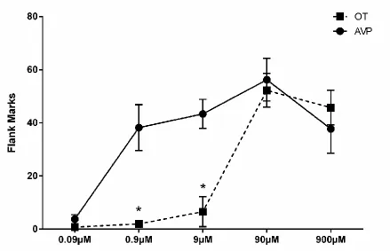
Hamsters were handled daily for four days before any behavioral testing. In Experiment 1, flank marking behavior was recorded in each hamster’s home cage for 5 minutes immediately following ICV injection of OT or AVP (0.09μM, 0.9μM, 9μM, 90μM, and 900μM). Each hamster was injected with OT and AVP at one of the above five concentrations in a counterbalanced order at 48 hour intervals (n=5 for doses 0.09μM, 0.9μM, and 9μM; n=4 for doses 90μM and 900μM). Flank marking was scored when a hamster pressed its flank gland region against a cage wall and moved forward (Ferris et al., 1984).
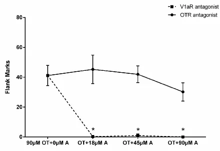
n=7, respectively). In Experiment 3, animals were given 90μM OT (n=5), mixed solutions of 90μM OT with OTA (n=5, n=9, n=7 for OT mixed with 90μM OTA, OT mixed with 45μM OTA, and OT with 18μM OTA, respectively), 90μM OT with V1aA (n=6, n=6, n=8 for OT mixed with 90μM V1aA, OT with 45μM V1aA, and OT with 18μM V1aA, respectively), OTA alone (n=6), and V1aA alone (n=7).
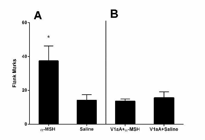
Experiment 4 was designed to examine whether α-MSH, which has been shown to be a potent releaser of endogenous OT in rats (Sabatier et al., 2003), would stimulate flank marking. Because ICV injections of α-MSH did not stimulate flank marking in hamsters tested in their home cages, the following experiments examined whether α-MSH would influence flank marking in hamsters tested in cages containing the flank gland scent of other hamsters (i.e., odor-stimulated flank marking). Immediately following ICV injections, hamsters were placed in a testing arena (23x43x20 cm) that contained flank odors from another male hamster, and flank marks were recorded for 10 minutes (n=9 for both α-MSH and saline). The flank odors were freshly deposited in the arena by a donor that had been injected ICV with AVP and allowed to mark the arena 50 times (Gutzler et al., 2011). In Experiment 4B, the V1aR (90μM) antagonist was injected ICV 1 hour before an injection of α-MSH or saline. Within subject design was used in the experiment and the order of α-MSH or saline injections was counter-balanced (n=5).
2.3.5 Data Analysis and Statistics
SPSS v21 was used to analyze all the data. The data are presented as mean ± standard error of the mean. Independent samples or paired t-tests were used for two-group comparisons and Analysis of Variance (ANOVA) was performed for comparisons among more than two groups. All comparisons were determined a priori; planned contrasts were performed following significant differences found in ANOVA tests. All tests were two tailed and differences were considered significant at p≤0.05.
2.4 Results
2.4.1 Dose-dependent effects of OT and AVP on Flank Marking
In order to examine the efficacies of OT and AVP in inducing flank marking, we microinjected OT and AVP ICV in a wide range of concentrations (0.09μM, 0.9μM, 9μM, 90μM, and 900μM). There was a significant drug by dose interaction
concentrations of AVP when injected into the same animals (paired t-tests: t(4)=4.691, p<0.01; t(4)=10.977, p<0.001; respectively).
2.4.2 Effects of OTR and V1aR Agonists on Flank Marking
This experiment employed highly selective OTR and V1aR agonists to investigate whether flank marking was induced by activation of OT or V1a receptors. There was a significant main effect of drug treatment on flank marking (F(5,33)=15.010, p<0.0001, Fig.2). Hamsters injected with low or high concentrations of the selective OTR agonist displayed significantly fewer flank marks than those injected with 90µM OT (p<0.005 and p<0.01, respectively). In fact, 10 of the 14 animals injected with either low or high concentrations of the OTR agonist marked less than three times during the five minute test. The numbers of flank marks induced by the low and intermediate concentrations of the selective V1aR agonist did not significantly differ from that induced by OT (p=0.616 and p=0.07 for low and intermediate, respectively). Hamsters injected with the high concentration of V1aR agonist flank marked significantly more than did those animals injected with OT (p<0.005).
2.4.3 Effects of OTR and V1aR Antagonists on OT-induced Flank Marking
Experiment 3 examined whether selective OTR and V1aR antagonists (OTA and V1aA, respectively) block OT-induced flank marking. There was a significant main effect of drug treatment on the number of flank marks produced (F(3, 22)=49.0, p< 0.0001; Fig.3). Hamsters injected with OT and any of the three concentrations of V1aA flank marked significantly less than those injected with OT alone (P<0.0001 for all comparisons). In fact, all three concentrations of the V1aR antagonist almost
the OTA did not significantly alter the amount of OT-induced flank marking (Fig.3). There were no significant differences in the number of flank marks among hamsters injected with OT and any of the three concentrations of OTA and those injected with OT alone (F(3, 22)=0.674, p=0.577). Hamsters in drug control groups injected with only V1aA (90μM) or OTA (90μM) did not differ from saline control animals in flank marking (flank marks: 90μM OTA: 0.0±0.0, n=7; 90μM V1aA: 0.9±1.3, n=6; saline: 0.1±0.2, n=7; F(2,17)=0.760, p=0.483).
2.4.4 Effects of Alpha-MSH on Odor-stimulated Flank Marking
This experiment examined whether α-MSH, a peptide that stimulates
endogenous OT release in rats, would enhance odor-induced flank marking. Hamsters injected with α-MSH displayed significantly more odor-induced flank marks than did hamsters injected with saline (independent samples t-test: t(16)=2.338, p<0.05; Fig.4A). This enhancement in flank marking by α-MSH was blocked by an injection of V1aR antagonist prior to testing. After administration of the V1aA, no differences in the number of flank marks were observed between groups of hamsters injected with α-MSH or saline (paired t-test: t(4)=0.611, p=0.574; Fig.4B).
2.5 Discussion
The results of the present study support the hypothesis that OT can induce flank marking by activating V1aRs and not OTRs. The data reveal that selective V1aR but not OTR agonists mimic the effects of OT in inducing flank marking, and that selective V1aR but not OTR antagonists block OT-induced flank marking. In addition, we
et al., 2003), enhances odor stimulated flank marking and that this enhancement by
α-MSH can be blocked by applying a V1aR antagonist prior to testing. Together, these findings suggest that the endogenous release of OT might contribute to the regulation of flank marking as well as other social behavior by acting on V1aRs and that the V1aR should be considered to be an OTR as well as an AVP receptor.
Comparison of OT and AVP induced flank marking reveals that OT stimulates flank marking at concentrations between 9 and 90 M, while AVP induces flank
marking at concentrations between 0.09 and 0.9 M, suggesting that OT is
approximately 100-fold less potent in activating V1aRs than is AVP. Because the levels of OT and AVP released within the hamster brain are not known, it is not clear whether OT released endogenously might contribute to the regulation of the expression of flank marking. In an attempt to investigate whether endogenously released OT might contribute to the induction of flank marking normally stimulated by exposure to flank gland secretions of conspecifics (i.e., odor-stimulated flank marking), we injected
-MSH, which has been shown in rats to induce endogenous OT, but not AVP release, from hypothalamic neurons (Sabatier et al., 2003). Although -MSH was not sufficient
to induce flank marking in the absence of conspecific odors, -MSH did significantly
enhance odor-stimulated flank marking, and this enhancement was blocked by a V1aR antagonist. The inability of -MSH to induce flank marking in the absence of
odors (Stoop, 2012). Even though -MSH, by itself, did not induce flank marking, it did
significantly enhance its expression when it was stimulated by relevant environmental cues.
In the present study, the effects of OTR/V1aR agonists and antagonists were investigated following their administration by ICV injection. Previous studies of the effects of AVP on flank marking examined its role in specific brain regions including the medial preoptic–anterior hypothalamus (MPOA-AH), periaqueductal gray, and lateral septum (Albers et al., 1986; Irvin et al., 1990; Hennessey et al., 1992; Albers & Cooper, 1995). The effects of AVP injections within these regions on flank marking are similar to the effects of ICV administration. In both cases, AVP-stimulated flank marking has an ED50 (the dose that elicits a half-maximal behavioral response) of 0.9µM, and the effect lasts for 5-10 minutes (Albers et al., 1986; Ferris et al., 1988; Caldwell & Albers, 2003; Ferris et al., 2013). Although several studies have examined the ability of OT to induce flank marking following its injection into the MPOA-AH, complete dose-response relationships are not available in most cases (Albers et al., 1986; Harmon et al., 2002). In general, the effects of different concentrations of OT into the MPOA-AH in inducing flank marking are consistent with the effects of OT given ICV in the present study (cf. Ferris et al., 1984).
by acting on V1aRs; OT rescues autistic-like deficits of OTR null mice in sociability, aggression, as well as cognitive flexibility by acting on V1aRs (Sala et al., 2011).
In summary, the present study demonstrated that OT induces flank marking by activating V1aRs. Our results raise the important possibility that endogenously released OT might influence other social behaviors by acting on V1aRs. These studies emphasize the importance of pharmacological profiling when studying the behavioral responses to OT and AVP.
2.6 Acknowledgements
2.7 Chapter 2 Figures
Figure 2.1 Effects of OTR and V1aR agonists on flank marking.
Figure 2.2 Effects of OTR and V1aR agonists on flank marking.
Figure 2.3 Effects of OTR and V1aR antagonists on OT-induced flank marking.
Figure 2.4 Effects of alpha-MSH on odor-stimulated flank marking and involvement of V1aR.
CHAPTER 3: OXYTOCIN (OT) AND ARGININE-VASOPRESSIN (AVP) ACT ON OT RECEPTORS AND NOT AVP V1A RECEPTORS TO ENHANCE
SOCIAL RECOGNITION IN ADULT SYRIAN HAMSTERS (MESOCRICETUS AURATUS)
Zhimin SONG, Tony E. LARKIN, II, Maureen O’ MALLEY, H. Elliott ALBERS
Neuroscience Institute, Center for Behavioral Neuroscience, Georgia State University, Atlanta, GA USA
Submitted to Hormones & Behavior, in revision
3.1 Abstract
Social recognition is a fundamental requirement for all forms of social
relationships. A majority of studies investigating the neural mechanisms underlying social recognition in rodents have investigated relatively neutral social stimuli such as juveniles or ovariectomized females over short time intervals (e.g., 2 hrs). The present study developed a new testing model to study social recognition among adult males using a potent social stimulus. Flank gland odors are used extensively in social communication in Syrian hamsters and convey important information such as dominance status. We found that the recognition of flank gland odors after a brief exposure lasted for at least 24 hrs, substantially longer than the recognition of other social cues in rats and mice. Intracerebroventricular injections of OT and AVP
V1aR agonists, mimicked these enhancing effects of OT and AVP. Similarly, selective OTR but not V1aR antagonists blocked recognition of the odors. In contrast, the recognition of non-social stimuli was not blocked by either the OTR or the V1aR antagonists. Our findings suggest both OT and AVP enhance social recognition via acting on OTRs and not V1aRs and that the recognition enhancing effects of OT and AVP are limited to social stimuli.
Keywords: social behavior; nonapeptides; neuropeptides; neurohypophyseal hormones; social communication; flank marking; chemosensory, olfaction;; odor recognition; memory
3.2 Introduction
The ability to recognize one individual from another of the same species is a fundamental requirement for nearly all forms of social relationships including pair bonding, maternal behavior and dominance (Petrulis, 2009; Gabor et al., 2012). Social recognition is expressed in many different levels of complexity from the relatively simple ability to discriminate between familiar and unfamiliar social cues to the true
To date, a majority of social recognition studies have focused on the ability of an adult rodent to recognize a juvenile or ovariectomized female of the same species (Le Moal et al., 1987; Ferguson et al., 2000; Ferguson et al., 2001; Winslow & Insel, 2004; Veenema et al., 2012). This approach was taken to investigate the social recognition of a relatively “neutral” social stimulus. Rats and mice fail to recognize a juvenile or an ovariectomized female within 2~3 hrs after an initial encounter in most behavioral tests (Ferguson et al., 2001; Winslow & Insel, 2004; Veenema et al., 2012), perhaps because of the neutral nature of the social stimulus. In the present study we investigated the duration of social recognition in adult male Syrian hamsters (Mesocricetus auratus) using odors obtained from flank glands as the social cue. Flank gland odor is a powerful social cue that serves to communicate a variety of different types of social information in hamsters including dominance status (Johnston & Lee, 1976; Ferris et al., 1984; Ferris et al., 1987). As such, investigating the recognition of flank gland odors in adult males
provides a social recognition test with a high degree of relevance and validity for understanding of social memory.
The nonapeptides oxytocin (OT) and arginine vasopressin (AVP) are both
functionally significant responses by activating AVP V1a receptors (V1aRs) (Schorscher-Petcu et al., 2010; Sala et al., 2011; Ramos et al., 2013; Qiu et al., 2014; Albers, 2015). For example, both OT and AVP can induce communicative behavior in hamsters when injected into the lateral ventricle and do so by activating V1a receptors and not OT receptors (OTRs) (Song et al., 2014).
In the present study, we demonstrate that adult male hamsters can recognize social odors from other adult male hamsters for at least 24 hrs and that centrally administered OT and AVP significantly enhance the duration of social recognition in this species. Further, we demonstrate for the first time that OT and AVP can produce behavioral effects by acting on OTRs and not V1aRs using highly selective OT and AVP1a receptor agonists and antagonists.
3.3 Materials and Methods
3.3.1 Animals
Institutes of Health Guidelines for the Use of Animals and were approved by the Georgia State University Animal Care and Use Committee.
3.3.2 Surgery, microinjections, and histology
Hamsters were deeply anesthetized via 5% isoflurane in an induction chamber and maintained with 3.75% isoflurane throughout all surgical procedures. Each subject was implanted with a 4mm, 26-gauge cannula guide aimed at the left lateral ventricle. The skull was leveled and the guide cannula was implanted using the stereotaxic
coordinates: +0.8mm anterior to bregma, -1.1mm from the midline, and -2.2mm below dura. Hamsters were allowed one week to recover from surgery before behavioral testing.
Introcerebroventricular (ICV) injections were administered over the course of 1min into the lateral ventricle using an infusion pump (Harvard Apparatus), a 5μl
Hamilton syringe, and a 14mm, 32-gauge needle. The volume of all microinjections was 1μl. Post-injection, the needle was left in the cannula guide for an additional minute to allow drug diffusion into the ventricle. Hamsters were sacrificed by lethal injection of sodium pentobarbital after testing and were injected with ink to verify the injection sites.
3.3.3 Drugs
90μM d(CH2)5[Tyr(Me)2]AVP (a selective V1aR antagonist known as Manning Compound, a gift of Dr. Maurice Manning). The concentration of OT and AVP
administered was based on the concentrations used in previous studies that were found effective in altering other social behavior in hamsters and other studies on social
recognition in rats (Dantzer et al., 1988; Engelmann & Landgraf, 1994; Song et al., 2014). The concentrations of the OT and AVP agonists were based on their relative efficacies compared to those of OT and AVP in binding to OTR and V1aR in rats, respectively (Manning et al., 2012). The concentration of both OTR and V1aR
antagonists were based on the concentration used in previous studies that was found to block social behavior in hamsters and rats (Ferris et al., 1988; Nephew & Bridges, 2008). All control animals were given a 1μl injection of saline.
3.3.4 Behavioral Testing
3.3.4.1Recognition of social odors
The goal of Experiment 1 was to determine how long hamsters can recognize a conspecific odor. Hamsters were placed in a cage (23x43x20 cm) with a glass
wall and another slide taped to the opposite cage wall for 3min (Trial 2). One slide was scented with fresh flank gland odor of the same conspecific from Trial 1 (familiar odor) and the other slide with fresh flank gland odor of a novel male conspecific (novel odor). The familiar and novel stimulus hamsters were from two groups of animals that were different in age by 1-3 months; half of each of the two groups formed familiar hamsters and the other half formed novel hamsters. Each of the experimental hamsters had one stimulus hamster from each group as familiar and novel hamsters. Thus, the familiar and novel odors were not from the same litters for each subject and the intrinsic properties of each group that might affect investigation time were counterbalanced. Testing took place in a room with red dim lights and within the first 3 hr of the dark phase of the light-dark cycle. Time spent sniffing the odor was recorded and scored later by a trained experimenter blind to experimental conditions.
exposure. Forty-eight hours later, they were again placed in a cage to allow for
investigation of an odor from the previously encountered conspecific (familiar odor) and an odor from a novel conspecific (novel odor) for 3min (Trial 2).
Experiment 4 determined whether OTR and V1aR antagonists injected into the ventricle could block social recognition 20min after an initial odor exposure. The
protocol for Experiment 4 was the same as in Experiments 2&3 except the time between the first odor exposure and the odor test was 20 min. The OTR (n = 11), V1aR (n = 8) antagonists or saline (n = 10) were injected before the initial odor exposure in order to allow time for the antagonists to act before the first odor exposure.
3.3.4.2Recognition for non-social stimulus
the cocktail were placed on opposite sides of the cage and their locations were
counterbalanced. Experiment 5 examined how long hamsters could recognize a non-social odor after an initial exposure. The odor test occurred 20 min (n = 6), 60 min (n = 6), and 24 hr (n = 6) after the initial odor exposure. Because this initial experiment found that the lemon scent could be recognized for 20 min and 60 min, but not 24 hr, the ability of the OTR and V1aR antagonists to block recognition was tested after 20 min. In Experiment 6, hamsters were randomly divided into 3 groups and were injected with 90uM OTR antagonist (n =12), 90uM V1aR antagonist (n=6), or saline (n = 9) immediately before the initial odor exposure. The odor test took place 20 min after the initial odor exposure.
3.3.5 Data analyses and statistics
significant differences found in ANOVA tests. All tests were two tailed and differences were considered significant at p ≤ 0.05.
3.4 Results
3.4.1 Recognition of social odors
Experiment 1: How long do hamsters recognize a previously encountered social odor?
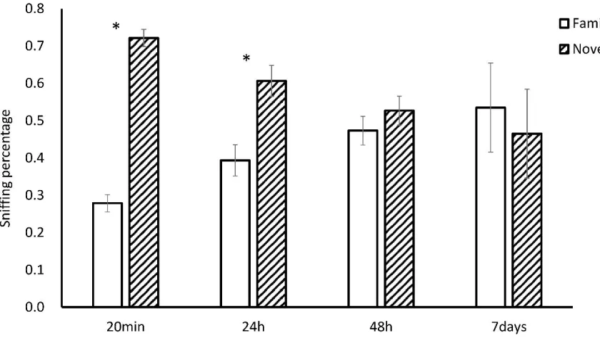
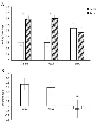
Hamsters were first exposed to a flank gland odor for 3 min and then tested 20 min, 24 hr, 48 hr, or 7 days later, in a social discrimination test with the same flank gland odor and a novel flank gland odor. Hamster that were tested 20 min and 24 hr after the initial odor exposure spent more time sniffing the novel odors compared to the familiar odors (20 min, t(8) = 5.070, p < 0.05; 24 hr, t(8) = 2.561, p < 0.05, Fig. 1), whereas those tested 48 hr or 7 days later spent similar amounts of time sniffing both odors (48 hr, t(7) = 1.214, p = 0.270; 7 days, t(6) = -0.293, p = 0.780, Fig. 1). These data indicate hamsters can recognize a conspecific odor 20 min and 24 hr, but not 48 hr or 7 days after an initial 3 min exposure.
Experiment 2: Do OT and AVP increase the duration of recognition of social odors?
of time sniffing the novel odor and the odor presented initially (t(5) = 0.278, p = 0.792); this is consistent with the outcomes from Experiment 1 (Fig. 2A). In contrast, hamsters that were injected with OT or AVP spent more time investigating the novel odor
compared to the odor presented initially (OT: t(7) = 4.202, p<0.05; AVP: t(6) = 2.643, p<0.05, Fig. 2A). Thus, OT or AVP administration after exposure to the initial odor increased the time hamsters were able to discriminate between familiar and novel odors from 24 to 48 hr. ANOVA indicated that there was a significant difference in the
Difference Percentages among the three groups (F(2,18) = 4.152, p < 0.05, Fig. 2B). Planned post hoc contrasts indicated that the Difference Percentages in OT and AVP injected hamsters were higher than that in saline injected hamsters (p < 0.05) and there was no difference of the Difference Percentages between the OT and AVP injected
hamsters (p = 0.524).
Experiment 3: Do selective OTR and V1aR agonists increase the duration of recognition of social odors?
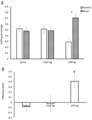
sniffing the novel odor than the odor presented initially (t(8) = 3.452, p < 0.05, Fig. 3A). There was a difference in the Difference Percentages among the three groups (F(2,24) = 3.921, p < 0.05, Fig. 3B). Post hoc tests indicated the hamsters injected with the OTR agonist had a significantly higher Difference Percentage than the hamsters injected with V1aR agonist (p < 0.05) or the hamsters injected with (p < 0.05). No significant
differences were observed in Difference Percentages between the groups injected with the V1aR agonist and the group injected with saline (p = 0.760).
Experiment 4: Do OTR and V1aR antagonists reduce the duration of recognition of social odors?
Hamsters were initially exposed to a flank gland odor immediately following ICV injections of OTA, V1aA, or saline. Twenty min later hamsters were then exposed to flank gland odor from the same individual used in the initial odor exposure and a flank gland odor from a novel hamster. Hamsters injected with saline spent significantly more time sniffing the novel odor than the familiar odor (t(9) = 3.547, p < 0.01, Fig. 4A), consistent with data obtained in Experiment 1. Hamsters injected with the selective V1aR antagonist also spent significantly the time spent sniffing the novel odor than the odor presented initially did not was not significantly (t(7) = 3.189, p < 0.05, Fig. 4A). In contrast, hamsters injected with the OT antagonist spent similar amounts of time
significant differences were observed in Difference Percentages between the groups injected with the V1aR antagonist and the group injected with saline (p = 0.760).
3.4.2 Recognition of non-social odors
Experiment 5: How long do hamsters recognize a previously encountered non-social scent?
Hamsters were first exposed to a lemon odor for 3 min and then were tested 20 min, 60 min or 24 hr later in an odor discrimination test with the lemon odor and an odor composed of a cocktail of lemon and vanilla together. As a control for an initial preference for the scents, another group of hamsters was not pre-exposed to the lemon odor but was directly exposed to the lemon odor and the cocktail odor. Control
hamsters preferred the lemon odor over the cocktail (initial preference). To determine the retention of the recognition of the lemon odor during the pre-exposure, the
percentage of time on the cocktail odor versus on the lemon odor was compared
hamsters can recognize non-social scents 20min and 60min, but not 24h after an initial 3min encounter.
Experiment 6: Do OTR and V1aR antagonists reduce the duration of recognition of non-social odors?
Hamsters were initially exposed to lemon odor immediately following ICV
injections of OTA, V1aA, or saline. Twenty minutes later hamsters were then exposed to lemon odor and the cocktail odor. Another group of hamsters was not initially exposed to lemon odor but was directly exposed to the lemon odor and the cocktail odor as an initial preference control (there was no difference in the duration of sniffing between these hamsters and those used for the same purpose in Experiment 5, so they were pooled together for data analysis). Again, to detect the recognition of the lemon odor during the pre-exposure, the percentage of time on the cocktail odor versus on the lemon odor was compared between hamsters that were pre-exposed to the lemon and those that were not pre-exposed. There was a significant difference in the percentages of time spend sniffing the novel cocktail odor compared to the lemon odor among the 4 hamster groups (F(3,34) = 14.930, p < 0.001, Fig. 6). All the hamsters that were
3.5 Discussion
In the present study adult male hamsters were found to recognize the social odors of other adult males for 20 min and 24 hr, but not 48 hr or 7 days. The duration of this social recognition is substantially longer than that seen in many previous tests of social recognition that employed more neutral social stimuli, e.g., juvenile rats. An advantage of using a social odor as opposed to another animal as the social stimulus is that it eliminates the possibility that the properties of the stimulus will change as the result of social interactions between the stimulus animal and the test animal. As such, this social recognition test provides a simple and robust approach with a great deal of relevance and validity that can be used to investigate neurobiological mechanisms of social recognition.
The injection of either AVP or OT in the lateral ventricle prolonged recognition of flank gland odors to at least 48 hr. The OTR agonist but not the V1aR agonist mimicked these recognition enhancing effects of OT and AVP. Similarly, the OTR but not the V1aR antagonist blocked recognition of the odor. These findings indicate that both OT and AVP can significantly increase the duration of social recognition and do so by acting on OTRs and not V1aRs. Importantly, our studies also demonstrated that the effects of AVP and OT do not extend to the recognition of non-social odors. Hamsters recognized lemon scent for up to 60 min but not 24 hr. Unlike in the social recognition
experiments, neither the OTR nor V1aR antagonists reduced the duration of the recognition of lemon scent. These data support the hypothesis that OT and AVP enhance the recognition of social but not non-social stimuli.
studies in rats and mice (2~3 hrs) (Dantzer et al., 1987; Winslow & Insel, 2004; Choleris et al., 2009; Engelmann et al., 2011; Gabor et al., 2012). The recognition of social odors
may be particularly important for species like Syrian hamsters because individuals do not encounter other individuals frequently in the wild (Gattermann et al., 2008). The longer duration of social recognition may also result from using flank gland odors as the social cue because these odors represent a powerful social stimulus that serves to
communicate important social information such as dominance status (Ferris et al., 1987). The recognition and memory of social stimuli can be quite sophisticated in rodents. Social recognition can be influenced by social and environmental context including the sex of the stimulus animal, the place where the initial exposure occurs (home cage, clean cage, or cage of another individual) (Zheng et al., 2013), and whether the stimulus animal was infected with parasites (Kavaliers et al., 2005). Rodents are also capable of kin recognition and the true recognition of specific individuals (e.g. Mateo & Johnston, 2003; Johnston & Peng, 2008; Petrulis, 2009). It will be important to determine if OT and AVP may be involved in mediating some of the more complex forms of social recognition.
social recognition. Medial amygdala (MeA) has been reported as an essential site for OT’s effects on social recognition. Antagonism of OTRs in the MeA impairs social recognition of juveniles and OT injection into the MeA rescues the deficits in social recognition seen in OT knock-out mice (Ferguson et al., 2000; Ferguson et al., 2001). Endogenous OT release in the main olfactory bulb (MOB) can enhance the retention of social memory and this enhancement can be blocked by an OTR antagonist (Larrazolo-Lopez et al., 2008). AVP appears to influence social recognition by acting on the lateral septum (LS) and the MOB. Administration of AVP in the LS extends the recognition of juveniles and antagonism of V1aRs in this area impairs the recognition (Dantzer et al., 1988; Veenema et al., 2012). AVP injected into the LS restores social recognition in AVP-deficit of Brattleboro rats (Engelmann & Landgraf, 1994). AVP injection in the MOB also prolongs the recognition of juvenile rats (Dluzen et al., 1998) and V1aR antagonism in the MOB impairs recognition (Tobin et al., 2010). Taken together, these studies show there are multiple regions in the CNS where OT and AVP can influence social recognition.
habituate to the presence of ovariectomized females, suggesting an impairment in social recognition (Bielsky et al., 2004; Bielsky et al., 2005). In contrast, another study using V1aR knock-out mice from the same source in a similar habituation and dishabituation paradigm found a subtle olfactory deficit but normal social recognition (Wersinger et al., 2007). Our data are consistent with the later study suggesting that OTRs and not
V1aRs mediate social recognition. The reasons for the discrepancy in studies regarding V1aRs are unknown. It is possible that the V1aR is involved in other behaviors such as anxiety-related behavior that may indirectly affect social recognition in a less profound way. It could be that different testing models such as using a juvenile animal versus social odors may underlie these different results. It is also possible, of course, that the role of V1aRs in social recognition may vary across species.
The present study provides the first evidence that OT and AVP act on OTRs and not V1aRs to produce behavioral effects. Recently, another study from our lab found that both OT and AVP induce social communication via acting on V1aRs and not OTRs in hamsters (Song et al., 2014). Other studies have also found that both OT and AVP can act on V1a receptors to influence behavioral and physiological responses
(Schorscher-Petcu et al., 2010; Sala et al., 2011; Ramos et al., 2013; Qiu et al., 2014; Albers, 2015). Taken together, these data suggest that while OT and AVP can activate both OTRs and V1aRs to influence social behavior OTRs regulate some social behaviors and V1aRs regulate other social behaviors. At present there is no evidence that a
Freund-Mercier, 1997). As such, the activation of small anatomically separable populations of OTRs or V1aRs by the local synaptic release of neuropeptide may influence a single social behavior while the release of large amounts of AVP and/or OT by volume transmission could potentially activate large numbers of both OTRs and V1aRs and thereby influence the expression of multiple behaviors.
3.6 Conclusion
The present study employed a new test of social recognition of adult conspecifics wherein flank gland odors of adult male hamsters were used as the social cue to
investigate neurobiological mechanisms underlying social recognition. Centrally
administered OT and AVP significantly prolong the recognition of social cues from other adult individuals but these enhancing effects of OT and AVP do not extend to
recognition of non-social cues. Further, both OT and AVP were found to act on OTRs but not V1aRs to influence social recognition, thus providing the first evidence that AVP and OT can act on OTRs to regulate social behavior. These findings emphasize the potential significance of the interactions between OT, AVP and their receptors in regulating complex social behavior in the central nervous system.
3.7 Acknowledgements
3.8 Chapter 3 Figures
Figure 3.1 Duration of social recognition in hamsters
Figure3.2 OT and AVP enhance social recognition
Hamsters injected with OT or AVP spent less time in the familiar odor 48hr after their initial exposure, whereas control hamsters spent the same amount of time on the
Figure 3.3 Effects of V1aR and OTR agonists on social recognition
Figure 3.4 Effects of V1aR and OTR antagonists on social recognition
Figure 3.5 Duration of recognition for non-social cues
Hamsters that were not pre-exposed to lemon (the Initial group) spent more time with lemon when presented with a lemon and vanilla cocktail, whereas hamsters that were pre-exposed to lemon did not. B. Hamsters that were pre-exposed 20min and 60min earlier had significantly greater Difference Percentages than hamsters in the Initial group. Hamsters that were pre-exposed 24h earlier had similar Difference Percentages than the Initial group. * indicates a significant difference between Lemon and Cocktail. # indicates a significant difference compared with Initial.
Figure 3.6 Neither V1aR nor OTR antagonist blocked non-social recognition
Hamsters that were not pre-exposed (Initial group) spent more time with lemon; whereas hamsters that were injected Sal, OTA, or V1aA did not. B. Hamsters that were injected with Saline, OTA, or V1aA had greater Difference Percentages than hamsters in the Initial group. OTA: OTR antagonist; V1Aa: V1aR antagonist. * indicates a
significant difference between Lemon and Cocktail. # indicates a significant difference compared with Initial.
CHAPTER 4 ACTIVATION OF OXYTOCIN RECEPTORS IN THE
VENTRAL TEGMENTAL AREA IS ESSENTIAL FOR THE REWARDING PROPERTIES OF SOCIAL INTERACTIONS
Zhimin SONG, Tony E. LARKIN, II, Maureen O’ MALLEY, H. Elliott ALBERS
Neuroscience Institute, Center for Behavioral Neuroscience, Georgia State University, Atlanta, GA USA
4.1 Abstract
Interestingly, OT and AVP injected into the VTA induced a two-fold increase in the place preference when compared to control injections of saline. Finally, because OT and AVP can act on each other’s receptors to influence social behavior, we also determined which receptor is responsible for mediating these effects by applying highly selective OTR and V1aR agonists and antagonists. Our results not only showed that OT and AVP activate OTRs and not V1aRs to enhance social reward, they also showed the OTR activation in the VTA is essential for the expression of social reward.
Keywords: social interaction, social behavior, social salience, conditioned place preference, neuropeptides
4.2 Introduction
being alone as long as the loss is not too severe (Gil et al., 2013b). The mechanisms underlying social reward are critical for the expression of adaptive social behavior and dysfunctions in the regulation of social reward can result in the development of
devastating psychiatric disorders (Bora et al., 2009).
Oxytocin (OT) and arginine-vasopressin (AVP) are evolutionarily conserved mammalian neuropeptides that play essential roles in regulating a variety of motivated social behaviors including aggression, social recognition, parental behavior and social communication (Ishak et al., 2011; Albers, 2012; 2015). The effects of OT and AVP in the brain are mediated primarily through the activation of OT receptors or V1a AVP receptors. Many of the behavioral effects of OT and AVP are the result of their actions in structures that have been proposed to form a social behavior neural network (SBNN) (Newman, 1999). There is increasing evidence that the SBNN, composed of neural groups or “nodes” including, but not limited to, the extended amygdala, the bed nucleus of the stria terminalis (BNST), lateral septum (LS), periaqueductal gray (PAG), medial preoptic area (MPOA), ventromedial hypothalamus (VMH), and anterior hypothalamus (AH), controls the expression of social behavior (Crews, 1997; Albers, 2012; Goodson & Kingsbury, 2013; Albers, 2015).
Motivated social behaviors also require the activity of the mesolimbic dopamine (DA) system, a network of reciprocally connected brain regions that determine the salience of stimuli, assign motivational value, and initiate appropriate action (Love, 2014; Caldwell & Albers, 2015). The ventral tegmental area (VTA) is a key region in this motivational network providing many of the dopamine containing projections that innervate a variety of cortical and limbic structures. While the SBNN and the
making about the expression of motivated social behaviors (O'Connell & Hofmann, 2011a; b).
While OT and AVP can act within the mesolimbic DA system to influence the expression of various forms of social behavior there is very little data on whether OT or AVP might contribute to the rewarding properties of social behavior by their action within these structures. The purpose of the present study was test the hypothesis that activation of OT and/or V1a receptors in the VTA mediates the rewarding properties of social interaction. Because OT and AVP can influence social behavior by acting on each other’s receptors (Sala et al., 2011; Song et al., 2014) we also determined which
receptors are responsible for inducing social reward by applying highly selective OT and AVP receptor agonists and antagonists.
4.3 Materials and Methods
4.3.1 Animals
Syrian hamsters (Mesocricetus auratus), purchased from Charles River Laboratories Inc., Wilmington, MA, USA, were used in all experiments. The