0095-1137/08/$08.00⫹0 doi:10.1128/JCM.02453-07
Copyright © 2008, American Society for Microbiology. All Rights Reserved.
Validation of Virulence and Epidemiology DNA Microarray for
Identification and Characterization of
Staphylococcus aureus
Isolates
䌤
†
Richard P. Spence, Victoria Wright, Dlawer A. A. Ala-Aldeen, David P. Turner,
Karl G. Wooldridge, and Richard James*
Centre for Healthcare Associated Infections, Institute of Infection, Immunity and Inflammation, CBS Building, University Park, University of Nottingham, Nottingham NG7 2RD, United Kingdom
Received 20 December 2007/Returned for modification 29 December 2007/Accepted 13 February 2008
The human pathogen Staphylococcus aureus is isolated and characterized using traditional culture and
sensitivity methodologies that are slow and offer limited information on the organism. In contrast, DNA microarray technology can provide detailed, clinically relevant information on the isolate by detecting the presence or absence of a large number of virulence-associated genes simultaneously in a single assay. We have developed and validated a novel, cost-effective multiwell microarray for the identification and characterization
ofStaphylococcus aureus. The array comprises 84 gene targets, including species-specific, antibiotic resistance,
toxin, and other virulence-associated genes, and is capable of examining 13 different isolates simultaneously,
together with a reference control strain. Analysis ofS. aureusisolates whose complete genome sequences have
been determined (Mu50, N315, MW2, MRSA252, MSSA476) demonstrated that the array can reliably detect
the combination of genes known to be present in these isolates. Characterization of a further 43S. aureus
isolates by the microarray and pulsed-field gel electrophoresis has demonstrated the ability of the array to
differentiate between isolates representative of a spectrum ofS.aureustypes, including methicillin-susceptible,
methicillin-resistant, community-acquired, and vancomycin-resistantS. aureus, and to simultaneously detect
clinically relevant virulence determinants.
Staphylococcus aureusis a common human pathogen respon-sible for a plethora of infections, from superficial skin infec-tions to life-threatening diseases such as endocarditis, sepsis, and pneumonia. Methicillin-resistant S. aureus (MRSA) is a major cause of morbidity and mortality in the hospital setting. The emerging threats of community-associated MRSA (CA-MRSA) and vancomycin-resistantS. aureus(VRSA) highlight the importance of rapid detection of such infections.
Most diagnostic microbiology laboratories continue to iden-tifyS. aureususing traditional culture and susceptibility meth-ods that are slow (48 to 72 h) and provide only limited infor-mation. Molecular assays based on PCR have been reported for the detection of MRSA (4, 6, 7, 9, 10, 30), the identification of staphylococcal species (17, 20, 21), or the identification of specific virulence genes (5, 11, 14, 15, 18, 19, 22, 24, 26, 33). DNA microarrays can identify, subtype, and detect acquired antibiotic resistance determinants simultaneously (1, 23, 32, 35); however, their clinical value has been limited by a com-plicated methodology that is unsuitable for routine use in di-agnostic microbiology laboratories.
We have developed an oligonucleotide-based microarray (designated VirEp, for virulence and epidemiology
microar-ray) incorporating 84 clinically relevant gene targets for the characterization and molecular typing of clinical isolates ofS. aureusin an economical, multiwell format enabling 13S. au-reusisolates to be analyzed simultaneously.
MATERIALS AND METHODS
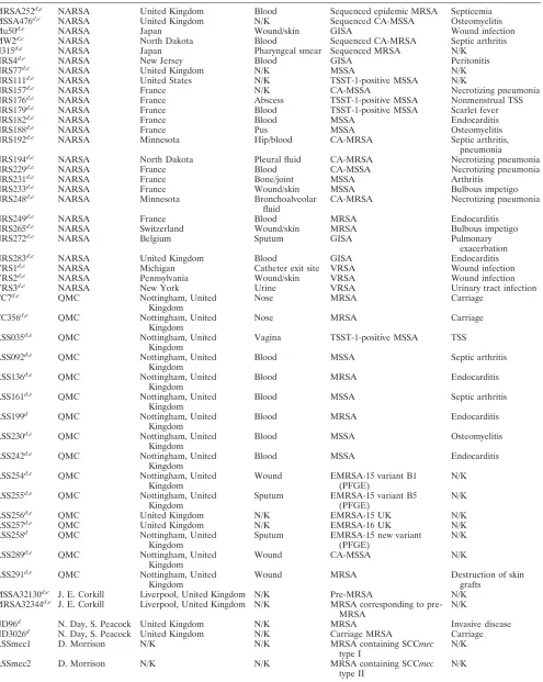
Bacterial isolates and culture conditions.The isolates used in this study (Table 1) were grown at 37°C for 16 h on brain heart infusion agar, except for the VRSA isolates, for which 6g/ml of vancomycin was added to the brain heart infusion agar.
Oligonucleotide probe design and synthesis. Oligonucleotide probes were designed using OligoArray 2.0 software (28) and were synthesized at a 10-nmol scale with amino C6 modification (Operon Biotechnologies). Three 45- to 46-mer oligonucleotides with calculated melting temperatures of 68 to 72°C and minimal internal structure were selected for each gene target (see Appendix S1 in the supplemental material). Where this was not possible, shorter or longer oligonucleotides were used.
Slide printing. Oligonucleotides were printed onto amine silane-coated UltraGAPS slides (Corning B.V. Life Sciences) at a concentration of 20M in spotting buffer (3⫻SSC [1⫻SSC is 0.15 M NaCl plus 0.015 M sodium citrate], 1.5 M betaine) along with Universal ScoreCard controls (GE Healthcare UK Ltd.) and appropriate positive and negative controls (see the supplemental material). Fourteen replicates of the array were printed on each microarray slide using a QArray Lite robotic arrayer (Genetix Ltd.).
DNA extraction and labeling.Genomic DNA (gDNA) was extracted fromS. aureuscultures using the DNeasy tissue kit (Qiagen) according to the manufac-turer’s instructions with the addition of 5l of lysostaphin (0.5 mg/ml) and 2l of RNase A (100 mg/ml) to the lysis buffer. The concentration of gDNA was determined using a Nanodrop ND-1000 spectrophotometer (Nanodrop Tech-nologies Inc.). gDNA and spike-in Universal controls (for details, see Appendix S3 in the supplemental material) were labeled with Cy3-dCTP using a protocol based on that described by Pearson et al. (27).
Hybridization.Microarray slides were incubated in prehybridization solution (5⫻SSC, 0.1% sodium dodecyl sulfate [SDS], 0.1 mg/ml bovine serum albumin) for 60 min at 60°C, washed twice in 0.1⫻SSC for 5 min and once in purified water for 30 s at room temperature, and then dried by centrifugation. Prior to
* Corresponding author. Mailing address: Centre for Healthcare Associated Infections, Institute of Infection, Immunity and Inflamma-tion, CBS Building, University Park, University of Nottingham, Not-tingham NG7 2RD, United Kingdom. Phone: 44 115 84667952. Fax: 44 115 8467951. E-mail: [email protected].
† Supplemental material for this article may be found at http://jcm .asm.org/.
䌤Published ahead of print on 20 February 2008.
1620
on May 16, 2020 by guest
http://jcm.asm.org/
TABLE 1. S. aureusisolates used in this study
Isolate Sourcea Originb Site of isolation Descriptionc Disease association
MRSA252d,e NARSA United Kingdom Blood Sequenced epidemic MRSA Septicemia
MSSA476d,e NARSA United Kingdom N/K Sequenced CA-MSSA Osteomyelitis
Mu50d,e NARSA Japan Wound/skin GISA Wound infection
MW2d,e NARSA North Dakota Blood Sequenced CA-MRSA Septic arthritis
N315d,e NARSA Japan Pharyngeal smear Sequenced MRSA N/K
NRS4d,e NARSA New Jersey Blood GISA Peritonitis
NRS77d,e NARSA United Kingdom N/K MSSA N/K
NRS111d,e NARSA United States N/K TSST-1-positive MSSA N/K
NRS157d,e NARSA France N/K CA-MSSA Necrotizing pneumonia
NRS176d,e NARSA France Abscess TSST-1-positive MSSA Nonmenstrual TSS
NRS179d,e NARSA France Blood TSST-1-positive MSSA Scarlet fever
NRS182d,e NARSA France Blood MSSA Endocarditis
NRS188d,e NARSA France Pus MSSA Osteomyelitis
NRS192d,e NARSA Minnesota Hip/blood CA-MRSA Septic arthritis,
pneumonia
NRS194d,e NARSA North Dakota Pleural fluid CA-MRSA Necrotizing pneumonia
NRS229d,e NARSA France Blood CA-MSSA Necrotizing pneumonia
NRS231d,e NARSA France Bone/joint MSSA Arthritis
NRS233d,e NARSA France Wound/skin MSSA Bulbous impetigo
NRS248d,e NARSA Minnesota Bronchoalveolar
fluid
CA-MRSA Necrotizing pneumonia
NRS249d,e NARSA France Blood MRSA Endocarditis
NRS265d,e NARSA Switzerland Wound/skin MRSA Bulbous impetigo
NRS272d,e NARSA Belgium Sputum GISA Pulmonary
exacerbation
NRS283d,e NARSA United Kingdom Blood GISA Endocarditis
VRS1d,e NARSA Michigan Catheter exit site VRSA Wound infection
VRS2d,e NARSA Pennsylvania Wound/skin VRSA Wound infection
VRS3d,e NARSA New York Urine VRSA Urinary tract infection
CC7d,e QMC Nottingham, United
Kingdom
Nose MRSA Carriage
CC356d,e QMC Nottingham, United Kingdom
Nose MRSA Carriage
RSS035d,e QMC Nottingham, United Kingdom
Vagina TSST-1-positive MSSA TSS
RSS092d,e QMC Nottingham, United Kingdom
Blood MSSA Septic arthritis
RSS136d,e QMC Nottingham, United Kingdom
Blood MRSA Endocarditis
RSS161d,e QMC Nottingham, United Kingdom
Blood MSSA Septic arthritis
RSS199d QMC Nottingham, United
Kingdom
Blood MRSA Endocarditis
RSS230d,e QMC Nottingham, United Kingdom
Blood MSSA Osteomyelitis
RSS242d,e QMC Nottingham, United Kingdom
Blood MSSA Endocarditis
RSS254d,e QMC Nottingham, United Kingdom
Wound EMRSA-15 variant B1 (PFGE)
N/K
RSS255d,e QMC Nottingham, United Kingdom
Sputum EMRSA-15 variant B5 (PFGE)
N/K
RSS256d,e QMC United Kingdom N/K EMRSA-15 UK N/K
RSS257d,e QMC United Kingdom N/K EMRSA-16 UK N/K
RSS258d QMC Nottingham, United
Kingdom
Sputum EMRSA-15 new variant (PFGE)
N/K
RSS289d,e QMC Nottingham, United Kingdom
Wound CA-MSSA N/K
RSS291d,e QMC Nottingham, United Kingdom
Wound MRSA Destruction of skin
grafts MSSA32130d,e J. E. Corkill Liverpool, United Kingdom N/K Pre-MRSA N/K MRSA32344d,e J. E. Corkill Liverpool, United Kingdom N/K MRSA corresponding to
pre-MRSA
N/K
ND96d N. Day, S. Peacock United Kingdom N/K MRSA Invasive disease
ND3026d N. Day, S. Peacock United Kingdom N/K Carriage MRSA Carriage
RSSmec1 D. Morrison N/K N/K MRSA containing SCCmec
type I
N/K
RSSmec2 D. Morrison N/K N/K MRSA containing SCCmec
type II
N/K
Continued on following page
on May 16, 2020 by guest
http://jcm.asm.org/
hybridization, ProPlate superstructures (Stratech Scientific Ltd.) were attached to the slides to create a multiwell format. Forty picomoles of Cy3-labeled gDNA in 50l of hybridization solution (5⫻SSC, 0.1% SDS, and 0.1 mg/ml herring sperm DNA) was denatured at 95°C for 5 min. Hybridization mixtures were then added to individual wells on the slide before wells were sealed. Slides were hybridized at 60°C for 16 h in the dark with gentle agitation before being washed in 2⫻SSC–0.1% SDS at 42°C once to remove the superstructure and once for 5 min. Slides were then washed twice for 5 min in 0.1⫻SSC–0.1% SDS, five times for 1 min in 0.1⫻SSC, and once for 10 s in 0.01⫻SSC. Arrays were dried by centrifugation at 1,600⫻gfor 2 min.
Data analysis.Hybridized slides were scanned with an Axon 4000B slide scanner (Molecular Devices Corporation) using a resolution of 10m, and images were analyzed with GenePix Pro 6.0 software. Spots with a signal-to-noise ratio ofⱖ1 and a total median fluorescence at 532 nm of⬎1,000 after subtrac-tion of the background fluorescence were classified as positive. For a gene target to be considered present, at least two-thirds of the spots corresponding to that gene target had to be positive. Microarray experiments were MIAME (minimum information about a microarray experiment) compliant, and experimental data were deposited in the ArrayExpress repository (2).
PFGE.S. aureuschromosomal SmaI digests were prepared with the GenePath group 1 reagent kit (Bio-Rad Laboratories), and pulsed-field gel electrophoresis (PFGE) patterns were obtained with a contour-clamped homogeneous electric field apparatus (Bio-Rad) as described previously (13). PFGE images were analyzed with BioNumerics 2.0 software (Applied Maths). Dendrograms were generated using the Dice coefficient and the unweighted-pair group method using average linkages (UPGMA) with 1% tolerance and 0.5% optimization. A similarity cutoff of 80% and a difference ofⱕ6 bands were used to define clusters (29, 31). The presence and absence of genes determined by microarray analysis were recorded in a binary format and processed using Bionumerics 2.0 software, and the results were presented as a dendrogram.
Statistical analysis.The reproducibility of microarray data was examined by calculating the coefficient of variation, which is the standard deviation divided by the normalized mean for replicates hybridized to different microarrays. The
discriminatory power of the VirEp (virulence and epidemiology) microarray as a typing method was determined by calculating Simpson’s index of diversity (8).
RESULTS
Validation of the complete multiwell formatS. aureus
oligo-nucleotide microarray by analysis of sequencedS. aureus
iso-lates. An initial list of 89 gene targets, including acquired
antibiotic resistance determinants, toxins, adhesins, proteases, and other virulence genes, was selected on the basis of the significance of these genes in clinical disease and epidemiol-ogy. Four replicates of S. aureus isolates MW2, N315, MRSA252, MSSA476, and Mu50 (Table 1) were hybridized to UltraGAPS microarray slides printed with the VirEp microar-ray. Oligonucleotides for 84/89 genes examined generated the expected results compared to sequencing data. Five gene tar-gets (sea,seg,sep,edin-B, andsdrD) gave discrepant results and were discarded. The specificity of oligonucleotides for staphy-lococcal cassette chromosomemec(SCCmec) types I to V were confirmed by hybridizing gDNA from isolates containing each SCCmecelement to the VirEp microarray (Table 1). The ex-perimental coefficient of variation for the VirEp microarray was found to be 0.14 (14%), indicating that the data generated by the VirEp microarray are reproducible.
Application of the VirEp microarray to the identification
and characterization of clinical isolates ofS. aureus.Labeled
[image:3.585.47.539.81.318.2]DNAs from a collection of 64 clinical isolates ofS. aureuswere
TABLE 1—Continued
Isolate Sourcea Originb Site of isolation Descriptionc Disease association
RSSmec3 D. Morrison N/K N/K MRSA containing SCCmec
type III
N/K
RSSmec4 D. Morrison N/K N/K MRSA containing SCCmec
type IV
N/K
WIS T. Ito N/K N/K MRSA containing SCCmec
V
N/K
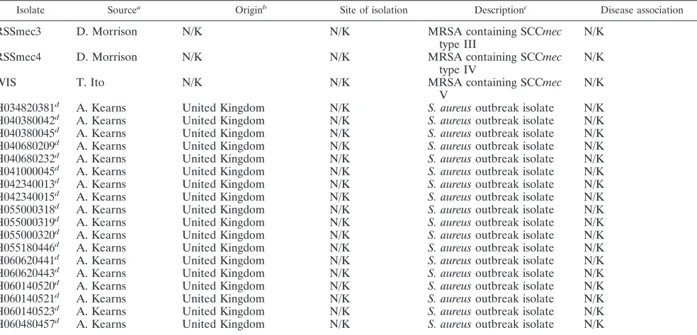
H034820381d A. Kearns United Kingdom N/K S. aureusoutbreak isolate N/K H040380042d A. Kearns United Kingdom N/K S. aureusoutbreak isolate N/K H040380045d A. Kearns United Kingdom N/K S. aureusoutbreak isolate N/K H040680209d A. Kearns United Kingdom N/K S. aureusoutbreak isolate N/K H040680232d A. Kearns United Kingdom N/K S. aureusoutbreak isolate N/K H041000045d A. Kearns United Kingdom N/K S. aureusoutbreak isolate N/K H042340013d A. Kearns United Kingdom N/K S. aureusoutbreak isolate N/K H042340015d A. Kearns United Kingdom N/K S. aureusoutbreak isolate N/K H055000318d A. Kearns United Kingdom N/K S. aureusoutbreak isolate N/K H055000319d A. Kearns United Kingdom N/K S. aureusoutbreak isolate N/K H055000320d A. Kearns United Kingdom N/K S. aureusoutbreak isolate N/K H055180446d A. Kearns United Kingdom N/K S. aureusoutbreak isolate N/K H060620441d A. Kearns United Kingdom N/K S. aureusoutbreak isolate N/K H060620443d A. Kearns United Kingdom N/K S. aureusoutbreak isolate N/K H060140520d A. Kearns United Kingdom N/K S. aureusoutbreak isolate N/K H060140521d A. Kearns United Kingdom N/K S. aureusoutbreak isolate N/K H060140523d A. Kearns United Kingdom N/K S. aureusoutbreak isolate N/K H060480457d A. Kearns United Kingdom N/K S. aureusoutbreak isolate N/K
aNARSA, Network on Antimicrobial Resistance inStaphylococcus aureus, Focus Technologies Inc., Herndon, VA; QMC, Diagnostic Microbiology Laboratory,
Queens Medical Centre, Nottingham, United Kingdom. The affiliations of individuals who supplied isolates are as follows: John E. Corkill, Department of Medical Microbiology, Royal Liverpool University Hospital, Liverpool, United Kingdom; Nick Day and Sharon Peacock, Faculty of Tropical Medicine, Mahidol University, Bangkok, Thailand; Donald Morrison, Scottish MRSA Reference Laboratory, Department of Microbiology, Stobhill Hospital, Glasgow, United Kingdom; Teruyo Ito, Department of Bacteriology, Juntendo University, Tokyo, Japan; and Angela M. Kearns,Staphylococcus Reference Laboratory, Centre for Infections, Health Protection Agency, London, United Kingdom.
bN/K, not known.
cTSST-1, toxic shock syndrome toxin 1.
dExamined using the completeS. aureusmicroarray. eExamined by microarray and PFGE.
on May 16, 2020 by guest
http://jcm.asm.org/
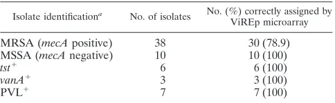
hybridized to the VirEp microarray with the remaining 84 gene targets (Table 1). All isolates were correctly identified asS. aureusbased on the presence of cap, coa, cpn60, femA, nuc, andtpigenes. Ten of ten (100%) isolates were correctly iden-tified as MSSA (Table 2), and 30 of 38 (78.9%) isolates were correctly identified as MRSA (Table 2). However, false-nega-tive results for themecAgene were obtained for eight MRSA isolates from the United Kingdom. Six of the eight isolates were confirmed to possess themecAgene by PCR. We assume that isolates RSS257 and RSS258, which weremecAnegative by PCR, had lost theirmecelement upon storage at⫺80°C, which has been reported to be common among MRSA isolates (34). All four CA-MRSA isolates and all three Panton-Valen-tine leukocidin (PVL)-positive MSSA isolates were correctly identified by the microarray, as were each of sixtst-positive MSSA isolates and each of three VRSA isolates tested.
Analysis of microarray results revealed that 36/84 gene targets were present in all 64 isolates analyzed while 12 gene targets were absent in all isolates examined (see Appendix S2 in the supple-mental material). The conserved genes included the identification genes (11%), genes encoding adhesins (25%), proteases (22%), and toxins (16.5%), acquired antibiotic resistance determinants (16.5%), and molecular typing genes (9%). Additional genes as-sociated with adherence, antibiotic resistance, gene regulation, or production of extracellular virulence factors were found in⬎96% of isolates examined (aapA,etc,hla,norA,sarA,spa). Of the 12 genes determined to be absent in all isolates tested, 42% encoded antibiotic resistance genes, 33% encoded SCCmec type specific genes, 17% encoded toxin genes, and 8% encoded biofilm-related genes. Eight additional genes representing antibiotic resistance determinants (ermB, msrB, smr, tet, tetM, and vanA[identified only in the known VRSA isolates]), SCCmectype I, and entero-toxin B were identified in⬍10% of the isolates studied.
Among the toxin genes, those encoding two exfoliative toxins (etaandetc), alpha-hemolysin (hla), beta-toxin (hlb), delta-toxin (hld), and gamma-hemolysin (hlgA,hlgB,hlgC) were found to be present in almost all of the isolates examined. The leukocidin encoded bylukDandlukEwas present in 45.3% of isolates, and PVL, encoded by thelukSandlukFgenes, was identified in 10.9% of isolates. Each enterotoxin gene included on the microarray was identified in 6.3% to 67.2% of the isolates examined.
The frequency of antibiotic resistance genes in the isolates studied differed greatly. Genes involved in trimethoprim resis-tance (dfrAanddfrB), penicillin resistance (fmt), sulfonamide resistance (folP), streptogramin A resistance (lsa, vga), and macrolide resistance (msrA) were identified in all isolates
ex-amined. Some antibiotic resistance genes (ereA, ereB, ermC,
vat, and vgb) were absent from our study isolates, whereas others (e.g.,blaZ) were found in the majority of isolates ex-amined (87.5%). Of the three genes encoding components of multidrug efflux pumps screened,norAwas identified in 98.4% of the isolates examined. In contrast, qacA and qacB were found in only 11%, and smr in only ⬍2%, of the isolates. Although we have included a number of acquired antibiotic resistance genes among the gene targets used, we have not attempted at this stage to use them as predictors of antibiotic susceptibility, because considerable further work is required to confirm that the presence of a resistance gene is correlated with phenotypic resistance inS.aureusisolates.
Nine of eleven adhesins included on the microarray were identified in the study isolates. In addition,spa, encoding pro-tein A, was found in 98.4% of isolates, andcna, encoding a collagen adhesin protein, was found in 75% of isolates. All six proteases included on the microarray were present in all study isolates. Of the three gene targets involved in biofilm forma-tion, one (icaA) was present in all isolates, one (aapA) was present in 98.4% of isolates, and one (bap) was absent from the study isolates. The three genes linked to capsular polysaccha-ride synthesis (cap1A,cap5A, andcap8A) were identified in all the study isolates. The virulence gene regulatorsarAwas iden-tified in 63 of 64 (98.4%) isolates.
Among the MRSA isolates examined, there were a number of discrepancies with the SCCmectypes identified by the VirEp microarray. First, 19 of 38 MRSA isolates failed to hybridize with any of the SCCmecoligonucleotides included on the mi-croarray. Second, for three isolates, oligonucleotides specific for both SCCmectypes II and IVc were identified as positive (see Appendix S2 in the supplemental material). Third, five isolates determined to be mecA negative generated positive results for SCCmec type IVc. Finally, isolate MRSA32344, which was identified as possessing SCCmectype I (3), produced a positive result with oligonucleotides specific for SCCmectype IVa. The corresponding pre-MRSA isolate MSSA32130 was cor-rectly identified asmecAnegative, but the SCCmecelement was not detected.
Analysis of the population structure of clinical S. aureus
isolates using PFGE and the VirEp microarray.All replicates
[image:4.585.43.285.98.173.2]of the internal-controlS.aureusisolate (NCTC8325) included in the PFGE analysis produced identical restriction fragment profiles clustering at 100% similarity, demonstrating the repro-ducibility of the method. The 43 test isolates generated 34 PFGE profiles according to the criteria of Tenover et al. (31) and were separated into seven clusters containing 41 out of 43 isolates (Fig. 1). Control isolate NCTC8325 and test isolate NRS265 were the only isolates found to be outliers. Cluster 1 contained 14 isolates (33%) from the United Kingdom, the United States, and France associated with a variety of diseases and included VRSA, MRSA, MSSA, CA-MRSA, and PVL-positive MSSA. Cluster 2 contained two isolates (5%): one from Nottingham, United Kingdom, associated with septic ar-thritis and one from France linked to endocarditis. Cluster 3 contained seven isolates (16%) from France, Japan, and the United States that were associated with different disease out-comes and included VRSA, glycopeptide-intermediateS. au-reus(GISA), and MSSA. Cluster 4 consisted of two isolates (5%), one from France (MRSA) and one from Belgium
TABLE 2. Comparison of laboratory and VirEp microarray characterization for diagnosis of 64 clinical isolates
ofS. aureus
Isolate identificationa No. of isolates No. (%) correctly assigned by
ViREp microarray
MRSA (mecApositive) 38 30 (78.9)
MSSA (mecAnegative) 10 10 (100)
tst⫹ 6 6 (100)
vanA⫹ 3 3 (100)
PVL⫹ 7 7 (100)
a
Determined using standard phenotypic and genotypic techniques in the lab-oratories where the isolates were collected.
on May 16, 2020 by guest
http://jcm.asm.org/
(GISA). Cluster 5 was also made up of two isolates (5%), one from France and one from Nottingham, both MSSA. Cluster 6 contained eight isolates (19%) from France and the United Kingdom and included two epidemic MRSA-16 (EMRSA-16) isolates, as well as PVL-positive MSSA and MSSA isolates. Cluster 7 contained five EMRSA-15 isolates from the United Kingdom and one PVL-positive MSSA isolate from France.
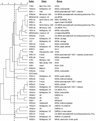
Replicates of sequenced isolates (Mu50, N315, MW2, MRSA252, and MSSA476) clustered together at 100% simi-larity when analyzed by the VirEp microarray, demonstrating the reproducibility of the assay. As expected, the closely re-lated isolates Mu50 and N315, as well as isolates MW2 and MSSA476, were found to group together (16). The population structure of the 43S. aureusclinical isolates as determined by the VirEp microarray is shown in Fig. 2. The similarity cutoff for distinguishing genotypes using the VirEp microarray
(93.5%) was established by determining the percentage of sim-ilarity that grouped the sequencedS. aureusisolates correctly, as elucidated by PFGE (Fig. 2). Genotype A contained 10 isolates (23%), mainly CA-MRSA and MSSA from the United Kingdom, the United States, and France. Genotype B com-prised eight MRSA and MSSA isolates (19%) from Notting-ham, France, and Switzerland. Genotype C was composed of two isolates (5%), a GISA isolate from Belgium and a MRSA isolate from France. Genotype D contained eight isolates (19%), includ-ing all three VRSA, GISA, and MSSA isolates from the United States, France, and Japan. Finally, genotype E contained 14 iso-lates (33%) from the United Kingdom and France, including both EMRSA-15 and -16 isolates, PVL-positive MSSA, and MSSA.
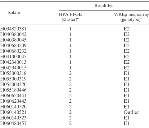
[image:5.585.124.459.69.492.2]Analysis of 18S. aureusoutbreak isolates (Table 1) alone, using an arbitrary cutoff of 96% similarity, indicated that the isolates fall into two genotypes with one outlier. Comparison of
FIG. 1. Dendrogram of 43S. aureusisolates examined by PFGE, produced with Bionumerics (version 2.0) software using the Dice coefficient and UPGMA. Isolates were clustered using the criteria of Tenover et al., where 80% similarity is the cutoff for differentiating closely related isolates (31). Clusters 1 through 7 are shown.
on May 16, 2020 by guest
http://jcm.asm.org/
microarray and PFGE results for these isolates revealed that 17 of the 18 isolates examined grouped in agreement (Table 3).
DISCUSSION
The VirEp microarray described in this paper represents a new tool for the identification and characterization ofS. aureus
isolates. The benefit of the VirEp microarray forS. aureuslies in its ability to simultaneously identify numerous virulence genes while avoiding the complexities of high-density microar-ray analysis. The VirEp microarmicroar-ray successfully identified all
PVL-positive isolates, all tst-positive isolates, and all VRSA isolates (Table 2), demonstrating that these clinically relevant
S. aureusvirulence genes can be detected by this assay. This contrasts with the limited number of genes that can be de-tected by PCR-based assays. While the presence of such genes does not necessarily equate with expression of the protein product, it does give a good indication to the clinician of the pathogenic potential of the isolate, which can be used to guide appropriate antimicrobial chemotherapy and infection control measures.
[image:6.585.134.454.65.561.2]Furthermore, the fact that six of the eight MRSA isolates
FIG. 2. Dendrogram of 43 clinicalS. aureusisolates examined by the VirEp microarray, produced with Bionumerics (version 2.0) software using the Dice coefficient and UPGMA. A cutoff value of 93.5% was used to distinguish genotypes. Genotypes A through E were distinguished.
on May 16, 2020 by guest
http://jcm.asm.org/
from the United Kingdom failed to hybridize to the mecA
probes on the VirEp microarray yet contained themecAgene by PCR indicated that the DNA sequences of mecA in the regions corresponding to the three probes are not conserved among all MRSA isolates. This suggests that for some genes, includingmecA, sequence variation may be much greater in the wider population than among the fewS. aureus isolates that have been sequenced to date. As a consequence, it is clear that further oligonucleotides are required to increase the robust-ness ofmecAdetection, a problem that is being encountered with all rapid MRSA detection systems.
The dendrogram generated from the VirEp microarray re-sults illustrates that in general, the grouping of isolates was highly congruent with that observed with PFGE (Fig. 1 and 2). However, all three VRSA isolates were assigned to the same genotype as the EMRSA-15 and -16 isolates (Fig. 1 and 2). It is noteworthy that the percentage of similarity required to differentiate the sequencedS. aureusisolates by using the mi-croarray results is 93.5%, whereas for PFGE it is 80% (31). This indicates that the microarray analysis provides slightly less discrimination than PFGE, probably due to the limited num-ber of gene targets included in the microarray. This was con-firmed when values for Simpson’s index of diversity were cal-culated for the VirEp microarray and PFGE (0.771 versus 0.811). Unlike PFGE, however, the microarray provides bio-logically meaningful data in addition to the typing data. When isolates from two epidemiologically distinct outbreaks were examined by the VirEp microarray and PFGE, only a single incongruent isolate was found. The differences observed with this isolate were due to a difference in gene content (lukD
positive,semnegative) (see Appendix S2 in the supplemental material) that could not be detected by PFGE. There were three differences in gene content between genotypes E1 and E2. Genotype E1 lacked the ermA, mupA, and tst genes,
whereas genotype E2 possessed these genes (see Appendix S2 in the supplemental material). These data provide evidence that the VirEp microarray may be capable of distinguishingS. aureusisolates from different outbreaks. The VirEp microarray could be refined by the inclusion of additional selected targets that could improve the discriminatory power of the microarray. A recent study has demonstrated the potentially superior re-solving power of microarrays compared to PFGE and multilo-cus sequence typing for typing of CA-MRSA isolates (12).
We anticipate that the VirEp assay could be used after presumptive staphylococci (gram-positive cocci in clusters) have been observed in a positive blood culture and species identification has been confirmed by an alternative methodol-ogy, such as a rapid PCR-based assay or fluorescence in situ hybridization with peptide nucleic acid probes (25). The inclu-sion in the array of oligonucleotides to detect coagulase-neg-ative staphylococci would indicate if the blood culture was a mixture ofS. aureusand coagulase-negative staphylococci. Our current development work has been successful in reducing the time taken to perform the VirEp assay to⬍24 h.
ACKNOWLEDGMENTS
Ph.D. studentship support of R.P.S. by the Medical Research Coun-cil, United Kingdom, and the University of Nottingham is gratefully acknowledged.
We thank Paddy Tighe, University of Nottingham, for helpful dis-cussions concerning microarray technology and Katrina Levi, Notting-ham University Hospitals NHS Trust, for assistance with PFGE and BioNumerics software. Nick Day, Angela Kearns, Sharon Peacock, Donald Morrison, John Corkill, Teruyo Ito, and the Network on An-timicrobial Resistance inStaphylococcus aureus(NARSA) are thanked for the provision ofS. aureusclinical isolates.
All authors declare no conflict of interest.
REFERENCES
1.Anthony, R. M., A. R. Schuitema, L. Oskam, and P. R. Klatser.2005. Direct detection ofStaphylococcus aureusmRNA using a flow through microarray. J. Microbiol. Methods60:47–54.
2.Brazma, A., P. Hingamp, J. Quackenbush, G. Sherlock, P. Spellman, C. Stoeckert, J. Aach, W. Ansorge, C. A. Ball, H. C. Causton, T. Gaasterland, P. Glenisson, F. C. Holstege, I. F. Kim, V. Markowitz, J. C. Matese, H. Parkinson, A. Robinson, U. Sarkans, S. Schulze-Kremer, J. Stewart, R. Taylor, J. Vilo, and M. Vingron.2001. Minimum information about a mi-croarray experiment (MIAME)—toward standards for mimi-croarray data. Nat. Genet.29:365–371.
3.Corkill, J. E., J. J. Anson, P. Griffiths, and C. A. Hart.2004. Detection of elements of the staphylococcal cassette chromosome (SCC) in a methicillin-susceptible (mecAgene negative) homologue of a fucidin-resistant MRSA. J. Antimicrob. Chemother.54:229–231.
4.Fang, H., and G. Hedin.2003. Rapid screening and identification of methi-cillin-resistantStaphylococcus aureusfrom clinical samples by selective-broth and real-time PCR assay. J. Clin. Microbiol.41:2894–2899.
5.Francois, P., G. Renzi, D. Pittet, M. Bento, D. Lew, S. Harbarth, P. Vaudaux, and J. Schrenzel.2004. A novel multiplex real-time PCR assay for rapid typing of major staphylococcal cassette chromosomemecelements. J. Clin. Microbiol.42:3309–3312.
6.Grisold, A. J., E. Leitner, G. Muhlbauer, E. Marth, and H. H. Kessler.2002. Detection of methicillin-resistantStaphylococcus aureusand simultaneous confirmation by automated nucleic acid extraction and real-time PCR. J. Clin. Microbiol.40:2392–2397.
7.Huletsky, A., R. Giroux, V. Rossbach, M. Gagnon, M. Vaillancourt, M. Bernier, F. Gagnon, K. Truchon, M. Bastien, F. J. Picard, A. van Belkum, M. Ouellette, P. H. Roy, and M. G. Bergeron.2004. New real-time PCR assay for rapid detection of methicillin-resistantStaphylococcus aureusdirectly from specimens containing a mixture of staphylococci. J. Clin. Microbiol.42:1875– 1884.
8.Hunter, P. R., and M. A. Gaston.1988. Numerical index of the discrimina-tory ability of typing systems: an application of Simpson’s index of diversity. J. Clin. Microbiol.26:2465–2466.
[image:7.585.43.283.88.295.2]9.Jaffe, R. I., J. D. Lane, S. V. Albury, and D. M. Niemeyer.2000. Rapid extraction from and direct identification in clinical samples of methicillin-resistant staphylococci using the PCR. J. Clin. Microbiol.38:3407–3412.
TABLE 3. Comparison of PFGE and VirEp microarray results for 18S. aureusoutbreak isolates
Isolate Result by: HPA PFGE (cluster)a ViREp microarray (genotype)b
H034820381 1 E2
H040380042 1 E2
H040380045 1 E2
H040680209 1 E2
H040680232 1 E2
H041000045 1 E2
H042340013 1 E2
H042340015 1 E2
H055000318 2 E1
H055000319 2 E1
H055000320 2 E1
H055180446 2 E1
H060620441 2 E1
H060620443 2 E1
H060140520 2 E1
H060140521 2 Outlier
H060140523 2 E1
H060480457 2 E1
aStandard PFGE methodology was used at the StaphylococcusReference
Laboratory, Health Protection Agency (HPA), London, United Kingdom. Clus-ters were defined using the criteria of Tenover et al. (31).
bGenotypes were differentiated using a 96% similarity cutoff.
on May 16, 2020 by guest
http://jcm.asm.org/
10.Jonas, D., M. Speck, F. D. Daschner, and H. Grundmann.2002. Rapid PCR-based identification of methicillin-resistantStaphylococcus aureusfrom screening swabs. J. Clin. Microbiol.40:1821–1823.
11.Klotz, M., S. Opper, K. Heeg, and S. Zimmermann.2003. Detection of Staphylococcus aureusenterotoxins A to D by real-time fluorescence PCR assay. J. Clin. Microbiol.41:4683–4687.
12.Koessler, T., P. Francois, Y. Charbonnier, A. Huyghe, M. Bento, S. Dharan, G. Renzi, D. Lew, S. Harbarth, D. Pittet, and J. Schrenzel.2006. Use of oligoarrays for characterization of community-onset methicillin-resistant Staphylococcus aureus. J. Clin. Microbiol.44:1040–1048.
13.Kumari, D. N., V. Keer, P. M. Hawkey, P. Parnell, N. Joseph, J. F. Rich-ardson, and B. Cookson.1997. Comparison and application of ribosome spacer DNA amplicon polymorphisms and pulsed-field gel electrophoresis for differentiation of methicillin-resistant Staphylococcus aureus strains. J. Clin. Microbiol.35:881–885.
14.Lapierre, P., A. Huletsky, V. Fortin, F. J. Picard, P. H. Roy, M. Ouellette, and M. G. Bergeron.2003. Real-time PCR assay for detection of fluoro-quinolone resistance associated withgrlAmutations inStaphylococcus au-reus. J. Clin. Microbiol.41:3246–3251.
15.Letertre, C., S. Perelle, F. Dilasser, and P. Fach.2003. Detection and geno-typing by real-time PCR of the staphylococcal enterotoxin genesseatosej. Mol. Cell. Probes17:139–147.
16.Lindsay, J. A., and M. T. Holden.2004.Staphylococcus aureus: superbug, super genome? Trends Microbiol.12:378–385.
17.Louie, L., J. Goodfellow, P. Mathieu, A. Glatt, M. Louie, and A. E. Simor.
2002. Rapid detection of methicillin-resistant staphylococci from blood cul-ture bottles by using a multiplex PCR assay. J. Clin. Microbiol.40:2786–2790. 18.Løvseth, A., S. Loncarevic, and K. G. Berdal.2004. Modified multiplex PCR method for detection of pyrogenic exotoxin genes in staphylococcal isolates. J. Clin. Microbiol.42:3869–3872.
19.Martineau, F., F. J. Picard, L. Grenier, P. H. Roy, M. Ouellette, and M. G. Bergeron.2000. Multiplex PCR assays for the detection of clinically relevant antibiotic resistance genes in staphylococci isolated from patients infected after cardiac surgery. The ESPRIT Trial. J. Antimicrob. Chemother.46:527– 534.
20.Martineau, F., F. J. Picard, D. Ke, S. Paradis, P. H. Roy, M. Ouellette, and M. G. Bergeron.2001. Development of a PCR assay for identification of staphylococci at genus and species levels. J. Clin. Microbiol.39:2541–2547. 21.Mason, W. J., J. S. Blevins, K. Beenken, N. Wibowo, N. Ojha, and M. S. Smeltzer.2001. Multiplex PCR protocol for the diagnosis of staphylococcal infection. J. Clin. Microbiol.39:3332–3338.
22.Monday, S. R., and G. A. Bohach.1999. Use of multiplex PCR to detect classical and newly described pyrogenic toxin genes in staphylococcal iso-lates. J. Clin. Microbiol.37:3411–3414.
23.Monecke, S., and R. Ehricht.2005. Rapid genotyping of methicillin-resistant
Staphylococcus aureus(MRSA) isolates using miniaturised oligonucleotide arrays. Clin. Microbiol. Infect.11:825–833.
24.Oliveira, D. C., and H. de Lencastre.2002. Multiplex PCR strategy for rapid identification of structural types and variants of themecelement in methi-cillin-resistantStaphylococcus aureus. Antimicrob. Agents Chemother.46:
2155–2161.
25.Oliveira, K., G. W. Procop, D. Wilson, J. Coull, and H. Stender.2002. Rapid identification ofStaphylococcus aureusdirectly from blood cultures by fluo-rescence in situ hybridization with peptide nucleic acid probes. J. Clin. Microbiol.40:247–251.
26.Palladino, S., I. D. Kay, J. P. Flexman, I. Boehm, A. M. Costa, E. J. Lambert, and K. J. Christiansen.2003. Rapid detection ofvanAandvanBgenes directly from clinical specimens and enrichment broths by real-time multi-plex PCR assay. J. Clin. Microbiol.41:2483–2486.
27.Pearson, B. M., C. Pin, J. Wright, K. I’Anson, T. Humphrey, and J. M. Wells.
2003. Comparative genome analysis ofCampylobacter jejuni using whole genome DNA microarrays. FEBS Lett.554:224–230.
28.Rouillard, J. M., C. J. Herbert, and M. Zuker.2002. OligoArray: genome-scale oligonucleotide design for microarrays. Bioinformatics18:486–487. 29.Struelens, M. J., A. Deplano, C. Godard, N. Maes, and E. Serruys.1992.
Epidemiologic typing and delineation of genetic relatedness of methicillin-resistant Staphylococcus aureus by macrorestriction analysis of genomic DNA by using pulsed-field gel electrophoresis. J. Clin. Microbiol.30:2599– 2605.
30.Tan, T. Y., S. Corden, R. Barnes, and B. Cookson.2001. Rapid identification of methicillin-resistantStaphylococcus aureusfrom positive blood cultures by real-time fluorescence PCR. J. Clin. Microbiol.39:4529–4531.
31.Tenover, F. C., R. D. Arbeit, R. V. Goering, P. A. Mickelsen, B. E. Murray, D. H. Persing, and B. Swaminathan.1995. Interpreting chromosomal DNA restriction patterns produced by pulsed-field gel electrophoresis: criteria for bacterial strain typing. J. Clin. Microbiol.33:2233–2239.
32.Trad, S., J. Allignet, L. Frangeul, M. Davi, M. Vergassola, E. Couve, A. Morvan, A. Kechrid, C. Buchrieser, P. Glaser, and N. El-Solh.2004. DNA macroarray for identification and typing ofStaphylococcus aureusisolates. J. Clin. Microbiol.42:2054–2064.
33.Tristan, A., L. Ying, M. Bes, J. Etienne, F. Vandenesch, and G. Lina.2003. Use of multiplex PCR to identifyStaphylococcus aureusadhesins involved in human hematogenous infections. J. Clin. Microbiol.41:4465–4467. 34.van Griethuysen, A., I. van Loo, A. van Belkum, C. Vandenbroucke-Grauls,
W. Wannet, P. van Keulen, and J. Kluytmans.2005. Loss of themecAgene during storage of methicillin-resistantStaphylococcus aureusstrains. J. Clin. Microbiol.43:1361–1365.
35.Vernet, G., C. Jay, M. Rodrigue, and A. Troesch.2004. Species differentia-tion and antibiotic susceptibility testing with DNA microarrays. J. Appl. Microbiol.96:59–68.