0095-1137/07/$08.00⫹0 doi:10.1128/JCM.02023-06
Copyright © 2007, American Society for Microbiology. All Rights Reserved.
Typing and Subtyping of
Clostridium difficile
Isolates by Using
Multiple-Locus Variable-Number Tandem-Repeat Analysis
䌤
Renate J. van den Berg,
1Inge Schaap,
1Kate E. Templeton,
1†
Corne
´ H. W. Klaassen,
2and Ed J. Kuijper
1*
Department of Medical Microbiology, Center of Infectious Diseases, Leiden University Medical Center, Leiden,1and Department of
Medical Microbiology and Infectious Diseases, Canisius Wilhelmina Hospital, Nijmegen,2The Netherlands
Received 1 October 2006/Returned for modification 8 November 2006/Accepted 5 December 2006
Using the genomic sequence ofClostridium difficilestrain 630, we developed multiple-locus variable-number tandem-repeat analysis (MLVA) with automated fragment analysis and multicolored capillary electrophoresis as a typing method forC. difficile. All reference strains, representing 31 serogroups, 25 toxinotypes, and 7 known subtypes of PCR ribotype 001, could be discriminated from each other. Application of MLVA to 28 isolates from 7 outbreaks due to the emerging hypervirulent PCR ribotype 027–pulsed-field gel electrophoresis type NAP1 resulted in recognition of 13 clusters. Additionally, 29 toxin A-negative, toxin B-positive isolates belonging to PCR ribotype 017 from eight different countries revealed eight country-specific clusters. MLVA is a highly discriminatory genotyping method and a new tool for subtyping of newly emerging variants of C. difficile.
To study the epidemiology ofClostridium difficile, a typing method with higher discriminatory power, typeability, and re-producibility than currently available methods is required. Multiple-locus variable-number tandem-repeat analysis (MLVA) is a new candidate technique that has already been applied successfully to a number of bacterial and fungal species (5, 10). Recently, MLVA using automated sequence detection and subsequent manual determination of the number of repeat loci has been developed forC. difficile (12). For faster and easier application of the MLVA toC. difficile, we developed an MLVA method using smaller short tandem repeats (2 to 9 bp) to facilitate automated fragment analysis with multicolored capillary electrophoresis instead of sequencing. Subsequently, we applied MLVA to seven subtypes of a common PCR ri-botype, 001, and two other, emerging PCR ribotypes of C.
difficile. Since 2004, a new toxin-hyperproducing C. difficile
strain, characterized as PCR ribotype 027, toxinotype III, pulsed-field gel electrophoresis (PFGE) type NAP1, and re-striction endonuclease analysis (REA) group BI, has been rec-ognized in Canada, the United States, the United Kingdom, The Netherlands, Belgium, and France as an important cause of hospital outbreaks (3, 8, 9, 11, 13). Additionally, an increas-ing number of reports mention severe infections and outbreaks due to toxin A-negative, toxin B-positive isolates (1, 2, 7, 14). These toxin A-negative, toxin B-positive isolates belong to PCR ribotype 017, REA group CF, and toxinotype VIII and were first recognized as the cause of an outbreak in 1999 in Canada (1, 2).
Bacterial strains.Isolates included in the analysis were 57 reference strains, all seven subtypes of PCR ribotype 001, 27 toxin A-negative, toxin B-positive isolates belonging to PCR ribotype 017 from eight different countries, and 29 isolates belonging to PCR ribotype 027 from The Netherlands (Ta-ble 1) and the United Kingdom. Of these 29 PCR ribotype 027 strains, 28 strains were outbreak related, from six dif-ferent hospitals in The Netherlands and one in the United Kingdom, and 1 strain was a sporadic isolate from 2003 (8). The United Kingdom strain was obtained from Jon Brazier (Anaerobe Reference Laboratory, NPHS Microbiology Cardiff, Cardiff, United Kingdom). The outbreak strains of each hospital were randomly selected. DNA was isolated from colonies ofC. difficileby QiaAmp DNA isolation col-umns (QIAGEN, Hilden, Germany) according to the man-ufacturer’s recommendations. PCR ribotyping was performed as described previously (4), and the method of Rupnik et al. was used for toxinotyping (15).
MLVA. Seven regions with short tandem repeats spread over the genome, designated markers MLVAC.difficileA6 (A6Cd), B7Cd, C6Cd, E7Cd, F3Cd, G8Cd, and H9Cd, were
identified using Tandem Repeat Finder, version 3.21, on the genome ofC. difficilestrain 630 (http://www.sanger.ac.uk/Projects /C_difficile/) (16). Four of these, MLVA A6Cd, B7Cd, E7Cd, and
G8Cd, were identical to CDR4, CDR49, CDR48, and CDR9,
respectively, in the assay described recently by Marsh et al. (12). Primers were designed based on the flanking sequences of the repeats using the Primer3 program (http://www.broad .mit.edu/cgi-bin/primer/primer3_www.cgi). Three separate du-plex PCRs (MLVA A6Cd-H9Cd, B7Cd-F3Cd, and C6Cd-E7Cd)
and one singleplex PCR (MLVA G8Cd) were developed (Table
2). The repeats were amplified using a single PCR protocol. The amplification reactions were performed in a 50-l final volume containing 25l of HotStarTaqmaster mix (QIAGEN, Hilden, Germany), 1M each primer, 3 mM magnesium chlo-ride, and 5l of DNA. After an initial enzyme activation step
* Corresponding author. Mailing address: Department of Medical Microbiology, E4-67, Centre of Infectious Diseases, P.O. Box 9600, 2300 RC Leiden, The Netherlands. Phone: 5263931. Fax: 31-71-5248148. E-mail: [email protected].
† Present address: Specialist Virology Centre, Royal Infirmary Hos-pital, Edinburgh, United Kingdom.
䌤Published ahead of print on 13 December 2006.
1024
on May 16, 2020 by guest
http://jcm.asm.org/
of 15 min at 95°C, the protocol consisted of 35 cycles of 30 s at 94°C for denaturation, 30 s at 51°C for annealing, and 30 s at 72°C for elongation. A final elongation step was performed for 10 min at 72°C. The forward primer of each PCR was labeled at the 5⬘end with either carboxyfluorescein (FAM), hexachloro-fluorescein (HEX), 2⬘-chloro-7⬘ -phenyl-1,4-dichloro-6-carboxy-fluorescein (VIC), or 2⬘-chloro-5⬘-fluoro-7⬘,8⬘-fused phenyl-1,4-dichloro-6-carboxyfluorescein (NED). PCR fragments were analyzed using multicolored capillary electrophoresis on an ABI3100 genetic analyzer, with a ROX500 marker as an internal marker for each sample. The size of each marker was determined by Genescan software (Applied Biosystems). Markers from a selected number of isolates were sequenced to verify accurate assignment of repeat numbers. All sequence results were equal to the results of fragment analysis by the ABI system and to the calculated repeat numbers. The repeat numbers were analyzed using BioNumerics (version 3.5) software (Applied Maths, Kortrijk, Belgium) and the unweighted-pair group method with arithmetic averages (UPGMA) with the multi-state categorical similarity coefficient (MCSC). All markers
were given equal weight, irrespective of the number of repeats. The percentages in the dendrogram reflect the percentage of homology between the specific markers. Thus, if two strains have an equal number of repeats in six of seven markers, they are 86% identical.
C. difficilecontrol strain 630 revealed identical results in five
different experiments using both separate cultures and DNA extractions. The stability of the repeat numbers of the different markers was tested in duplicate after isolates belonging to PCR ribotypes 014 and 027 were subcultured a total of 10 and 30 times. The repeats from the isolate belonging to type 014 were stable in all experiments. An expansion of 1 repeat unit in marker A6Cdwas observed in one duplicate sample of the type
027 isolate after 10 subcultures, which subsequently returned to the original number of repeats after 30 subcultures. For marker C6Cd, a reduction of 1 repeat unit could be detected
[image:2.585.52.539.82.335.2]after 30 subcultures for this isolate. Based on the stability tests, we concluded that a difference of 1 repeat unit between strains should not be interpreted as indicative of separate types or subtypes. This conclusion is in complete concordance with the
TABLE 1. Isolates included in this study
Characteristic
No. of isolates (total, 120)
Isolate identification Outbreak or
sporadic Origin (reference)
Serogroups 31 A to I, K, X, A1 to A11, A13
to A17, S1 to S4
NAa Gift from M. Delme´e
Toxinotypes 25 I to XXII NA Gift from M. Rupnik
Strain 630 1 630 NA Gift from P. Mastrantonio
Subtypes of PCR ribotype 001, by REP-PCRb
7 001-1 to 001-7 NA Gift from J. Brazier
Toxin A-negative, toxin B-positive isolates, PCR ribotype 017
12 Arg28, -31, -32, -36 to -38, -77, -126, -127, -134, -143, -152
Outbreak Argentina; gift from C. Legaria
2 CD16, CD17 Outbreak Amsterdam, The Netherlands (18)
2 Can1, Can3 Outbreak Canada (18)
1 123825R Endemic Leiden, The Netherlands
2 1110/98, 1745/00 Endemic Poland (18)
2 60, 99-3050 Endemic France (18)
2 CF2, CF4 Endemic United States (18)
2 R10205, R10430 Endemic United Kingdom (18)
2 GAI95601, GAI95602 Endemic Japan (18)
Isolates belonging to PCR ribotype 027
5 AF1 to -5 Outbreak Amersfoort, The Netherlands (8) 7 AMC1 to -7 Outbreak Amsterdam-1, The Netherlands (8)
1 VUMC Endemic Amsterdam-2, The Netherlands (8)
3 SV1 to -3 Outbreak Amsterdam-3, The Netherlands (8)
4 HL1 to -4 Outbreak Haarlem, The Netherlands (8)
5 HW1 to -5 Outbreak Harderwijk, The Netherlands (8)
3 UMC1 to -3 Outbreak Utrecht, The Netherlands (8)
1 UK027 Outbreak United Kingdom (gift from J. Brazier)
aNA, not applicable.
bREP, repetitive extragenic palindromic.
TABLE 2. Characteristics of markers and primers
Marker Repeat motif Locationa
Forward primer sequence (label–5⬘33⬘) Reverse primer sequence (5⬘33⬘)
A6Cd AAGAGC 755721 FAM-TTAATTGAGGGAGAATGTTAAA AAATACTTTTCCCACTTTCATAA
B7Cd ATCTTCT 3688632 FAM-CTTAATACTAAACTAACTCTAACCAGTAA TTATATTTTATGGGCATGTTAAA
C6Cd TATTGC 3239736 HEX-GTTTAGAATCTACAGCATTATTTGA ATTGGAATTGAATGTAACAAAA
E7Cd ATAGATT 167124 FAM-TGGAGCTATGGAAATTGATAA CAAATACATCTTGCATTAATTCTT
F3Cd TTA 1954915 HEX-TTTTTGAAACTGAACCAACATA ACAAAAGACTGTGCAAATATACTAA
G8Cd TAAAAGAG 664660 NED-TGTATGAAGCAAGCTTTTTATT AATCCAGCAATCTAATAATCCA
H9Cd TCTTCTTCC 4116072 VIC-GTTTTGAGGAAACAAACCTATC GATGAGGAAATAGAAGAGTTCAA
aLocation on the genomic sequence of strain 630 (36).
on May 16, 2020 by guest
http://jcm.asm.org/
[image:2.585.42.550.630.715.2]study of the stability ofC. difficileMLVA loci by Marsh et al. (12). They found three pairs of serial isolates from individual patients with a single-locus variation of only 1 tandem repeat and one pair of isolates with a double-locus variation of 1 tandem repeat each. Therefore, they concluded that isolates with a summed tandem repeat difference ofⱕ2 are genetically related. MLVA discriminated between isolates belonging to all 31 serogroups, the 7 subtypes of PCR ribotype 001, and all 25 toxinotypes, except for toxinotypes XII, XIII, and XIV. An isolate belonging to serogroup A15 was completely identical (100%) to toxinotype V, as has been observed previously (15). Toxinotypes XII, XIII, and XIV were clustered into one MLVA type with 100% similarity, indicating that toxinotyping is a method that merely reflects the status of the toxin genes (15). With one marker difference, toxinotype XIb was comparable to the 100% cluster of toxinotypes XII to XIV. The similarity of isolate 630 to serogroup C (the closest match) was only 43% (3 of the 7 markers), although markers B7Cdand E7Cddiffered by only
1 repeat. PCR ribotype 001 isolates were quite stable in markers E7Cd(5 to 7 repeats), F3Cd(5 repeats), G8Cd(6 to 8 repeats), and
H9Cd(2 repeats). By using these characteristics, type 001 isolates
can be discriminated from most serogroups and toxinotypes. Con-sequently, MLVA is able to replace PCR ribotyping and PFGE for identification and recognition of subtypes of PCR ribotype 001. Until recently, strains belonging to PCR ribotype 001 were the most common in the United Kingdom, and the importance of the ability to subtype these strains is high (6, 17). A recent Health Protection Agency report (available at http://www.hpa.org.uk/) indicated that PCR ribotypes 106 and 027 are the most common in the United Kingdom, followed by type 001 (still approximately 25%) (6a).
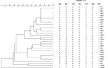
Among the isolates belonging to PCR ribotype 027 (n ⫽
29), 100% similarity (Fig. 1) was detected for isolate AF4 with HW3, for isolate SV1 with SV2, for isolate AF5 with AMC4, HW1, HW2, and HW5, and for isolate AMC3 with AMC6 and AMC7. With 86% similarity, 14 clusters were detected among the 29 isolates. Hospital-specific clusters were seen for SV, HW, HL, and AMC (Fig. 1). The sporadic endemic isolate recovered in 2003 was only 53% similar to the outbreak isolates and 71% similar to isolate HL3. The United Kingdom isolate was only 40% identical to all Dutch outbreak isolates. For all type 027 isolates, markers E7Cd,
F3Cd, and H9Cd were completely identical, except for the
United Kingdom isolate, which had 6 repeats for marker E7Cd (Fig. 1).
Toxin A-negative, toxin B-positive isolates (including the two reference strains belonging to serogroup F and toxinotype VIII) (n⫽29), belonging to PCR ribotype 017 and toxinotype VIII, could be divided into eight clusters at a similarity of 86% (6 markers identical) (Table 3, clusters G to N). Six clusters with 100% homology were recognized (Table 3, clusters A to F). All isolates with 100% similarity were country specific (clusters A to F), as were clusters H and I. Toxin A-negative, toxin B-positive isolates could be differentiated from all other types by using the combination of markers A6Cd(2 repeats),
F3Cd(5 repeats), G8Cd(fragment size,⬎400 bp), and H9Cd(2
repeats). For marker G8Cd, all PCR ribotype 017 isolates
showed the previously described larger fragment size, exceed-ing the 400 bp detectable by our system. MLVA discriminated toxin A-negative, toxin B-positive isolates better than ampli-fied fragment length polymorphism (18).
[image:3.585.83.509.72.339.2]Application of MLVA to C. difficile isolates was easy to
FIG. 1. Dendrogram based on profiles of seven markers for all PCR ribotype 027 isolates (n⫽29) tested in this study. Numbers of repeats for the specified markers in each strain are given on the right.
on May 16, 2020 by guest
http://jcm.asm.org/
perform and consisted of four separate PCR mixes and a single PCR protocol. Although MLVA has yet to show its value in longer-term epidemiology or phylogeny studies, MLVA can be widely applied in outbreak situations. Therefore, MLVA is an important new tool for study of the epidemiology of toxin A-negative, toxin B-positive PCR ribotype 017 isolates and PCR ribotype 027–PFGE NAP1–REA BI isolates, which are newly emerging worldwide. MLVA is a highly discriminatory genotyping method forC. difficileand is able to discriminate between isolates with identical PCR ribotypes belonging to types 001, 017, and 027. MLVA also clearly differentiated these PCR ribotypes from other PCR ribotypes included in this study. Future studies should be performed on all currently available PCR ribotypes to explore this in more detail.
This work was supported by a grant from the Foundation Microbi-ology Leiden, The Netherlands.
We thank, in alphabetical order, Michelle Alfa (University of Mani-toba, Winnipeg, Canada), Frederic Barbut (Centre Hospitalo-Univer-sitaire Saint-Antoine, Paris, France), Jon Brazier (University Hospital of Wales, United Kingdom), Stu Johnson (Northwestern University, Chicago, IL), Haru Kato (Gifu University School of Medicine, Gifu, Japan), Cristina Legaria (Hospital Tornu´, Buenos Aires, Argentina), Paola Mastrantonio (Instituto Superiore di Sanita, Rome, Italy), Hanna Pituch (Medical University of Warsaw, Warsaw, Poland), and Maja Rupnik (University of Maribor, Maribor, Slovenia) for kindly providing the isolates used in this study.
REFERENCES
1.al Barrak, A., J. Embil, B. Dyck, K. Olekson, D. Nicoll, M. Alfa, and A. Kabani.1999. An outbreak of toxin A negative, toxin B positiveClostridium difficile-associated diarrhea in a Canadian tertiary-care hospital. Can.
Com-mun. Dis. Rep.25:65–69.
2.Alfa, M. J., A. Kabani, D. Lyerly, S. Moncrief, L. M. Neville, A. al Barrak, G. K. Harding, B. Dyck, K. Olekson, and J. M. Embil.2000. Characterization
of a toxin A-negative, toxin B-positive strain ofClostridium difficile
respon-sible for a nosocomial outbreak ofClostridium difficile-associated diarrhea.
J. Clin. Microbiol.38:2706–2714.
3.Anonymous.2005. Outbreak ofClostridium difficilein a hospital in south east
England. CDR Weekly15:1–2.
4.Bidet, P., F. Barbut, V. Lalande, B. Burghoffer, and J. C. Petit.1999.
De-velopment of a new PCR-ribotyping method forClostridium difficilebased on
ribosomal RNA gene sequencing. FEMS Microbiol. Lett.175:261–266.
5.de Valk, H. A., J. F. Meis, I. M. Curfs, K. Muehlethaler, J. W. Mouton, and C. H. Klaassen.2005. Use of a novel panel of nine short tandem repeats for
exact and high-resolution fingerprinting of Aspergillus fumigatusisolates.
J. Clin. Microbiol.43:4112–4120.
6.Fawley, W. N., P. Parnell, P. Verity, J. Freeman, and M. H. Wilcox.2005.
Molecular epidemiology of endemicClostridium difficileinfection and the
significance of subtypes of the United Kingdom epidemic strain (PCR
ri-botype 1). J. Clin. Microbiol.43:2685–2696.
6a.Health Protection Agency.24 July 2006, posting date.Clostridium difficile: findings and recommendations from a review of the epidemiology and a survey of directors of infection prevention and control in England. Health Protection Agency, London, United Kingdom. [Online.] http://www.hpa .org.uk/infections/topics_az/clostridium_difficile/documents/Clostridium _difficile_survey_findings_recommendations.pdf.
7.Kuijper, E. J., J. de Weerdt, H. Kato, N. Kato, A. P. van Dam, E. R. van der Vorm, J. Weel, C. van Rheenen, and J. Dankert.2001. Nosocomial outbreak ofClostridium difficile-associated diarrhoea due to a clindamycin-resistant
enterotoxin A-negative strain. Eur. J. Clin. Microbiol. Infect. Dis.20:528–
534.
[image:4.585.36.546.89.388.2]8.Kuijper, E. J., R. J. van den Berg, S. Debast, C. E. Visser, D. Veenendaal, A. TABLE 3. MLVA results and cluster information for toxin A-negative, toxin B-positive isolates belonging
to PCR ribotype 017 isolatesatested in this study
Isolate Origin
No. of repeats detected for the following marker:
Cluster(s)b
A6Cd B7Cd C6Cd E7Cd F3Cd G8Cd H9Cd
Arg32 Argentina 2 7 18 7 5 ⬎400 2 G
Arg38 Argentina 2 7 17 7 5 ⬎400 2 G
CF2 United States 2 7 31 7 5 ⬎400 2 G
Can3 Canada 2 9 20 7 5 ⬎400 2 H
1110/98 Poland 2 10 23 7 5 ⬎400 2 I
1745/00 Poland 2 10 15 7 5 ⬎400 2 I
R10205 United Kingdom 2 8 26 8 5 ⬎400 2 A, J
R10430 United Kingdom 2 8 26 8 5 ⬎400 2 A, J
Can1 Canada 2 8 22 8 5 ⬎400 2 J
CF4 United States 2 8 30 8 5 ⬎400 2 J
F Reference strain 2 13 22 8 5 ⬎400 2 K
CD16 Amsterdam 2 10 30 8 5 ⬎400 2 B, L
CD17 Amsterdam 2 10 30 8 5 ⬎400 2 B, L
123825R Leiden 2 10 32 8 5 ⬎400 2 L
GAI95601 Japan 2 10 43 8 5 ⬎400 2 L
GAI95602 Japan 2 10 40 8 5 ⬎400 2 L
Arg77 Argentina 2 7 20 8 5 ⬎400 2 C, M
Arg134 Argentina 2 7 20 8 5 ⬎400 2 C, M
Arg152 Argentina 2 7 19 8 5 ⬎400 2 D, M
Arg37 Argentina 2 7 19 8 5 ⬎400 2 D, M
Arg127 Argentina 2 7 19 8 5 ⬎400 2 D, M
Arg31 Argentina 2 7 18 8 5 ⬎400 2 E, M
Arg126 Argentina 2 7 18 8 5 ⬎400 2 E, M
Arg28 Argentina 2 7 21 8 5 ⬎400 2 F, M
Arg36 Argentina 2 7 21 8 5 ⬎400 2 F, M
Arg143 Argentina 2 7 22 8 5 ⬎400 2 M
60 (fr) France 2 7 28 8 5 ⬎400 2 M
99-3050 France 2 7 24 8 5 ⬎400 2 M
VIII Reference strain 2 9 21 8 5 ⬎400 2 N
a n⫽29.
b
Clusters at 100% similarity: A to F. Clusters at 86% similarity: G to N.
on May 16, 2020 by guest
http://jcm.asm.org/
Troelstra, T. van der Kooi, S. van den Hof, and D. W. Notermans.2006.
Clostridium difficileribotype 027, toxinotype III, The Netherlands. Emerg.
Infect. Dis.12:827–830.
9.Kuijper, E. J., B. Coignard, and P. Tu¨ll.2006. Emergence ofClostridium difficile-associated disease in Canada, the United States of America and
Europe. Clin. Microbiol. Infect.12(Suppl. 6):2–18.
10.Lindstedt, B. A.2005. Multiple-locus variable number tandem repeats
anal-ysis for genetic fingerprinting of pathogenic bacteria. Electrophoresis26:
2567–2582.
11.Loo, V. G., L. Poirier, M. A. Miller, M. Oughton, M. D. Libman, S. Michaud, A. M. Bourgault, T. Nguyen, C. Frenette, M. Kelly, A. Vibien, P. Brassard, S. Fenn, K. Dewar, T. J. Hudson, R. Horn, P. Rene, Y. Monczak, and A. Dascal.2005. A predominantly clonal multi-institutional outbreak of Clos-tridium difficile-associated diarrhea with high morbidity and mortality.
N. Engl. J. Med.353:2442–2449.
12.Marsh, J. W., M. M. O’Leary, K. A. Shutt, A. W. Pasculle, S. Johnson, D. N. Gerding, C. A. Muto, and L. H. Harrison.2006. Multilocus variable-number
tandem-repeat analysis for investigation ofClostridium difficiletransmission
in hospitals. J. Clin. Microbiol.44:2558–2566.
13.Pepin, J., L. Valiquette, M. E. Alary, P. Villemure, A. Pelletier, K. Forget, K. Pepin, and D. Chouinard.2004.Clostridium difficile-associated diarrhea in a region of Quebec from 1991 to 2003: a changing pattern of disease severity.
CMAJ171:466–472.
14.Pituch, H., J. S. Brazier, P. Obuch-Woszczatynski, D. Wultanska, F. Meisel-Mikolajczyk, and M. Luczak. 2005. Prevalence and association of PCR
ribotypes ofClostridium difficileisolated from symptomatic patients from
Warsaw with macrolide-lincosamide-streptogramin B (MLSB) type
resis-tance. J. Med. Microbiol.55:207–213.
15.Rupnik, M., V. Avesani, M. Janc, C. Eichel-Streiber, and M. Delmee.1998. A novel toxinotyping scheme and correlation of toxinotypes with serogroups ofClostridium difficileisolates. J. Clin. Microbiol.36:2240–2247.
16.Sebaihia, M., B. W. Wren, P. Mullany, N. F. Fairweather, N. Minton, R. Stabler, N. R. Thomson, A. P. Roberts, A. M. Cerdeno-Tarraga, H. Wang, M. T. Holden, A. Wright, C. Churcher, M. A. Quail, S. Baker, N. Bason, K. Brooks, T. Chillingworth, A. Cronin, P. Davis, L. Dowd, A. Fraser, T. Feltwell, Z. Hance, S. Holroyd, K. Jagels, S. Moule, K. Mungall, C. Price, E. Rabbinowitsch, S. Sharp, M. Simmonds, K. Stevens, L. Unwin, S. Whithead, B. Dupuy, G. Dougan, B. Barrell, and J. Parkhill.2006. The
multidrug-resistant human pathogenClostridium difficilehas a highly mobile, mosaic
genome. Nat. Genet.38:779–786.
17.Stubbs, S. L., J. S. Brazier, G. L. O’Neill, and B. I. Duerden.1999. PCR
targeted to the 16S–23S rRNA gene intergenic spacer region ofClostridium
difficileand construction of a library consisting of 116 different PCR
ri-botypes. J. Clin. Microbiol.37:461–463.
18.van den Berg, R. J., E. C. Claas, D. H. Oyib, C. H. Klaassen, L. Dijkshoorn, J. S. Brazier, and E. J. Kuijper.2004. Characterization of toxin A-negative,
toxin B-positive Clostridium difficile isolates from outbreaks in different
countries by amplified fragment length polymorphism and PCR ribotyping.
J. Clin. Microbiol.42:1035–1041.