JOURNAL OFCLINICALMICROBIOLOGY, Aug. 2009, p. 2489–2495 Vol. 47, No. 8 0095-1137/09/$08.00⫹0 doi:10.1128/JCM.00290-09
Copyright © 2009, American Society for Microbiology. All Rights Reserved.
Specific Distribution within the
Enterobacter cloacae
Complex of
Strains Isolated from Infected Orthopedic Implants
䌤
Philippe C. Morand,
1,2,3* Annick Billoet,
2Martin Rottman,
4,5Vale
´rie Sivadon-Tardy,
5,6Luc Eyrolle,
7Luc Jeanne,
7Asmaa Tazi,
1,2,3Philippe Anract,
1,8Jean-Pierre Courpied,
1,8Claire Poyart,
1,2,3and Vale
´rie Dumaine
8Faculty of Medicine, Universite´ Paris Descartes, Paris, France1; Department of Bacteriology, Cochin Hospital (AP-HP), Paris, France2;
Institut Cochin, INSERM U567, Paris, France3; Laboratoire de Microbiologie, Hoˆpital Raymond Poincare´ (AP-HP),
Garches, France4; EA 3647, Faculte´ de Me´decine Paris-Ile de France-Ouest, Universite´ de Versailles-Saint-Quentin en Yvelines,
Garches, France5; Department of Microbiology, Ambroise Pare´ Hospital (AP-HP), Boulogne-Billancourt, France6;
Department of Anesthesiology, Cochin Hospital (AP-HP), Paris, France7; and Department of
Orthopedic Surgery, Cochin Hospital (AP-HP), Paris, France8
Received 9 February 2009/Returned for modification 14 April 2009/Accepted 4 June 2009
Bacteria belonging to theEnterobactergenus are frequently isolated from clinical samples but are unusual
causative agents of orthopedic implant infections. Twelve genetic clusters (clusters I to XII) and one sequence
crowd (sequence crowdxiii) can be distinguished within theEnterobacter cloacaenomenspecies on the basis of
hsp60sequence analysis, and until now, none of these clusters could be specifically associated with a disease. In order to investigate if specific genetic clusters would be involved in infections of orthopedic material, two
series of bacterial clinical isolates identified asE. cloacaeby routine phenotypic identification methods were
collected either from infected orthopedic implants (nⴝ 21) or from randomly selected samples of diverse
anatomical origins (control;nⴝ52). Analysis of thehsp60gene showed that genetic clusters III, VI, and VIII
were the most frequent genetic clusters detected in the control group, whereas cluster III was poorly
repre-sented among the orthopedic implant isolates (Pⴝ0.006). On the other hand,E. hormaechei(clusters VI and
VIII), but not cluster III, is predominantly associated with infections of orthopedic implants and, more
specifically, with infected material in the hip (Pⴝ0.019). These results support the hypothesis that, among the
isolates within theE. cloacaecomplex,E. hormaecheiandhsp60gene sequencing-based cluster III are involved
in pathogenesis in different ways and highlight the need for more accurate routineEnterobacteridentification
methods.
Prosthetic joint infection (PJI) is, after aseptic loosening, the second most frequent complication of prosthetic joint replace-ment. Improvements in surgical techniques and the prevention of infection have lowered the risk of infection for primary hip or knee replacement to less than 1 and 2%, respectively, but the incidence of infection can increase 10-fold in the case of surgical revision (30). The population at risk for PJI continues to steadily increase, with an estimated 1 million arthroplasties being carried out worldwide each year (2), and the socioeco-nomic burden could become considerable (14–16). The aver-age cost of combined medical and surgical treatment of an infected joint prosthesis is estimated to be $30,000, with im-portant discomfort and substantial economic consequences for the patient (4). Besides joint prosthetic implants, other im-plantable devices such as screws and internal fixation devices are also at risk for infection in a wide range of clinical situa-tions. Gram-positive cocci are the most frequent pathogens encountered, but members of the Enterobacteriaceae family can also be involved (29). Among these,Enterobacterspecies
are major nosocomial pathogens often found in intensive care settings, and their role in PJI remains to be documented.
Bacteria of theEnterobactergenus are widely encountered in nature. These microorganisms are saprophytic in the environ-ment and commensal in the enteric flora since they are found in soil and sewage, as well as in the human gastrointestinal tract (17, 22). The taxonomy of the Enterobacter genus has been iteratively updated (8–12, 19). Several species are de-scribed in this taxon (Enterobacter aerogenes,E. amnigenus,E. cancerogenus, E. cowanii,E. gergoviae,E. intermedius,E. pyri-nus), as is a genetic complex, referred to as the “E. cloacae
complex,” in which other species have been identified (E. as-buriae,E. kobei,E. ludwigii,E. hormaechei,E. nimipressuralis, andE. cloacae).E. sakazakiistrains, which belong to several genomospecies, were recently reassigned to the newly pro-posed genusCronobacter(12).
Enterobacterspecies can act as pathogens, and theE. cloacae
complex, commonly referred to as “E. cloacae,” represents the
Enterobacter group most frequently encountered in human clinical samples. Although identification as “E. cloacae” is rou-tinely performed by phenotypic methods in clinical laborato-ries, the accurate identification of isolates within this taxon is difficult. Analysis of the 16S rRNA gene is widely used for bacterial identification, but it is poorly discriminatory for closely related members of theEnterobacteriaceaefamily and, more specifically, for members of theEnterobactergenus (27).
* Corresponding author. Mailing address: Service de Bacte´riologie, Hoˆpital Cochin AP-HP, Universite´ Paris Descartes, 27 rue du Fau-bourg Saint-Jacques, 75014 Paris, France. Phone: 33 (0)1 58 41 27 91. Fax: 33 (0)1 58 41 15 48. E-mail: [email protected].
䌤Published ahead of print on 10 June 2009.
2489
on May 16, 2020 by guest
http://jcm.asm.org/
Other targets for use for the molecular identification of iso-lates within theEnterobactergenus have been described, such as theoriClocus (23),gyrB (6), rpoB(8, 21), andhsp60(8). Sequence analysis of a fragment of thehsp60gene showed that theE. cloacaenomenspecies could be divided into 12 genetic clusters (clusters I to XII) and one sequence crowd (sequence crowdxiii). Specific names could be attributed to some of the genetic clusters:E. asburiae(cluster I) (9),E. kobei(cluster II) (9),E. ludwigii(cluster V) (11),E. nimipressuralis(cluster X) (8),E. cloacaesubsp.cloacae(cluster XI) (9), andE. cloacae
subsp.dissolvens(cluster XII) (9). Although the nameE. hor-maechei was sometimes used as a generic name for strains belonging to different hsp60 gene sequencing-based clusters (20), clusters VI, VII, and VIII together formally constitute the
E. hormaecheispecies, which has three subspecies:E. hormae-cheisubsp.oharae(cluster VI),E. hormaecheisubsp. hormae-chei(cluster VII), andE. hormaecheisubsp.steigerwallti (clus-ter VIII) (10). Species names were not attributed to clus(clus-ters III, IV, and IX and to sequence crowdxiii, although at least cluster III is of significant clinical importance (8, 26).
The degree of genomic diversity within theE. cloacae com-plex was recently reassessed by more global genotypic meth-ods. Multilocus sequence analysis (MLSA) identifies seven clusters within theE. cloacaecomplex, and each of these cor-responds to one or morehsp60gene sequencing-based genetic clusters. Microarray-based comparative genomic hybridization analysis (CGH) showed two genetically distinct clades (21). Most strains associated with clinical disease belong to the youngest CGH-based clade, to which strains of hsp60 gene sequencing-based clusters III, VI, and VIII also belong. The second, and older, CGH-based clade comprises heterogeneous strains, some of which are associated with commensalism.
Single-locus-based molecular methods, as well as global ap-proaches, such as MLSA or CGH, suggested that some genetic clusters are more prone to cause infection, although no specific epidemiological association could be demonstrated (8, 21). The repeated occurrence in our clinical laboratory of orthope-dic implant infections due toE. cloacaeisolates prompted us to further investigate these strains. We analyzed strains isolated from infected orthopedic devices and compared them to ran-domly selected strains isolated from clinical specimens taken from diverse anatomical sites. Analysis of the hsp60 gene showed that among the genetic clusters of the Enterobacter cloacaecomplex, clusters VI and VIII, but not cluster III, are predominantly associated with infections of orthopedic im-plants and, more specifically, with hip imim-plants. These data support the hypothesis that genetic clusters of theE. cloacae
complex are involved in pathogenesis in different ways and highlight the need for more accurate routine methods for the identification ofEnterobacterspecies.
MATERIALS AND METHODS
Bacterial strains and epidemiological data.For the study group, strains were collected from three large academic hospitals in the Paris, France, area
(Am-broise Pare´ Hospital, Raymond Poincare´ Hospital, and Cochin Hospital,
AP-HP) involved in the management of osteoarticular infectious conditions. In each hospital, all bacterial strains isolated from orthopedic device-related surgi-cal samples are systematisurgi-cally and prospectively collected and stored, irrespective of the bacterial species, the anatomical site of isolation, or the type of infection. For the study group, the collections were submitted to identical screening criteria
(1999 to 2006 for Ambroise Pare´ Hospital, 2000 to 2006 for Raymond Poincare´
Hospital, 2005 to 2007 for Cochin Hospital) for the selection of strains belonging
to theE. cloacaecomplex that were responsible for the infection of the
ortho-pedic implants. The following criteria were applied for inclusion of the strains in
the study: (i) identification of the infecting organism asE. cloacaewith routine
phenotypic identification systems (the API 20E or the Vitek 2 system; Bio-Merieux, Marcy l’Etoile, France), (ii) clinical evidence of infection of the
orthopedic implant in an adult patient, (iii) isolation of theE. cloacaestrain from
surgical samples collected at the point of contact between surrounding tissue and the implanted material (or from the implant itself) at the time of surgical
treatment, and (iv) involvement of theE. cloacaestrain as a probable causative
agent for the infection and clinical management as such. Isolation of anE.
cloacaestrain from a wound, sinus tract, or drainage was not sufficient to ascer-tain its involvement in the implant infection; and such strains were not included in the study. One strain per patient was included, and in the case of the isolation of multiple isolates, the most clinically relevant isolate (i.e., from the implant itself rather than from the surrounding tissue) was used. Twenty-one strains were selected and are referred to as strains isolated from orthopedic implanted ma-terial (Table 1). Since different types of implants were involved, only the ana-tomical site of infection was considered for data analysis. All selected strains
recovered from storage at⫺80°C were viable for subculture. Personal data (age,
sex, site of infection, mono- or polymicrobial infection) as well as the hospital and the date of isolation were anonymously collected for the purposes of this study.
For the control group (Table 2), 52 randomly selected clinical strains routinely
identified asE. cloacaein the clinical laboratory at the academic Cochin Hospital
by use of the API 20E or the Vitek 2 identification system were prospectively collected from clinical samples taken for diagnostic purposes from adult patients during the year 2006; environmental isolates were excluded. Thus, the control
group was representative ofE. cloacaestrains from an adult population routinely
identified in a clinical laboratory. One isolate was included per patient, and the strains collected were registered on the basis of the anatomical site of isolation,
as follows: skin and soft tissue (n⫽14), upper and lower respiratory tract (n⫽
15), urine (n⫽12), joint or bone (in the absence of infected material;n⫽3),
intravascular catheter (n⫽2), blood (n⫽2), and gastrointestinal suppuration
with exclusion of feces (n⫽4). Personal data (age, sex, site of isolation) were
anonymously collected for the purpose of this study.
Identification methods.Bacterial DNA from bacterial colonies grown over-night at 37°C on 5% horse blood agar plates was prepared for PCR analysis by using the Instagene nucleic acid purification method (Bio-Rad, Marnes la
Co-quette, France). Partial sequencing of thehsp60gene was performed by a
pre-viously described protocol (8). Briefly, oligonucleotide primers Hsp60-F (5⬘-GG
TAGAAGAAGGCGTGGTTGC-3⬘) and Hsp60-R (5⬘-ATGCATTCGGTGGT
GATCATCAG-3⬘) were used for genomic amplification of a 341-bp fragment of
thehsp60gene. A negative control containing all reagents except the target DNA
(which was replaced by H2O) was included in each series. PCR was performed
on a GeneAmp PCR system 9700 apparatus (Applied Biosystems) for 30 cycles by using the following conditions: 30 s at 94°C for denaturation, 30 s at 57°C for annealing, and 60 s at 72°C for elongation. Both strands of the purified amplified DNA fragment were sequenced by the BigDye Terminator cycle sequencing protocol with the same primers used for the PCR. Chromatograms of the com-plementary strands obtained with an ABI 313 apparatus (Applied Biosystems) were assembled by using the VectorNTi suite of programs (Invitrogen Corp.). A
272-bp fragment of thehsp60gene was obtained for the 73 strains, and the
sequence of the fragment was compared to reference sequences from strains previously described in taxonomic studies (8) by using the Clustal W algorithm (www.align.genome.jp). Sequence comparisons were exported as an unrooted neighbor-joining tree with proportional branch lengths.
Statistical analysis.Epidemiological associations were analyzed by use of the
Fisher exact test and the corresponding two-tailedPvalue.
Nucleotide sequence accession numbers.The sequences of the following type strains were retrieved from the GenBank database (the information in
paren-theses is the strain designation, GenBank accession number):E. asburiae(ATCC
35953, AJ417141),E. kobei(ATCC BAA260, AJ567899),E. cloacaesubsp.
dissolvens(ATCC 23373, AJ417143) (9),E. ludwigii(EN-119, AJ417114) (11),
E. hormaecheisubsp.oharae(EN-314, AJ543782),E. hormaecheisubsp. hor-maechei (ATCC 49162, AJ417108), E. hormaechei subsp. steigerwaltii
(CIP108489, AJ543908) (10),E. nimipressuralis(ATCC 9912, AJ567900),E.
cancerogenus (ATCC 33241, AJ567895), E. amnigenus (ATCC 3072,
AJ567894),E. cowanii (ATCC 107300T, AJ567896),E. gergoviae (ATCC
33028, AJ567897),E. pyrinus(ATCC 49851, AJ567901),C. sakazaki(ATCC
29544, AJ567902) (12), andE. aerogenes(AB008141). ForE. cloacaesubsp.
cloacae, nohsp60sequence is available for strain ATCC 13047, but one can be found under GenBank accession number AJ417142 (strain ATCC 13049),
on May 16, 2020 by guest
http://jcm.asm.org/
which was used in another study (13). The GenBank accession numbers for previously described strains (8) are listed in the Fig. 1 legend. The GenBank accession numbers for the clinical strains described in this work are FJ595719 to FJ595791.
RESULTS
Distribution of strains isolated from infected orthopedic
implants within the genetic clusters of theEnterobacter cloacae
complex.Enterobacteris an uncommon causative agent of
or-thopedic implant infections. Investigation of the databases of three large academic orthopedic surgical centers in the Paris, France, area yielded 21 cases of orthopedic device infections due toE. cloacaeon the basis of routine phenotypic identifi-cation methods (Table 1). The infections were found to be polymicrobial in 9 cases (43%), whereas isolates phenotypi-cally identified asE. cloacaewere found as the sole causative agent in 12 patients (57%). Partial sequencing of thehsp60
gene allowed the identification of all isolates as part of one of the clusters that form theE. cloacaecomplex (Fig. 1). Some
genetic clusters appeared to account for most of the cases: 15 strains (71%) belonged to theE. hormaecheispecies (clusters VI and VIII), 2 (9%) belonged to cluster III, and 2 (9%) belonged to cluster V (E. ludwigii).E. cloacaesubsp.cloacae
and E. asburiae(clusters XI and I, respectively) were found only once; and clusters II (E. kobei), IV, VII (E. hormaechei
subsp. hormaechei), IX, X (E. nimipressuralis), and XII (E. cloacaesubsp.dissolvens) were absent from among the isolates in the study group.
Predominance ofE. hormaecheiin hip prosthetic infections.
Various anatomical sites were involved in orthopedic implant infections. Surprisingly,E. hormaechei (clusters VI and VIII) was the species involved in all cases (n⫽9) of prosthetic hip infections, whereas strains isolated from other anatomic loca-tions also belonged to other clusters (Fig. 2). The epidemio-logical association of E. hormaechei with hip implants was statistically significant compared to its association with other anatomical implant sites (9/9 cases at hip implant sites versus 6/12 cases at other anatomical implant sites;P⫽0.019), butE. hormaechei infection of hip implants could not be linked to other demographic or clinical factors, such as age, a history of wound infection, or an association with other causative agents of infection.E. hormaecheisubsp.steigerwaltii(cluster VII) was the taxon that was more frequently involved (six of nine cases) thanE. hormaechei subsp.oharae (cluster VI; three of nine cases). The subspeciesE. hormaechei subsp.hormaechei was not found in this series.
Molecular identification of routinely identified clinical E.
cloacaeisolates.In order to investigate the species distribution among the strains isolated from routine clinical samples, we performed sequence analysis of thehsp60gene from the con-trol group, which consisted of 52 randomly selected clinical strains (Table 2). Similar to strains isolated from infected os-teoarticular implants,hsp60sequencing showed that all strains
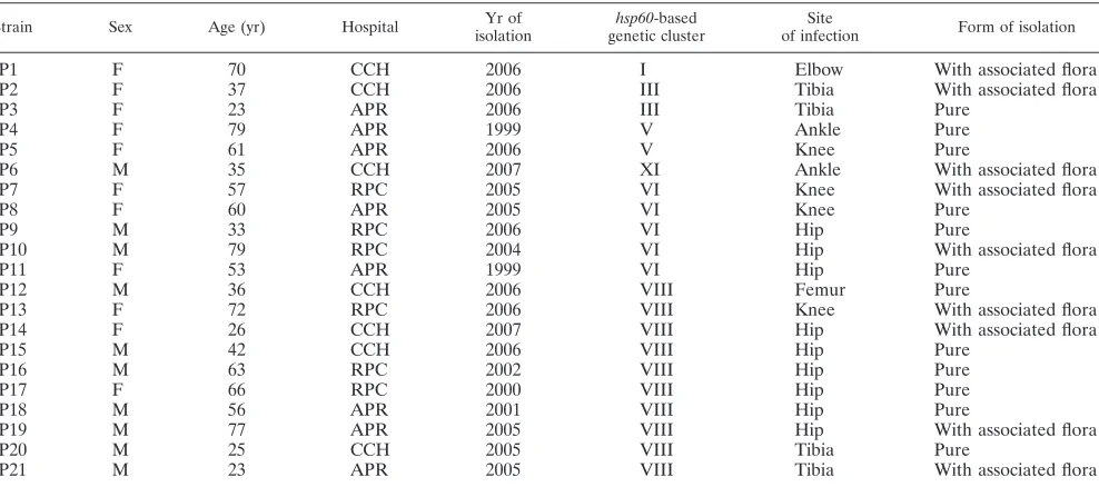
TABLE 1. Characteristics of strains isolated from orthopedic implanted materiala
Strain Sex Age (yr) Hospital Yr of
isolation
hsp60-based genetic cluster
Site
of infection Form of isolation
P1 F 70 CCH 2006 I Elbow With associated flora
P2 F 37 CCH 2006 III Tibia With associated flora
P3 F 23 APR 2006 III Tibia Pure
P4 F 79 APR 1999 V Ankle Pure
P5 F 61 APR 2006 V Knee Pure
P6 M 35 CCH 2007 XI Ankle With associated flora
P7 F 57 RPC 2005 VI Knee With associated flora
P8 F 60 APR 2005 VI Knee Pure
P9 M 33 RPC 2006 VI Hip Pure
P10 M 79 RPC 2004 VI Hip With associated flora
P11 F 53 APR 1999 VI Hip Pure
P12 M 36 CCH 2006 VIII Femur Pure
P13 F 72 RPC 2006 VIII Knee With associated flora
P14 F 26 CCH 2007 VIII Hip With associated flora
P15 M 42 CCH 2006 VIII Hip Pure
P16 M 63 RPC 2002 VIII Hip Pure
P17 F 66 RPC 2000 VIII Hip Pure
P18 M 56 APR 2001 VIII Hip Pure
P19 M 77 APR 2005 VIII Hip With associated flora
P20 M 25 CCH 2005 VIII Tibia Pure
P21 M 23 APR 2005 VIII Tibia With associated flora
a
The ratio of males (M) to females (F) was 0.9. The mean age⫾standard deviation was 51⫾20 years. CCH, Cochin Hospital; APR, Ambroise Paré Hospital; RPC,
[image:3.585.47.541.80.299.2]Raymond Poincaré Hospital.
TABLE 2. Characteristics of control strainsa
Characteristic Value
No. of patients... 52
M/Fbratio ... 1.4 Mean age at time of isolation⫾SD (yr) ...65⫾18 Anatomical site of isolation (no. of isolates) ... Skin and soft tissue... 14
Upper and lower respiratory tract ... 15
Urine... 12
Joint or bone, in the absence of infected material... 3
Intravenous catheter ... 2
Blood ... 2
Gastrointestinal tract ... 4
a
One isolate from each patient was studied.
b
M, male; F, female.
VOL. 47, 2009 E. CLOACAE COMPLEX AND ORTHOPEDIC IMPLANTS 2491
on May 16, 2020 by guest
http://jcm.asm.org/
2492
on May 16, 2020 by guest
phenotypically identified asE. cloacaebelonged to one of the molecular clusters of theE. cloacaecomplex (Fig. 1).
Without distinction by the site of isolation (Fig. 2), three clusters (clusters III, VI, and VIII) accounted for 90% of all
strains. Similar to the implant-associated strains, genetic clus-ters belonging to theE. hormaecheispecies (clusters VI and VIII) were predominant (48% of control strains). Interest-ingly, cluster VII (E. hormaecheisubsp.hormaechei) was
[image:5.585.134.450.67.481.2]ab-FIG. 1. Neighbor-joining unrooted tree resulting from analysis of thehsp60gene sequences of 73 clinical strains and previously reported sequences. Isolation site of clinical strains: P, infected orthopedic implant; B, blood; J, joint or bone, in the absence of implanted material; G, gastrointestinal tract; R, respiratory tract; S, skin and soft tissue; U, urine; K, intravascular catheter. For the previously described strains, type strains are indicated (8, 10–12). Strains labeled EN were reported previously (8) and correspond to sequences with GenBank accession numbers AJ417125, AJ417127, AJ543819, AJ567887, AJ543876, AJ543894, AJ567893, AJ543787, AJ543803, AJ543806, AJ543829, AJ543864, AJ543882, AJ543804, AJ543789, AJ543781, AJ543784, AJ567846, AJ543776, AJ543808, AJ543877, AJ543807, AJ543866, AJ543775, AJ543878, AJ543881, AJ543820, AJ567878, AJ567885, AJ543857, AJ543831, AJ543821, AJ543816, AJ543798, AJ567847, AJ543777, AJ543861, AJ543768, AJ543855, AJ543870, AJ567881, and AJ543837. EH,E. hormaechei; EC,E. cloacae. The bar indicates the number of substitutions per site. Genetic clusters are numbered according to the description provided previously (8).
FIG. 2. Distribution of clinical strains within the genetic clusters of theE. cloacaecomplex. All strains could be assigned to one of the previously reportedhsp60gene sequencing-based genetic clusters of theE. cloacaecomplex. (A) Strains isolated from implanted orthopedic material at different anatomical sites (n⫽21). Hip-associated strains exclusively belonged to theE. hormaecheispecies (clusters VI and VIII). (B) Randomly selected clinical strains of diverse anatomical origins (n⫽52). S, skin and soft tissue; R, upper and lower respiratory tract; U, urine; J, joint or bone, in the absence of infected material; K, intravenous catheter; B, blood; G,gastrointestinal tract. Irrespective of the site if isolation, cluster III accounted for 42% of the control isolates.
VOL. 47, 2009 E. CLOACAE COMPLEX AND ORTHOPEDIC IMPLANTS 2493
on May 16, 2020 by guest
http://jcm.asm.org/
sent from our study, and E. hormaechei subsp. steigerwaltii
(cluster VIII, n ⫽ 18) was predominant overE. hormaechei
subsp.oharae(cluster VI,n⫽8).
Cluster III accounted for 42% of control clinical isolates; thus, it occurred statistically more frequently within the control group of isolates than within the isolates from infected ortho-pedic implants (22/52 isolates versus 2/21 isolates;P⫽0.006). These strains were recovered from various anatomical sites (respiratory, urinary and gastrointestinal tract, skin or soft tissue, and blood). The three bone and joint strains obtained in the absence of material (Fig. 2, series J [joint or bone]) were isolated from knee synovial fluid, knee soft tissue, and hallux valgus and belonged to clusters III, VI, and VIII, respectively; they thus represented each of the most frequently isolated clusters in the control group. The other clusters, cluster II (E. kobei,n⫽1), cluster IV (n⫽1), cluster V (E. ludwigii,n⫽1), cluster IX (n⫽1), and cluster XI (E. cloacaesubsp.cloacae,
n⫽1), were poorly represented. Clusters I, VII, X, and XII and sequence crowdxiiiwere not found among the isolates in the control group. Taken together, these results show that cluster III, together with clusters VI and VIII, accounts for most of the strains routinely isolated from clinical specimens.
DISCUSSION
In this work, we investigated the distribution of strains in-volved in infections of osteoarticular implanted material within the genetic clusters of theE. cloacaecomplex. On the basis of
hsp60 analysis, we show that the cluster distribution for in-fected orthopedic implant-associated strains is different from that for randomly selected clinical strains of various anatomical origins. Our observation that all genetic clusters are not equally involved in pathogenesis highlights the need for more accurate routine bacterial identification tools and for a better understanding of the pathogenesis of theE. cloacaecomplex. All strains evaluated in this study (n⫽73) could be assigned to 1 of the 12 genetic clusters (clusters I to XII) of theE. cloacaecomplex. Only one strain was found to belong to clus-ter XI, the type strain of which isE. cloacaesubsp.cloacae, which suggests that the widely used name E. cloacae is not representative of most clinical strains of the taxon. Preliminary identification asE. cloacaeby conventional phenotypic identi-fication methods (performed with the API 20E or the Vitek 2 system) was a prerequisite for the inclusion of strains in either the control or the study group. Although they might have led to possible underrepresentation of misidentified strains that authentically belong to theE. cloacaecomplex, the phenotypic identification methods performed with the API 20E and the Vitek 2 systems can be considered reliable tools for identifi-cation, as long as identification as “E. cloacae” is understood as “belonging to theE. cloacaecomplex.”
Although patient populations vary from one hospital to an-other, the results of our studies of the distribution of the control strains within genetic clusters are concordant with those published previously, since we observed that clusters III, VIII, and VI account for most of the clinical isolates (8). Strains belonging tohsp60sequence analysis-based cluster III were shown to gather into the previously described MLSA-based cluster 1 as well as in CGH-MLSA-based clade 2, with the latter being associated with strains that are the most frequently
cul-tured in hospitals (21). Similarly, hsp60 sequence analysis-based cluster VI (E. hormaechei subsp. oharae) and cluster VIII (E. hormaecheisubsp. steigerwaltii) were shown to gather in MLSA-based cluster 2 but also to belong to the clinically relevant CGH-based clade 2. Thus, our data showing a pre-dominance of cluster III, VI, and VIII isolates among control clinical strains of different anatomical origins are consistent with data presented in previous reports on the genetic di-versity of the strains within the E. cloacae complex and support the congruence of CGH-based clade 2 with clini-cally relevant samples.
Some genetic clusters were absent from our study. Cluster VII (E. hormaechei subsp. hormaechei) harbors the original species type strain and was also poorly represented in other studies (8). Cluster X (E. nimipressuralis) is found in potable water reservoirs but, to our knowledge, has never been asso-ciated with human disease (13). Cluster XII (E. cloacaesubsp.
dissolvens), formerly part of the genusErwinia, was reassigned to the Enterobacter genus and forms a subspecies of theE. cloacaespecies. It is associated with plants (maize, coffee), but no human infections have been reported (7, 9).
Although it was the largest cluster within the group of con-trol strains (42%), cluster III was poorly represented within the group of orthopedic implant-associated strains (9%), and this difference was statistically significant (P⫽0.006). Low num-bers of cluster III isolates emphasize the large proportion of cluster VI (E. hormaecheisubsp.oharae) and cluster VIII (E. hormaecheisubsp.steigerwaltii) isolates, both of which belong to theE. hormaechei species. Hip joint prosthesis infections appeared to be specifically associated with E. hormaechei, since, in our series, all cases of infections of implants at this site were due to isolates of either cluster VIII (n⫽6) or cluster VI (n ⫽ 3). The third subspecies of the taxon, E. hormaechei
subsp.hormaechei(cluster VII), was not found in our analysis. The low prevalence of cluster III isolates within the group of isolates from infected orthopedic implants compared to their prevalence within the group of isolates from clinical samples of other origins suggests a specific pathogenicity and reinforces the need for a robust and discriminatory tool for the accurate identification of isolates within theE. cloacaecomplex. In this regard,hsp60gene sequencing-based identification appeared to be both discriminatory and easily implementable, whereas other sequence-based molecular methods for the identification ofEnterobacter were not as accurate. The absence of a con-sensus for the analysis of anrpoBDNA fragment (1 kb or 500 bp) has led to contradictory results, particularly for the genetic discrimination of isolates withinhsp60gene sequencing-based clusters III, VI, and VIII (8, 21), as well as to the confusing use of the species name E. hormaechei for strains that do not belong to hsp60 gene sequencing-based clusters VI to VIII (20). Similarly, sequence analysis of the gene encoding the DNA gyrase subunit B (gyrB) led to the hypothesis that most clinical isolates assigned to theE. cloacaecomplex by pheno-typic semiautomated methods would belong to theE. hormae-cheispecies (6). Analysis of thehsp60gene sequence was not included in this study, but the absence of cluster III as a specific group within the group of clinical strains evaluated in this study might suggest thatgyrBsequencing is not as discriminatory as
hsp60gene sequence analysis. Further investigation is needed to elucidate the latter point.
on May 16, 2020 by guest
http://jcm.asm.org/
The epidemiological association betweenE. hormaecheiand hip prosthetic implants allows new insights into previous ob-servations to be made. First, the E. hormaecheispecies was reported in a case of prosthetic hip infection, although it was initially misidentified as Escherichia coli (25). This strain ex-hibited a phenotype of small-colony-variant formation, which was shown to be associated with regulation of the hemin up-take system (24). Although we did not systematically search for it, at least one of the strains involved in prosthetic infection displayed such a phenotype, with each step of subculture on solid medium leading to the emergence of fast- and slow-growing bacterial colonies. Second, although the study did not specifically refer toE. hormaechei, the ability ofEnterobacter
species to participate in biofilm formation on orthopedic im-plants was reported (1). Third, the ability ofE. hormaecheito colonize implanted catheters as a biofilm and to be responsible for systemic infection was described (3, 5). The ability to grow as small-colony variants and the formation of biofilms are features frequently associated with the causative agents of or-thopedic implant infections and contribute to the increased difficulty of diagnosis and treatment of such infections (18, 28). Taken together, the findings from the previous reports rein-force our observation showing the predominance ofE. hormae-chei as the cause of orthopedic implant infections. Further work is needed in order to identify the bacterial and host factors specifically involved in the bacterial colonization of the implanted material and in the pathophysiology of these infec-tions.
ACKNOWLEDGMENTS
We thank David Biau for precious advice on the statistical analysis. This work was supported by Universite´ Paris Descartes, Institut Cochin, and Assistance Publique—Hoˆpitaux de Paris.
REFERENCES
1.Bartoszewicz, M., A. Rygiel, M. Krzeminski, and A. Przondo-Mordarska.
2007. Penetration of a selected antibiotic and antiseptic into a biofilm formed
on orthopedic steel implants. Ortop. Traumatol. Rehabil.9:310–318.
2.Bernard, L., P. Hoffmeyer, M. Assal, P. Vaudaux, J. Schrenzel, and D. Lew.
2004. Trends in the treatment of orthopaedic prosthetic infections. J.
Anti-microb. Chemother.53:127–129.
3.Campos, L. C., L. F. Lobianco, L. M. Seki, R. M. Santos, and M. D. Asensi.
2007. Outbreak ofEnterobacter hormaecheisepticaemia in newborns caused
by contaminated parenteral nutrition in Brazil. J. Hosp. Infect.66:95–97.
4.Darouiche, R. O. 2004. Treatment of infections associated with surgical
implants. N. Engl. J. Med.350:1422–1429.
5.da Silva, C. L., L. E. Miranda, B. M. Moreira, D. Rebello, L. A. Carson, M. E. Kellum, M. C. de Almeida, J. L. Sampaio, and C. M. O’Hara.2002.
Enterobacter hormaecheibloodstream infection at three neonatal intensive
care units in Brazil. Pediatr. Infect. Dis. J.21:175–177.
6.Delmas, J., F. Breysse, G. Devulder, J. P. Flandrois, and M. Chomarat.2006.
Rapid identification ofEnterobacteriaceaeby sequencing DNA gyrase
sub-unit B encoding gene. Diagn. Microbiol. Infect. Dis.55:263–268.
7.Frank, H. A., N. A. Lum, and A. S. Delacruz.1965. Bacteria responsible for
mucilage-layer decomposition in Kona coffee cherries. Appl. Microbiol.13:
201–207.
8.Hoffmann, H., and A. Roggenkamp.2003. Population genetics of the
no-menspeciesEnterobacter cloacae. Appl. Environ. Microbiol.69:5306–5318.
9.Hoffmann, H., S. Stindl, W. Ludwig, A. Stumpf, A. Mehlen, J. Heesemann, D. Monget, K. H. Schleifer, and A. Roggenkamp.2005. Reassignment of En-terobacter dissolvenstoEnterobacter cloacaeasE. cloacaesubspecies
dissol-venscomb. nov. and emended description ofEnterobacter asburiaeand
En-terobacter kobei. Syst. Appl. Microbiol.28:196–205.
10.Hoffmann, H., S. Stindl, W. Ludwig, A. Stumpf, A. Mehlen, D. Monget, D.
Pierard, S. Ziesing, J. Heesemann, A. Roggenkamp, and K. H. Schleifer.
2005.Enterobacter hormaechei subsp.oharaesubsp. nov., E. hormaechei
subsp.hormaecheicomb. nov., andE. hormaecheisubsp.steigerwaltiisubsp.
nov., three new subspecies of clinical importance. J. Clin. Microbiol.43:
3297–3303.
11.Hoffmann, H., S. Stindl, A. Stumpf, A. Mehlen, D. Monget, J. Heesemann, K. H. Schleifer, and A. Roggenkamp. 2005. Description ofEnterobacter ludwigiisp. nov., a novelEnterobacterspecies of clinical relevance. Syst. Appl.
Microbiol.28:206–212.
12.Iversen, C., N. Mullane, B. McCardell, B. D. Tall, A. Lehner, S. Fanning, R. Stephan, and H. Joosten.2008.Cronobacter gen. nov., a new genus to
accommodate the biogroups of Enterobacter sakazakii, and proposal of
Cronobacter sakazakiigen. nov., comb. nov.,Cronobacter malonaticussp.
nov., Cronobacter turicensis sp. nov., Cronobacter muytjensii sp. nov.,
Cronobacter dublinensissp. nov.,Cronobactergenomospecies 1, and of three
subspecies, Cronobacter dublinensis subsp. dublinensis subsp. nov.,
Cronobacter dublinensissubsp.lausannensissubsp. nov. andCronobacter dub-linensissubsp.lactaridisubsp. nov. Int. J. Syst. Evol. Microbiol.58:1442–1447. 13.Kampfer, P., A. Nienhuser, G. Packroff, F. Wernicke, A. Mehling, K. Nix-dorf, S. Fiedler, C. Kolauch, and M. Esser.2008. Molecular identification of coliform bacteria isolated from drinking water reservoirs with traditional
methods and the Colilert-18 system. Int. J. Hyg. Environ. Health211:374–
384.
14.Kurtz, S., K. Ong, E. Lau, F. Mowat, and M. Halpern.2007. Projections of primary and revision hip and knee arthroplasty in the United States from
2005 to 2030. J. Bone Joint Surg. Am.89:780–785.
15.Kurtz, S. M., E. Lau, J. Schmier, K. L. Ong, K. Zhao, and J. Parvizi.2008. Infection burden for hip and knee arthroplasty in the United States. J.
Arthroplasty23:984–991.
16.Kurtz, S. M., K. L. Ong, J. Schmier, F. Mowat, K. Saleh, E. Dybvik, J. Karrholm, G. Garellick, L. I. Havelin, O. Furnes, H. Malchau, and E. Lau.
2007. Future clinical and economic impact of revision total hip and knee
arthroplasty. J. Bone Joint Surg. Am.89(Suppl. 3):144–151.
17.Mazari-Hiriart, M., S. Ponce-de-Leon, Y. Lopez-Vidal, P. Islas-Macias, R. I. Amieva-Fernandez, and F. Quinones-Falconi.2008. Microbiological impli-cations of periurban agriculture and water reuse in Mexico City. PLoS One
3:e2305.
18.Neut, D., H. C. van der Mei, S. K. Bulstra, and H. J. Busscher.2007. The role of small-colony variants in failure to diagnose and treat biofilm infections in
orthopedics. Acta Orthop.78:299–308.
19.O’Hara, C. M., A. G. Steigerwalt, B. C. Hill, J. J. Farmer III, G. R. Fanning, and D. J. Brenner.1989.Enterobacter hormaechei, a new species of the family
Enterobacteriaceaeformerly known as enteric group 75. J. Clin. Microbiol.
27:2046–2049.
20.Paauw, A., M. P. Caspers, M. A. Leverstein-van Hall, F. H. Schuren, R. C. Montijn, J. Verhoef, and A. C. Fluit.2009. Identification of resistance and
virulence factors in an epidemicEnterobacter hormaecheioutbreak strain.
Microbiology155:1478–1488.
21.Paauw, A., M. P. Caspers, F. H. Schuren, M. A. Leverstein-van Hall, A. Deletoile, R. C. Montijn, J. Verhoef, and A. C. Fluit.2008. Genomic diversity
within theEnterobacter cloacaecomplex. PLoS One3:e3018.
22.Quintanilha, A. G., B. Zilberstein, M. A. Santos, D. Pajecki, E. G. Moura, P. R. Alves, F. Maluf-Filho, and I. Cecconello. 2007. A novel sampling
method for the investigation of gut mirobiota. World J. Gastroenterol.13:
3990–3995.
23.Roggenkamp, A.2007. Phylogenetic analysis of enteric species of the family
Enterobacteriaceaeusing theoriC-locus. Syst. Appl. Microbiol.30:180–188. 24.Roggenkamp, A., H. Hoffmann, and M. W. Hornef.2004. Growth control of
small-colony variants by genetic regulation of the hemin uptake system.
Infect. Immun.72:2254–2262.
25.Roggenkamp, A., A. Sing, M. Hornef, U. Brunner, I. B. Autenrieth, and J. Heesemann.1998. Chronic prosthetic hip infection caused by a
small-colony variant ofEscherichia coli. J. Clin. Microbiol.36:2530–2534.
26.Stumpf, A. N., A. Roggenkamp, and H. Hoffmann.2005. Specificity of en-terobacterial repetitive intergenic consensus and repetitive extragenic palin-dromic polymerase chain reaction for the detection of clonality within the
Enterobacter cloacaecomplex. Diagn. Microbiol. Infect. Dis.53:9–16. 27.Tang, Y. W., N. M. Ellis, M. K. Hopkins, D. H. Smith, D. E. Dodge, and D. H.
Persing.1998. Comparison of phenotypic and genotypic techniques for iden-tification of unusual aerobic pathogenic gram-negative bacilli. J. Clin.
Mi-crobiol.36:3674–3679.
28.Trampuz, A., and A. F. Widmer.2006. Infections associated with orthopedic
implants. Curr. Opin. Infect. Dis.19:349–356.
29.Trampuz, A., and W. Zimmerli.2005. Prosthetic joint infections: update in
diagnosis and treatment. Swiss Med. Wkly.135:243–251.
30.Zimmerli, W., A. Trampuz, and P. E. Ochsner.2004. Prosthetic-joint
infec-tions. N. Engl. J. Med.351:1645–1654.
VOL. 47, 2009 E. CLOACAE COMPLEX AND ORTHOPEDIC IMPLANTS 2495