0095-1137/11/$12.00 doi:10.1128/JCM.02509-10
Copyright © 2011, American Society for Microbiology. All Rights Reserved.
Use of
bexB
To Detect the Capsule Locus in
Haemophilus influenzae
䌤
Gregg S. Davis,
1Sara A. Sandstedt,
1Mayuri Patel,
2Carl F. Marrs,
1and Janet R. Gilsdorf
1,2*
Department of Epidemiology, University of Michigan School of Public Health, Ann Arbor, Michigan,1and Department of
Pediatrics and Communicable Diseases, University of Michigan Medical School, Ann Arbor, Michigan2
Received 10 December 2010/Returned for modification 11 January 2011/Accepted 19 April 2011
Haemophilus influenzaestrains are classified as typeable or nontypeableH. influenzae(NTHI) based upon the
presence or absence of capsule. In addition to serotyping, which is subject to false-positive results, typeable strains can be identified through the detection of the capsular export genebexAand one of six capsule-specific genes, but this method is resource intensive, especially in characterizing large numbers of strains. To address these challenges, we developed abexB-based method to differentiate true NTHI strains from typeable strains. We validated a PCR-based method to detectbexBin 10 strains whose capsule status was well defined. Among 40 strains that were previously serotype positive in clinical microbiology laboratories, 5 lackedbexA,bexB, and capsule type-specific genes by PCR analysis and thus likely represent false-positive serotyping results. Among 94 additional otitis media, commensal, and serotype b-negative invasive strains, 85 werebexAandbexBnegative and 9 contained either a complete or partial capsule locus, i.e., 8 werebexAandbexBpositive and 1 wasbexA
negative butbexBpositive. Finally, we adapted the method for use in a high-throughput DNA hybridization-based microarray method, which showed 98.75 and 97.5% concordance to the PCR methods forbexAandbexB, respectively. In addition,bexBshowed 84% or greater nucleotide identity among strains containing the capsule locus. In this study, we demonstrate thatbexBis a reliable proxy for the capsule locus and that its detection provides a simple and reliable method for differentiating strains that lack the entire capsule locus from those containing a partial or complete capsule locus.
Haemophilus influenzae strains, which are Gram-negative bacteria that commonly reside as commensals within the hu-man pharynx and may cause respiratory or invasive infections, are variable for the presence of a polysaccharide capsule. His-torically,H. influenzaestrains lacking a capsule were detected by their failure to react with antibodies directed against the six immunologically distinct capsular polysaccharides and were designated nontypeable, i.e., serotype negative,H. influenzae
(NTHI) (33). Nontypeable strains predominate among com-mensal organisms (9, 29) and those causing respiratory infec-tions such as acute otitis media, sinusitis, or bronchitis, while serotypeable strains are isolated more often from clinical sam-ples of patients with invasive infections such as bacteremia, meningitis, facial cellulitis, and septic arthritis. In addition, multilocus sequence typing has shown that NTHI strains are genetically widely variable, while serotypeable strains are more clonal (25).
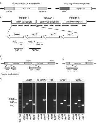
The polysaccharide capsule of typeableH. influenzaestrains is encoded by thecaplocus, which is composed of three distinct regions, designated regions I to III (Fig. 1B). The genes con-tained within regions I and III, designated bexDCBA and
hcsAB, respectively, are highly conserved across all capsular types and are required for transport of capsule constituents across the outer membrane (20, 21, 23, 37, 40). Region II genes encode capsule type a- through f-specific proteins and thus vary by serotype. The organization and genetics of the cap
locus are complex; duplications, partial loss, and complete loss
of thecaplocus have been documented (3, 4, 12, 14–17, 19, 21, 22, 32, 37, 38, 44).
Differentiating typeable from nontypeable H. influenzae
strains can be challenging. Type-specific serum agglutination has classically been used to confirm the presence and specific-ity of theH. influenzaecapsule. However, a strain may fail to react with typing sera and thus be classified as “nontypeable” for several reasons. First, inaccuracies in performing and in-terpreting slide agglutination tests have been well documented (30, 45). Second, strains with one copy of the capregion in whichbexAis partially deleted (3, 4, 12, 14, 15, 17, 19) are referred to as capsule-deficient variants because they contain a majority of thecaplocus; genetically, they arecaplocus posi-tive, but they show an altered ability to produce functional capsule and fail to agglutinate with typing serum, i.e., they are nonserotypeable. Third, a previously serotypeable strain could have a deletion of the entire capsule locus, as apparently oc-curred with strain Rd, a nontypeable variant of a type d strain that has been passaged extensively in the laboratory (37). Fi-nally, a strain may lack the entirecaplocus as a consequence of long-past evolutionary events, i.e., may be a true NTHI strain.
Detection ofbexAand capsule-specific genes by PCR is the most commonly used method of capsule genotyping. PCR methods based on detection of bexAalone, however, fail to identify capsule-deficient variants because the 5⬘primer of the standard primer pair hybridizes in the deleted region ofbexA. This problem was overcome by the addition of PCR analyses to detect individual type a to f capsule-specific regions, along with
bexA, for each strain (8, 26). This approach, however, requires up to seven separate PCRs, making it both labor- and re-source-intensive for studying a large number of isolates, espe-cially when clinical or epidemiological data suggest that a
sig-* Corresponding author. Mailing address: University of Michigan Medical Center, D5101, Medical Professional Building Spc 5718, Ann Arbor, MI 48109-5718. Phone: (734) 763-2440. Fax: (734) 936-7635. E-mail: [email protected].
䌤Published ahead of print on 27 April 2011.
2594
on May 16, 2020 by guest
http://jcm.asm.org/
nificant portion of the strains may be NTHI. This study was motivated by the need for a simple and accurate method to differentiate true NTHI strains from genetically cap locus-positiveH. influenzaestrains.
We hypothesized thatbexB, which is located within region I of thecaplocus (Fig. 1), could serve as a more reliable marker for the capsule locus thanbexA, based on the observations that region I genes are present across all capsular types (20, 23) and that there are no reports ofbexBpartial deletions analogous to those observed inbexA. We first validated the PCR method to detectbexBby usingH. influenzaestrains whose capsule status had been well defined. We then tested 40H. influenzaestrains that were serotype positive when tested in clinical microbiology
laboratories. To better understand the prevalence of geneti-cally cap region-positive strains that fail to produce a bexA
amplicon, we then tested thebexB PCR method on invasive strains that were serotype negative in clinical microbiology laboratories and on strains likely to be nontypeable based upon their site of isolation, i.e., commensal and otitis media isolates. Finally, we adapted the molecular capsular typing method for use in a high-throughput hybridization format and tested it with a microarray containing the genomic DNAs of 546 H. influenzae strains. We demonstrate here that screening for
bexBprovides a simple and reliable method for distinguishing
[image:2.585.135.449.64.461.2]H. influenzaestrains containing a complete or partial capsule locus from those lacking the capsule locus.
FIG. 1. Schematic representations of the capsule locus. (A) The capsule locus can be categorized based upon its location in theH. influenzae genome relative to either an IS1016element orsodC. In one arrangement, thecaplocus is flanked by IS1016insertion element sequences. In the second arrangement,sodCis upstream of thecaplocus. (B) Thecaplocus is divided into three regions, namely, regions I, II, and III; bothbexA andbexBare located within region I. Previously describedbexA-specific primers produce a 343-bp amplicon (8), and thebexBprimers used in this study amplify a 567-bp product. (C) Representativecaplocus arrangements. (i) Duplicated IS1016-caplocus harboring abexApartial deletion in one copy. (ii) Single-copy IS1016-caparrangement.ⴱ, partialbexAdeletion precludes annealing with the standardbexA5⬘primer. (D) Repre-sentative PCR results. Eagan is a type b strain, containing intactbexA,bexB, and the serotype b-specificcapgene; 86-028NP is an NTHI strain lacking bexA and bexB; strain Rd is a type d strain in which the entire cap region has been deleted; AAr64 is abexA-negative, type b capsule-deficient variant; and F22477 is abexA-negative, type f capsule-deficient variant. Invitrogen 1-kb Plus (Invitrogen Corporation, Carlsbad, CA) served as the DNA ladder.
on May 16, 2020 by guest
http://jcm.asm.org/
MATERIALS AND METHODS
Bacterial strains.All strains used in this study were defined asH. influenzaeby use of current clinical laboratory criteria.H. influenzaeisolates, maintained with minimal passaging, were stored in skim milk at⫺80°C and were cultured for testing on chocolate II agar (BD Diagnostics) at 37°C in the presence of 5% CO2.
H. influenzaetype a, c, and d strains were obtained from the ATCC; the fully sequencedH. influenzaestrain Rd is a lab-adapted strain that lacks the entire capsule locus; the well-studied strain Eagan (1) was the source of chemically purified type b capsule; two type b capsule-deficient variant strains were previ-ously described (15, 27); and three fully sequenced nontypeable otitis media strains, 86-028NP (a kind gift from Lauren Bakaletz, Ohio State University) and R2866 and R2846 (kind gifts from Arnold Smith, University of Washington), lack capsule genes. The capsule status of these strains is considered to be well defined (see Table 2). AHaemophilus haemolyticusstrain served as a non-H. influenzae control.
H. influenzaestrains used for PCR- and microarray-based analyses were cho-sen from our large collection ofH. influenzaeclinical and commensal isolates collected over 30 years from Michigan, California, Kentucky, Pittsburgh, Min-nesota, and Georgia (a kind gift of the Georgia Emerging Infections Program) and included 40H. influenzaestrains previously found to be serotype positive by clinical microbiology laboratories (see Table 3). Also included were throat iso-lates from healthy children attending day care (9, 39) and middle ear isoiso-lates from children with acute otitis media from Finland (a kind gift of Tehri Kilpi) and Israel (a kind gift of Ron Dagan), all considered likely to be NTHI based on the site of isolation, as well as invasive blood or cerebrospinal fluid isolates that failed to react with the type b serotyping serum in the clinical microbiology laboratory (see Table 3).
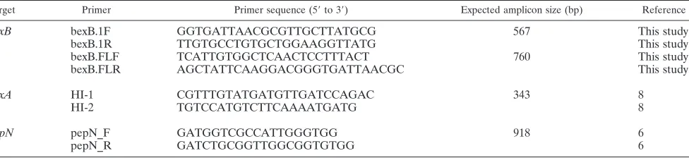
PCR-based studies.The design of PCR primers targeting a 567- or 760-bp region ofH. influenzae bexB(Table 1) was based on sequence data available in GenBank. Primers targetingbexA,pepN(which served as a DNA positive con-trol), and serotype-specific capsule genes have been described previously (Table 1) (6, 8, 28). Whole-cell lysate, i.e., crude lysate, served as the DNA template source for PCRs and was prepared by suspending several bacterial colonies, collected with a sterile cotton swab, from a fresh plate into Tris-EDTA (TE; pH 7.5), heating them for 10 min at 100°C in a thermocycler, centrifuging them for 10 min, and removing the supernatant, which was stored at⫺20°C until it was used for PCR. For routine screening, PCR was performed withTaqpolymerase (New England BioLabs, Ipswich, MA). Each 20-l PCR mixture consisted of 1⫻ ThermoPol reaction buffer, a 0.2 mM concentration of each deoxynucleoside triphosphate (dNTP), a 0.2M concentration of each primer, 2.0 UTaq, and 1
l crude lysate (template). Reaction mixtures were subjected to an initial dena-turation step of 2 min at 95°C followed by 30 amplification cycles of 95°C for 30 s, 54°C for 30 s, and 72°C for 45 s. AllbexB-positive isolates were subjected to further analysis with region II serotype-specific primers (Table 1). Amplicons were separated in 1.5% agarose gels in 1⫻Tris-acetate-EDTA (TAE) buffer and visualized by ethidium bromide staining.
Microarray studies.H. influenzaelibrary-on-a-slide (LOS) microarrays, con-sisting ofH. influenzaegenomic DNAs arrayed on a glass slide, were prepared, tested, and analyzed as previously described (36, 46). In brief, total genomic DNA from eachH. influenzaestrain was isolated and spotted in duplicate onto membrane-coated slides. First, a digoxigenin-labeled genomic DNA quantity control probe consisting of equal amounts of each of the sevenH. influenzae multilocus sequence typing (MLST) genes (http://Haemophilus.mlst.net) and pepNwas hybridized to the microarray (36). The intensity of control probe hybridization to each spot was determined using Spotfinder v.3.1.1 (http://www .tm4.org/spotfinder.html), and duplicate spots were averaged using MIDAS
v.2.19 (http://www.tm4.org/midas.html) (34). The slides were stripped and then rehybridized with either abexAorbexBfluorescein-labeled probe (18). ThebexA andbexBprobes were “mixed probes” in that they contained equal concentra-tions of eitherbexAorbexBgenes amplified from strains representing each of the six capsular serotypes (type a, strain ATCC 9006; type b, strain AA194; type c, strain ATCC 9007; type d, strain ATCC 9008; type e, strains AA52 and M4; and type f, strain F2243.7). The mixed probes were PCR amplified from genomic DNA with primers HI-1 and HI-2 (bexAprobe) or with bexB.1 primers (bexB probe) (Table 1). Signal intensities were calculated as described above, the ratio of the log-transformedbexAorbexBsignal to the MLST-pepNconcentration control signal was calculated for each spot, and the distribution of intensities was modeled using programs written in the statistical software R (http://www.r -project.org). Spots were classified as positive, negative, or uncertain as described by Sandstedt et al. (36).
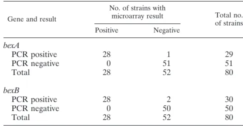
Concordance of thebexAandbexBresults between the PCR-based method and the microarray method was estimated by testing 80 strains for these two genes by both PCR and hybridization of thebexAandbexBprobes on the microarray.
DNA sequence analysis.To test for the magnitude of DNA sequence identity among a relatively large number of capsule gene-containingH. influenzaestrains, DNA sequence analysis was conducted on 45bexB-positive isolates by first amplifying a 760-bp internal region ofbexBwith the Expand high-fidelity system (Roche, Indianapolis, IN), using 1⫻Expand high-fidelity buffer with 1.5 mM MgCl2, a 0.2 mM concentration of each dNTP, a 0.2M concentration of each
primer (bexB.FLF and bexB.FLR), 1.3 U Expand high-fidelity enzyme mix, and 1l crude lysate (template) per 25-l reaction mix. The resultant amplicon was purified with a Qiagen QIAquick PCR purification kit (Qiagen, Valencia, CA) following the manufacturer’s protocol. Sequencing was performed directly from the purified PCR product, in both directions, at the University of Michigan DNA Sequencing Core. Sequences were analyzed with DNASTAR Lasergene (v8). After removal of primers and poor-quality sequences, 666 nucleotides from each strain were aligned using Clustal W (43) and then adjusted manually where necessary. Phylogenetic analyses using all codon positions were conducted in MEGA4 (41). Whenever two or more sequences from strains of the same capsule type were 100% identical at the nucleotide level, a single consensus sequence was used for subsequent analyses.
Nucleotide sequence accession numbers.ThebexBsequences obtained in this study were deposited in GenBank under accession numbers HQ699679 to HQ699723.
RESULTS
bexBPCR test results.Both pairs ofbexBprimers, bexB.1F/ bexB.1R and bexB.FLF/bexB.FLR, produced amplicons of the expected sizes (567 bp and 760 bp, respectively), and thebexA
[image:3.585.52.540.82.194.2]and pepN primers also produced amplicons of the expected sizes (343 bp and 918 bp, respectively). Figure 1D shows the PCR products for 5 representative strains: the type b strain Eagan, from which the type b capsule was isolated chemically and characterized (1); the true nontypeable strain 86-028NP (13), which lacks the capsule locus by DNA sequence analysis and thus isbexAandbexBnegative; strain Rd, initially char-acterized as a type d strain, which lacks the entire capsule locus
TABLE 1. PCR primers used in this study
Target Primer Primer sequence (5⬘to 3⬘) Expected amplicon size (bp) Reference
bexB bexB.1F GGTGATTAACGCGTTGCTTATGCG 567 This study
bexB.1R TTGTGCCTGTGCTGGAAGGTTATG This study
bexB.FLF TCATTGTGGCTCAACTCCTTTACT 760 This study
bexB.FLR AGCTATTCAAGGACGGGTGATTAACGC This study
bexA HI-1 CGTTTGTATGATGTTGATCCAGAC 343 8
HI-2 TGTCCATGTCTTCAAAATGATG 8
pepN pepN_F GATGGTCGCCATTGGGTGG 918 6
pepN_R GATCTGCGGTTGGCGGTGTGG 6
on May 16, 2020 by guest
http://jcm.asm.org/
due to a deletion (11) by DNA sequence analysis and thus is
bexA,bexB, and capd negative; and two previously described strains that arecapregion positive by gene analysis but sero-type negative (i.e., capsule-deficient variants) (15, 27) and that, as expected, arebexAnegative andbexBpositive and contain their respectivecaptype-specific genes.
bexBPCR validation results.Table 2 shows the results of the study to validate thebexBPCR method, using well-definedH. influenzaestrains that were known to be capsule positive or negative by gene analysis or capsule isolation or were obtained as serotypeable strains from the ATCC. As expected, all four serotypeable strains (the ATCC type a, c, and d strains and type b strain Eagan) were bexAand bexB positive. The two strains previously known to be capsule-deficient variants (15, 27) werebexAnegative andbexBpositive, as expected, while the four strains known by genomic sequence analysis to lack
thecapregion werebexAand bexBnegative. TheH. haemo-lyticusstrain wasbexAandbexBnegative.
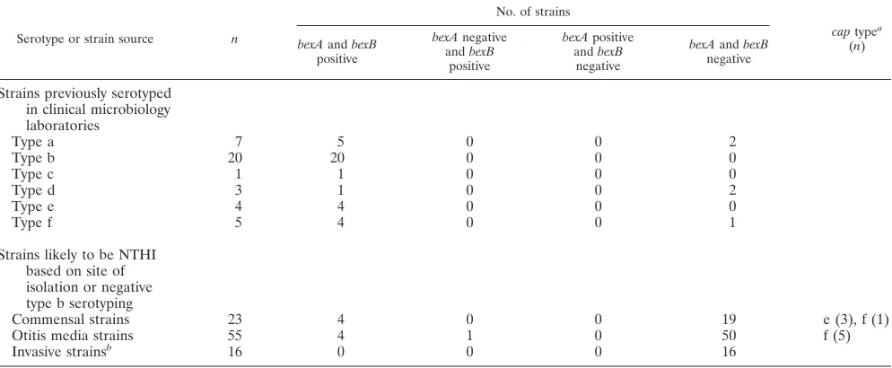
Correlation betweenbexBPCR and serotyping.Among the 40 strains previously serotyped in clinical microbiology labora-tories (Table 3), 35 werebexA and bexB positive, consistent with the serotyping results performed at the time of isolation. Five werebexAandbexBnegative and lacked region II capsule-specific genes, strongly suggesting that these strains are in fact true NTHI strains, i.e., they lack the entire capsule locus. This finding is consistent with other reports of false-positive sero-typing ofH. influenzae(2, 24, 31, 32), even in reference labo-ratories.
Prevalence ofbexBgene among strains likely to be NTHI.To identify the prevalence of capsule-deficient variants amongH. influenzaepopulations in which NTHI isolates predominate, a total of 94H. influenzaestrains likely to be nontypeable (Table 3) were tested by PCR withbexA-,bexB-, and region II capsule-specific PCR primers. Among 23 commensal H. influenzae
strains from the throats of healthy children, 19 werebexAand
bexB negative (true NTHI strains), 3 were bexA and bexB
positive and PCR positive for the type e-specific region II, and 1 wasbexAandbexB positive and PCR positive for the type f-specific region II. Among 55 otitis media isolates, 50 were
bexAandbexBnegative (true NTHI strains), 4 werebexAand
bexBpositive and PCR positive for the type f-specific region II, and 1 was abexA-negative,bexB-positive, type f capsule-defi-cient variant because it was PCR positive for the type f-specific region II but was unable to export capsule because of thebexA
deletion. All 16 invasive blood or cerebrospinal isolates found to be serotype b negative in clinical microbiology laboratories werebexAandbexBnegative, confirming that they were true NTHI strains (Table 3).
[image:4.585.41.283.91.226.2]bexA andbexBmicroarray results. To assess the ability of thebexBprobe to hybridize withH. influenzaegenomic DNA and to expand the capsule characterization to a large number
TABLE 2. Results ofbexAandbexBdetection by PCR amongH. influenzaestrains with known capsule status in validation study
Strain Type
PCR resulta
bexA bexB
ATCC 9006 a ⫹ ⫹
Eaganb b ⫹ ⫹
ATCC 9007 c ⫹ ⫹
ATCC 9008 d ⫹ ⫹
Rd (KW20)c capnegative 0 0
AAr117d b negative 0 ⫹
AAr64d b negative 0 ⫹
86-028NPc NTHI 0 0
R2846c NTHI 0 0
R2866c NTHI 0 0
H. haemolyticus Non-H. influenzae 0 0
a⫹, gene is present; 0, gene is absent.
bThe type b capsule has been isolated and chemically characterized. cComplete genomic sequences are available.
dLacks cap region by gene analysis, as previously reported (15, 27).
TABLE 3. Results ofbexAandbexBdetection by PCR amongH. influenzaestrains
Serotype or strain source n
No. of strains
captypea (n) bexAandbexB
positive
bexAnegative andbexB
positive
bexApositive andbexB
negative
bexAandbexB negative
Strains previously serotyped in clinical microbiology laboratories
Type a 7 5 0 0 2
Type b 20 20 0 0 0
Type c 1 1 0 0 0
Type d 3 1 0 0 2
Type e 4 4 0 0 0
Type f 5 4 0 0 1
Strains likely to be NTHI based on site of isolation or negative type b serotyping
Commensal strains 23 4 0 0 19 e (3), f (1)
Otitis media strains 55 4 1 0 50 f (5)
Invasive strainsb 16 0 0 0 16
a
Determined by capsule-specific PCR. b
Non-serotype b.
on May 16, 2020 by guest
http://jcm.asm.org/
[image:4.585.44.541.504.709.2]of strains in a high-throughput assay, a total of 546H. influen-zaestrains were screened by whole-genome DNA microarray analysis for hybridization withbexAandbexB(Table 4). Only 10 of the 546 (1.8%) strains tested by microarray hybridization showed indeterminate results due to technical difficulties, e.g., damage to the array surface, too little genomic DNA, etc. Among all strains tested, no bexA-positive, bexB-negative strains were identified. Of 135 strains originally serotyped as types a to f, 2 were misclassified by microarray analysis—one was a bexA- andbexB-positive commensal strain that lacked serotype-specific genes, and the other was abexA-and
bexB-negative invasive strain that possessed a serotype b-specific gene, i.e., a genetically type b capsule-deficient variant, and thus would have been misclassified as NTHI bybexAPCR or hybridization alone or by serotyping.
bexA andbexBPCR-microarray concordance. The concor-dance betweenbexAandbexBdetection by microarray hybrid-ization using specific probes and by PCR using gene-specific primers was assessed using 80 strains that were analyzed by both methods. One strain was discordant forbexA
(98.7% concordance), and two strains were discordant forbexB
(97.5% concordance), one of which was thebexA-discordant strain, suggesting a possible strain mix-up (Table 5).
bexBsequencing results and phylogenetic analyses.To gain insight intobexBnucleotide diversity across all capsule types, a 760-bp region ofbexBwas sequenced in both directions for 45
bexB-positive strains as tested by PCR. Following sequence cleanup, a total of 666bexBbase pairs from each strain, with no gaps, were available for analysis (GenBank accession numbers HQ699679 to HQ699723). All 23 type bbexBsequences were 100% identical at the nucleotide level. There were three unique type f sequences (types f.1, f.2, and f.3), with three vari-able sites among 666 bp (0.45%), and three unique type e sequences (types e.1, e.2, and e.3), with five variable sites among 666 bp (0.75%). Nucleotide sequence similarities and genetic distances were estimated for a 666-bp region ofbexB
for 45bexB-positive strains (Table 6). Pairwise nucleotide sim-ilarities ranged from 84.2 to 100% and averaged 91.7% across all sequences. Evolutionary distances, estimated using the max-imum composite likelihood method (41, 42), and percent nu-cleotide similarities were estimated for all unique sequences. Maximum composite likelihood distances ranged from 0 to 0.199 base substitution per site, with an overall average dis-tance across sequences of 0.101.
To address the potential for nucleotide diversity within the bexB.1F/bexB.1R primer annealing regions, the nucleotide
se-quences within the primer annealing regions were analyzed. Four and two substitutions were identified in the bexB.1F and bexB.1R primers, respectively. With the exception of the for-ward primer (bexB.1F) and type abexB, none of the substitu-tions were localized to the 3⬘end of the primer (Fig. 2).
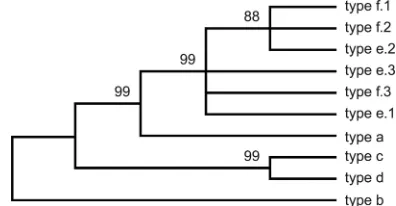
Maximum parsimony phylogenetic analysis produced 45 equally most parsimonious unrooted trees (length⫽148) that varied only slightly in topology. Based upon the 100% consen-sus tree (Fig. 3), type e and f sequences were closely related and remained unresolved; bootstrap branch support for the type e and type f cluster was 99% (based upon 5,000 repli-cates). Type abexBwas a sister to the group composed of type e and fbexBsequences, and the branch support for the type a, e, and f grouping was 99%. Type c and dbexBsequences were 100% identical to each other at the nucleotide level.
DISCUSSION
Current molecular capsule typing methods rely in part on
bexA, which encodes a protein important in capsule exporta-tion, as a marker for thecaplocus; these methods are reliable for the many strains that possess two copies of thecaplocus (one complete copy and a second copy with a deletion inbexA). SomeH. influenzaestrains, however, contain a singlecaplocus with a partially deletedbexAgene (Fig. 1C) that is not detected with currently usedbexAprimers. Accurate determination of the capsule locus status of these strains requires seven PCRs for each strain, one forbexAand then, for negative samples, testing for eachcap-specific region II gene. We have demon-strated thatbexB, which is located adjacent tobexAin region I of the capsule locus and encodes another protein important in capsule exportation, is a more reliable marker of the capsule locus because it can be detected inH. influenzaestrains that possess a singlecaplocus and abexAmutation in that locus. Importantly, none of the 76capregion-positive strains in this study werebexApositive andbexBnegative, demonstrating a high level of concordance between bexB positivity and the presence of type-specificcapregion genes. The ability to iden-tify true NTHI strains by using a singlebexBPCR, as demon-strated in this study, is especially useful for laboratories spe-cifically interested in capsule characterization of large H. influenzaestrain collections.
[image:5.585.42.284.81.190.2]The utility ofbexBas a capsule type-independent marker for the capsule locus was confirmed in this study by using 40H. TABLE 4. Results ofbexAandbexBmicroarray hybridization study
Strain group (n)
No. of strains (captype by PCR)
bexAandbexB positive
bexA and bexB negative
bexA negative andbexB positive
Indeterminatea
Commensal strains (322)
15 (10 type e, 4 type f, 1 NTHI)
300 0 7 (NTHI)
Otitis media strains (107)
4 (f) 100 0 3 (NTHI)
Invasive strains (117)
115 1 (b) 1 (b) 0
a
Indeterminate results withbexAandbexBprobes.
TABLE 5. Concordance ofbexAandbexBresults by PCR and microarray hybridization
Gene and result
No. of strains with
microarray result Total no. of strains Positive Negative
bexA
PCR positive 28 1 29
PCR negative 0 51 51
Total 28 52 80
bexB
PCR positive 28 2 30
PCR negative 0 50 50
Total 28 52 80
on May 16, 2020 by guest
http://jcm.asm.org/
[image:5.585.297.540.90.216.2]influenzaestrains previously serotyped in clinical microbiology laboratories. We identified strains possessingcapregion genes, regardless of capsule type orbexAstatus, based upon the pres-ence of a single bexB amplicon. The capsule type of each
bexB-positive strain was then determined bycap-specific PCR. Two previously described type b capsule-deficient variants, AAr117 and AAr64 (15, 27), were correctly identified asbexA
negative andbexB positive in this study (Fig. 1D; Table 2). Previous studies (2, 24, 31, 32) have clearly documented the poor sensitivity of serotyping to distinguishcapregion-positive fromcapregion-negativeH. influenzaestrains, even in refer-ence laboratories. The most common serotyping error appears to be positive serotype reactions for strains lacking the cap
locus. We identified fivebexA-andbexB-negative strains that
were previously serotype positive in a clinical microbiology laboratory. While a portion of the cap loci of these strains could conceivably have been deleted subsequent to serotype analysis in the clinical microbiology laboratory, we feel that this is unlikely given that these strains were minimally passaged. Furthermore, these strains also lacked region II capsule-spe-cific genes, as assessed by PCR, and thus would be unable to express capsule. Thus, we concluded that these five strains (Table 3) reflect false-positive serotyping results.
The bexBPCR-based test identified an otitis media type f capsule-deficient variant that lackedbexAand would not have been identified using onlybexAprimers for the PCR (Table 3). In addition, thebexBmicroarray study (Table 4) identified an invasive type b capsule-deficient variant that lackedbexA. Sim-ilarly, Cerquetti et al. (5) described 4 of 41 non-type-b invasive strains that failed to generate an amplicon usingbexAprimers but contained type b-specific regions. Similarly, Nelson and Smith (31), using a multiplex PCR to amplify bexAand the
[image:6.585.44.542.93.217.2]cap-specific gene regions fromH. influenzaeinvasive isolates, identified three strains with an absence ofbexAbut the pres-ence of cap b-specific regions. While such capsule-deficient
FIG. 2. Substitutions withinbexB primer annealing regions. The top row of each sequence set represents the actual PCR primer used (Table 1); dots represent agreement with the primer sequence. Primers were designed using publicly available type b nucleotide sequences. The bexB.1R and bexB.1F primer set amplified bexB from all six capsular types. There is a nucleotide substitution localized to the 3⬘ end of the forward primer (bexB.1R) relative to type abexB, but there were no substitutions in the 3⬘-most region of either primer.
[image:6.585.57.264.440.652.2]FIG. 3. The evolutionary history of bexB was inferred using the maximum parsimony method (7). The 100% consensus tree from the 45 most parsimonious trees (tree length⫽148) is shown. The consis-tency index is 0.965217, the retention index is 0.981651, and the com-posite index is 0.947507 for all sites and the 98 parsimony-informative sites. The percentages of replicate trees in which the associated taxa clustered together in the bootstrap test (5,000 replicates) are shown next to the branches (10). A total of 666 nucleotides were included in the final data set, and 98 nucleotides were parsimony informative. Phylogenetic analyses were conducted in MEGA4 (41), using all codon positions; there were no gaps in the sequence alignment.
TABLE 6. Pairwise percent nucleotide identities and maximum composite likelihood evolutionary distances for a 666-bp region ofH. influenzae bexB
H. influenzae type
% Nucleotide identity or maximum composite likelihood distance for typea
a b c d e.1 e.2 e.3 f.1 f.2 f.3
a 88.1 86.2 87.5 90.7 90.2 90.5 90.4 90.2 90.7
b 0.141 96.7 98.8 86.2 85.7 86.0 85.9 85.7 86.2
c 0.170 0.035 97.6 84.7 84.2 84.5 84.4 84.2 84.7
d 0.150 0.012 0.025 85.6 85.1 85.4 85.3 85.1 85.6
e.1 0.107 0.170 0.192 0.178 99.4 99.8 99.7 99.5 100
e.2 0.113 0.177 0.199 0.186 0.006 99.3 99.7 99.6 99.4
e.3 0.109 0.172 0.194 0.180 0.002 0.008 99.5 99.4 99.8
f.1 0.111 0.175 0.197 0.183 0.003 0.003 0.005 99.8 99.7
f.2 0.113 0.177 0.199 0.185 0.005 0.005 0.006 0.002 99.5
f.3 0.107 0.170 0.192 0.178 0.000 0.006 0.002 0.003 0.005
a
Percent nucleotide identities are listed above the shaded diagonal, and maximum composite likelihood distances (41, 42) are listed below the diagonal. Of the 45 bexBsequences analyzed, 10 were unique.
on May 16, 2020 by guest
http://jcm.asm.org/
[image:6.585.319.517.520.623.2]variants were rare, their identification is important for fully characterizing disease-causing or commensal H. influenzae
strains.
PCR-based screening methods dependent upon “univer-sal” primers may not yield an amplicon if nucleotide poly-morphisms, particularly those localized to the 3⬘ primer annealing regions, preclude adequate annealing of the prim-ers to the target gene region. Indeed, sequence divprim-ersity of
bexAhas been shown to compromise its detection by PCR (35, 47). To assess this potential inbexB, a 666-bp internal region ofbexB, which encompassed the entire bexB.1F and bexB.1R primer annealing sites, was PCR amplified from 45
bexB-positive strains, and the sequences were analyzed. Nu-cleotide substitutions were observed within the primer an-nealing regions across serotypes, but with the exception of the type abexBgene, none of the substitutions were local-ized to the 3⬘end of the primer (Fig. 2). Thus, we concluded that given our current knowledge ofbexBsequence diversity, the bexB.1F-bexB.1R primer pair used in this study will amplifybexBacross all six capsule types.
For testing large numbers of H. influenzae strains, high-throughput hybridization-based methods present an attractive and efficient alternative to PCR screening. Furthermore, probe-based hybridization methods, such as dot blots and microarrays, have the potential to improve the sensitivity of the test because they rely on a much larger region of probe-target complementarity than do PCR-based approaches (567 versus 44 bp in the case of bexB) and thus are less sensitive to sequence variation. In theory, hybridization-based approaches can detect sequences that vary by as much as 15% (L. Zhang, personal communication). To increase the probability of detectingbexB-positive strains across all serotypes, we employed a mixed probe composed of bexB
sequences representing each of the six capsule types (a to f). Using the mixed bexB probe, we demonstrated the utility and accuracy (97.5% bexB concordance with detection by PCR) of this method by screening a large collection of H. influenzae isolates simultaneously, using a genomic DNA microarray hybridization technique.
DNA sequence analyses revealed two distinctbexBsequence groups based upon nucleotide identity, with one containing type c, d, and b strains and the other containing type e and f strains; these groupings are similar to those forbexA(35, 47). Our finding of 100% nucleotide identity inbexBgenes of type b strains is consistent with the 100% identity ofbexAgenes among type b strains reported by Zhou et al. and Sam and Smith (35, 47). In contrast to the 100% nucleotide identity of
bexAsequences among both type e and type f strains reported previously (35, 47), we identified a small amount of nucleotide diversity among bexB sequences of both type e and type f strains (5/666 bp [0.75%] and 3/666 bp [0.45%], respectively). WhilebexBappears to be a reliable marker for the capsule locus, this method is not without limitations. The sequence diversity within thebexBprimer annealing sites across all six serotypes could compromise its use in PCR assays. We noted, however, that all of the 28 strains that hybridized with the mixedbexBprobe on the microarray were also amplified using the bexB.1 PCR primers (Table 5), suggesting that thebexB
primers we used were sufficient to amplify most, if not all,H. influenzae bexBsequences. A second limitation is that targeting
bexBalone cannot differentiate true NTHI strains from strains, such as Rd (21), from which the entire capsule locus has been deleted. The magnitude of this limitation is difficult to assess, as the frequency of naturally occurring, complete cap locus deletions is simply not known. This limitation is, however, not unique to the approach presented here, as none of the avail-able molecular typing methods can detect complete capsule deletions.
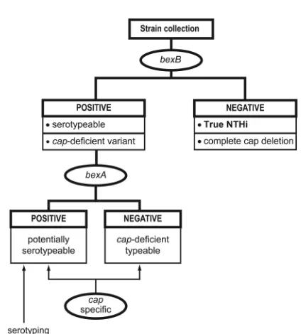
In conclusion, we have described abexB-targeted molecular strategy that rapidly and reliably distinguishes true nontype-ableH. influenzaestrains (i.e., those lacking the capsule locus) from those containing a complete or partialcaplocus (Fig. 4). Ongoing surveillance forH. influenzaeis necessary for assess-ing the organisms associated withH. influenzaetype b vaccine failure and for tracking changes in disease patterns. The ability to accurately classify strains, while challenging, is essential for any successfulH. influenzaesurveillance program, and incor-poratingbexB-based methods into molecular typing schemes can simplify strain characterization while simultaneously im-proving the ability to detect genetically typeableH. influenzae
[image:7.585.313.525.58.293.2](i.e., improved sensitivity). Thus,bexB used as a standalone marker for thecaplocus provides a valuable addition to the armamentarium of H. influenzae molecular characterization methods aimed at differentiating genetically typeable strains from true NTHI strains.
FIG. 4. Suggested workflow for characterizingH. influenzaestrain collections with regard to the capsule locus. Gene names listed within an oval represent detection of that gene via a PCR- or probe-based technique. Identification of “true” NTHI strains can be achieved by screening for the presence ofbexB; strains lackingbexBrepresent true NTHI strains or typeable strains in which the entire capsule locus has been deleted. In the second step, the presence ofbexAdifferentiates potentially serotypeable strains (bexApositive) from capsule-deficient but genetically typeable strains (nonserotypeable,bexAnegative,cap locus positive). Among bexB-positive strains, capsular type can be assigned by screening for capsule-specific genes residing within region II of the capsule locus (8). Finally, if necessary, production of a functional capsule can be verified with traditional type-specific serotyping (8).
on May 16, 2020 by guest
http://jcm.asm.org/
ACKNOWLEDGMENTS
We thank Sarah Satola and Monica Farley for providing type a strains.
This work was supported with funding from the Interdisciplinary Training Program in Infectious Disease (TA32AI049816), the Molec-ular Mechanisms of Microbial Pathogenesis Training Program (AI007528), and the National Institutes of Health (R01-DC05840 and R01-HL083893).
REFERENCES
1.Anderson, P., J. Pitt, and D. H. Smith.1976. Synthesis and release of polyribophosphate byHaemophilus influenzaetype b in vitro. Infect. Immun.
13:581–589.
2.CDC.2002. Serotyping discrepancies inHaemophilus influenzaetype b dis-ease, United States, 1998–1999. MMWR Morb. Mortal. Wkly. Rep.51:706– 707.
3.Cerquetti, M., et al.2005. Presence of multiple copies of the capsulation b locus in invasiveHaemophilus influenzaetype b (Hib) strains isolated from children with Hib conjugate vaccine failure. J. Infect. Dis.192:819–823. 4.Cerquetti, M., et al.2006. Genetic diversity of invasive strains of
Haemophi-lus influenzaetype b before and after introduction of the conjugate vaccine in Italy. Clin. Infect. Dis.43:317–319.
5.Cerquetti, M., et al. 2000. Characterization of non-type bHaemophilus influenzaestrains isolated from patients with invasive disease. The HI Study Group. J. Clin. Microbiol.38:4649–4652.
6.Ecevit, I. Z., et al.2004. Prevalence of thehifBC,hmw1A,hmw2A,hmwC, andhiagenes inHaemophilus influenzaeisolates. J. Clin. Microbiol.42:3065– 3072.
7.Eck, R., and M. O. Dayhoff.1966. Atlas of protein sequence and structure. National Biomedical Research Foundation, Silver Spring, MD.
8.Falla, T. J., et al.1994. PCR for capsular typing ofHaemophilus influenzae. J. Clin. Microbiol.32:2382–2386.
9.Farjo, R. S., et al.2004. Diversity and sharing ofHaemophilus influenzae strains colonizing healthy children attending day-care centers. Pediatr. In-fect. Dis. J.23:41–46.
10.Felsenstein, J.1985. Confidence limits on phylogenies: an approach using the bootstrap. Evolution39:783–791.
11.Fleischmann, R. D., et al.1995. Whole-genome random sequencing and assembly ofHaemophilus influenzaeRd. Science269:496–512.
12.Giufre, M., R. Cardines, P. Mastrantonio, and M. Cerquetti.2006. Variant IS1016 insertion elements in invasiveHaemophilus influenzaetype b isolates harboring multiple copies of the capsulation b locus. Clin. Infect. Dis.43:
1225–1226.
13.Harrison, A., et al.2005. Genomic sequence of an otitis media isolate of nontypeableHaemophilus influenzae: comparative study withH. influenzae serotype d, strain KW20. J. Bacteriol.187:4627–4636.
14.Hoiseth, S. K., C. J. Connelly, and E. R. Moxon.1985. Genetics of sponta-neous, high-frequency loss of b capsule expression inHaemophilus influen-zae. Infect. Immun.49:389–395.
15.Hoiseth, S. K., and J. R. Gilsdorf.1988. The relationship between type b and nontypableHaemophilus influenzaeisolated from the same patient. J. Infect. Dis.158:643–645.
16.Hoiseth, S. K., E. R. Moxon, and R. P. Silver.1986. Genes involved in Haemophilus influenzaetype b capsule expression are part of an 18-kilobase tandem duplication. Proc. Natl. Acad. Sci. U. S. A.83:1106–1110. 17.Kapogiannis, B. G., S. Satola, H. L. Keyserling, and M. M. Farley.2005.
Invasive infections withHaemophilus influenzaeserotype a containing an IS1016-bexA partial deletion: possible association with virulence. Clin. In-fect. Dis.41:e97–e103.
18.Kong, F., M. Brown, A. Sabananthan, X. Zeng, and G. L. Gilbert.2006. Multiplex PCR-based reverse line blot hybridization assay to identify 23 Streptococcus pneumoniae polysaccharide vaccine serotypes. J. Clin. Micro-biol.44:1887–1891.
19.Kroll, J. S., I. Hopkins, and E. R. Moxon.1988. Capsule loss inH. influenzae type b occurs by recombination-mediated disruption of a gene essential for polysaccharide export. Cell53:347–356.
20.Kroll, J. S., B. Loynds, L. N. Brophy, and E. R. Moxon.1990. Thebexlocus in encapsulatedHaemophilus influenzae: a chromosomal region involved in capsule polysaccharide export. Mol. Microbiol.4:1853–1862.
21.Kroll, J. S., B. M. Loynds, and E. R. Moxon.1991. TheHaemophilus influ-enzaecapsulation gene cluster: a compound transposon. Mol. Microbiol.
5:1549–1560.
22.Kroll, J. S., and E. R. Moxon.1988. Capsulation and gene copy number at the cap locus ofHaemophilus influenzaetype b. J. Bacteriol.170:859–864.
23.Kroll, J. S., S. Zamze, B. Loynds, and E. R. Moxon.1989. Common orga-nization of chromosomal loci for production of different capsular polysac-charides inHaemophilus influenzae. J. Bacteriol.171:3343–3347.
24.LaClaire, L. L., et al.2003. Identification ofHaemophilus influenzae sero-types by standard slide agglutination serotyping and PCR-based capsule typing. J. Clin. Microbiol.41:393–396.
25.Lacross, N. C., C. F. Marrs, M. Patel, S. A. Sandstedt, and J. R. Gilsdorf.
2008. High genetic diversity of nontypeableHaemophilus influenzaeisolates from two children attending a day care center. J. Clin. Microbiol.46:3817– 3821.
26.Maaroufi, Y., J. M. De Bruyne, C. Heymans, and F. Crokaert.2007. Real-time PCR for determining capsular serotypes ofHaemophilus influenzae. J. Clin. Microbiol.45:2305–2308.
27.McCrea, K. W., J. L. Sauver, C. F. Marrs, D. Clemans, and J. R. Gilsdorf.
1998. Immunologic and structural relationships of the minor pilus subunits amongHaemophilus influenzaeisolates. Infect. Immun.66:4788–4796. 28.McCrea, K. W., et al.2010. Prevalence of thesodCgene in nontypeable
Haemophilus influenzaeandHaemophilus haemolyticusby microarray-based hybridization. J. Clin. Microbiol.48:714–719.
29.Mukundan, D., Z. Ecevit, M. Patel, C. F. Marrs, and J. R. Gilsdorf.2007. Pharyngeal colonization dynamics ofHaemophilus influenzaeand Haemophi-lus haemolyticusin healthy adult carriers. J. Clin. Microbiol.45:3207–3217. 30.Murphy, T. F., and M. A. Apicella.1987. NontypableHaemophilus influen-zae: a review of clinical aspects, surface antigens, and the human immune response to infection. Rev. Infect. Dis.9:1–15.
31.Nelson, K. L., and A. L. Smith.2010. Determination of capsulation status in Haemophilus influenzaeby multiplex polymerase chain reaction. Diagn. Mi-crobiol. Infect. Dis.66:235–240.
32.Ogilvie, C., et al.2001. Capsulation loci of non-serotype b encapsulated Haemophilus influenzae.J. Infect. Dis.184:144–149.
33.Pittman, M.1931. Variation and type specificity in the bacterial species Haemophilus influenzae. J. Exp. Med.53:471–492.
34.Saeed, A. I., et al.2003. TM4: a free, open-source system for microarray data management and analysis. Biotechniques34:374–378.
35.Sam, I. C., and M. Smith.2005. Failure to detect capsule genebexAin Haemophilus influenzaetypes e and f by real-time PCR due to sequence variation within probe binding sites. J. Med. Microbiol.54:453–455. 36.Sandstedt, S. A., et al.2008. Comparison of laboratory-based and
phyloge-netic methods to distinguish betweenHaemophilus influenzaeandH. hae-molyticus. J. Microbiol. Methods75:369–371.
37.Satola, S. W., P. L. Schirmer, and M. M. Farley.2003. Complete sequence of the cap locus ofHaemophilus influenzaeserotype b and nonencapsulated b capsule-negative variants. Infect. Immun.71:3639–3644.
38.Satola, S. W., P. L. Schirmer, and M. M. Farley.2003. Genetic analysis of the capsule locus ofHaemophilus influenzaeserotype f. Infect. Immun.71:7202– 7207.
39.St. Sauver, J., et al. 2000. Risk factors for otitis media and carriage of multiple strains ofHaemophilus influenzaeandStreptococcus pneumoniae. Emerg. Infect. Dis.6:622–630.
40.Sukupolvi-Petty, S., S. Grass, and J. W. St. Geme III.2006. TheHaemophilus influenzaetype bhcsAandhcsBgene products facilitate transport of capsular polysaccharide across the outer membrane and are essential for virulence. J. Bacteriol.188:3870–3877.
41.Tamura, K., J. Dudley, M. Nei, and S. Kumar.2007. MEGA4: molecular evolutionary genetics analysis (MEGA) software version 4.0. Mol. Biol. Evol.
24:1596–1599.
42.Tamura, K., M. Nei, and S. Kumar.2004. Prospects for inferring very large phylogenies by using the neighbor-joining method. Proc. Natl. Acad. Sci. U. S. A.101:11030–11035.
43.Thompson, J. D., D. G. Higgins, and T. J. Gibson.1994. CLUSTAL W: improving the sensitivity of progressive multiple sequence alignment through sequence weighting, position-specific gap penalties and weight matrix choice. Nucleic Acids Res.22:4673–4680.
44.Ueno, K., J. Nishi, N. Imuta, K. Tokuda, and Y. Kawano.2010. Presence of multiple copies of capsulation loci in invasiveHaemophilus influenzaetype b (Hib) strains in Japan before introduction of the Hib conjugate vaccine. Microbiol. Immunol.54:160–163.
45.Wallace, R. J., Jr., et al.1981.Haemophilus influenzaeinfections in adults: characterization of strains by serotypes, biotypes, and beta-lactamase pro-duction. J. Infect. Dis.144:101–106.
46.Zhang, L., et al.2004. Library on a slide for bacterial comparative genomics. BMC Microbiol.4:12.
47.Zhou, J., D. K. Law, M. L. Sill, and R. S. Tsang.2007. Nucleotide sequence diversity of thebexAgene in serotypeableHaemophilus influenzaestrains recovered from invasive disease patients in Canada. J. Clin. Microbiol.45:
1996–1999.