Journal of Chemical and Pharmaceutical Research, 2019, 11(2): 22-31
Research Article
CODEN(USA): JCPRC5
ISSN: 0975-7384
22
Synthesis, Characterisation and Biomedical Application of Random
Copolyester Using 1,4-Dithiane 2,5-Diol
Kalpana B
*and Nanthini R
Department of Chemistry, Pachaiyapps's college, Chennai, India
____________________________________________________________________________
ABSTRACT
A New random linear copolyester Poly(1,4-dithiane 2,5-diol succinate-co-1,10 decane diol succinate), PDDS was synthesized by direct melt polycondensation method using 1,4-dithiane 2,5-diol and 1,10 decane diol and Succinic acid with Titanium tetra isopropoxide as catalyst. As the polymers with sulphur atom in the main chain has excellent properties and wider applications especially in making lens, it led us to synthesis a copolyester with sulphur moiety in main chain and study its biomedical property. A thorough literature survey revealed that 1,4-dithiane 2,5-diol is used as a monomer in the synthesis of polyurethane sealing compound and used as an biocontrol agent, crosslinking agent and chain extenders in polymerisation technique. The Synthesised copolyester PDDS was characterized by determining its inherent viscosity, Solubility, Glass transistion temperature (DSC), crystalline nature (XRD), FTIR, 1H-NMR and 13C-NMR spectroscopy. The biomedical properties such as antioxidant, antimicrobial and in vitro cytotoxicity against normal (Vero cell line) and cancer (A549 lung cancer cell line) by MTT Assay of synthesized copolyester PDDS were also studied.
Keywords: Copolyester; Polycondensation; 1,4-Dithiane 2,5-diol; Cytotoxic activity
_____________________________________________________________________________
INTRODUCTION
Polyester with sulphur atom in main chain was produced by reaction between sulphonic diols such as 4,4‘
thiodiphenol with aliphatic diacid chlorides having methylene groups from 2 to 10. These polyesters possess high
molecular weight and high thermo resistance [1-4]. The introduction of sulphur in main chain of linear polyester has
a remarkable increase in adhesive property [5]. A polymerisable composition comprising of polythiol with high
sulphur content and alicyclic polyisocyanate containing 1,4-dithiane derivative provides an optical material with
23
polysulphide based resin having polythiol and a compound containing iso(thio)cyanato group has been produced
using 1,4-dithiane derivates and the resin had extremely low dispersion properties and high refractive index and find
wide application as a optical, glazing, coating and adhesive material [7]. A polyurethane (Diol+Isocyanate)
biomaterial with sacrificial moiety susceptible to oxidation especially Sulphur containing moiety (1,4-dithiane
2,5-diol as a 2,5-diol) in backbone of the polymer chain has increased tensile strength and modulus of elasticity and also
degrade over time in an oxidizing environment or body, hence these biomaterial are used in making medical devices
particularly for use as insulation on pacing leads [8]. It was further noticed that polyurethanes are widely used in
implantable devices such as artificial hearts, cardiovascular catheters, pace maker lead insulations etc. [9]. As the
literature survey reveals that polymers with sulphur atom has excellent properties, a copolyester using 1,4-dithiane
2,5-diol was prepared by direct melt polycondensation method and its characteristics and biomedical properties such
as antioxidant, antimicrobial and cytotoxicity against normal and A549 cell line were studied.
EXPERIMENTAL SECTION
Materials and Methods
1,4-Dithiane 2,5-diol (Sigma Aldrich), Succinic acid (Sigma Aldrich), 1,10 decane diol (Sigma Aldrich) and
Titanium Tetra isopropoxide (Lancaster) were purchased and used. All other solvents and chemicals (AR Grade)
were used as such. The inherant viscosity was determined using ubbelohde viscometer. Perkin Elmer 883
spectrophotometer was used to record the FT-IR Spectra of synthesised copolyester. Using CDCl3 as a solvent the
1H-NMR and 13C-NMR spectra for copolyester was recorded on a Bruker 400 MHz and Bruker 100 MHz
Spectrometer respectively. DSC thermogram was recorded on DSC Q200 V23.10 Build 79 Differential Scanning
Calorimeter. To study the crystalline nature of the synthesised copolyester Wide angle XRD was taken by Bruker
B8 wide angle XRD with Cu/30 kv/15 mA. The In vitro cytotoxicity against normal (Vero cell line) and cancer (A549
lung cancer cell line) by MTT assay [10] and antioxidant activity by DPPH Scavenging Assay [11] and
antimicrobial activity by Well diffusion method [12] have been determined.
Synthesis of Copolyester
In a three necked round bottom flask 1,4-dithiane 2,5-diol (0.01 mole), Succinic acid (0.02 mole) and 1,10 decane
diol (0.01 mole) is taken and the left inlet is connected to nitrogen cylinder, middle inlet to the guard tube and the
right inlet is closed with stopper. Then the mixture heated in an oil bath, after complete melting of mixture about 0.8
ml of Titanium Tetra isopropoxide is added and the temperature is maintained at 185°C for one hour with stirring.
The temperature is then increased to about 194°C and maintained at that temperature for two hours. The crude
copolyester obtained is dissolved in chloroform and poured in ice cold methanol to reprecipitate the pure polyester
as shown in Figure 1.
RESULTS AND DISCUSSION
Solubility and Viscosity Measurement
Solubility of the copolyester PDDS in solvents like DMF, DMSO, THF and CHCl3 was determined and it has been
fouud to be soluble in almost all the solvents. The inherent viscosity of the Copolyester PDDS was determined using
Ubbelohde viscometer. By determining the flow time of the solvent and 1% solution of the copolyester in CHCl3 at
24
FT-IR Spectral Analysis
The IR spectra recorded for copolyester are presented in Figure 1. The IR spectra of the synthesized copolyester
showed a strong absorption band at around 1701.12 cm-1, which is characteristic absorptions of carbonyl stretching
vibration of ester groups and thus confirmed the formation of polyesters. The absorption band at 2986.07 was
assigned to methylene (-CH2-) groups for the diacids/diols while the band at 1122, 573.15 and 947 cm-1 was
attributed to C-O-C asymmetric stretching, C-S stretching and aliphatic C-C stretching respectively.
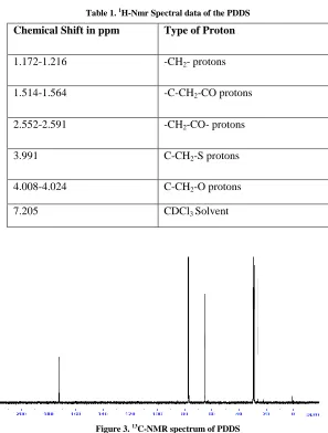
1H-NMR and 13C-NMR Spectral Analysis
The 1H-NMR and 13C-NMR Spectra of the copolyester PDDS are shown in Figures 2 and 3. The assignments of
characteristic peak in the 1H-NMR and 13C-NMR spectra of the copolymer are given the Tables 1 and 2 respectively.
All these chemical shift values show the random distribution of monomers in the synthesized copolyester.
DSC Analysis
The DSC thermogram of the synthesied copolyester PDDS are presented in Figure 4. The thermogram shows the
glass transition temperature as – 50°C and the melting point temperature as 66.13°C.
X-ray Diffraction Studies
The Wide angle XRD determines the degree of crystallinity of the polymer. The diffractogram of the synthesized
PDDS are shown in Figure 5. The diffractogram shows that the synthesized copolyester is amorphous in nature.
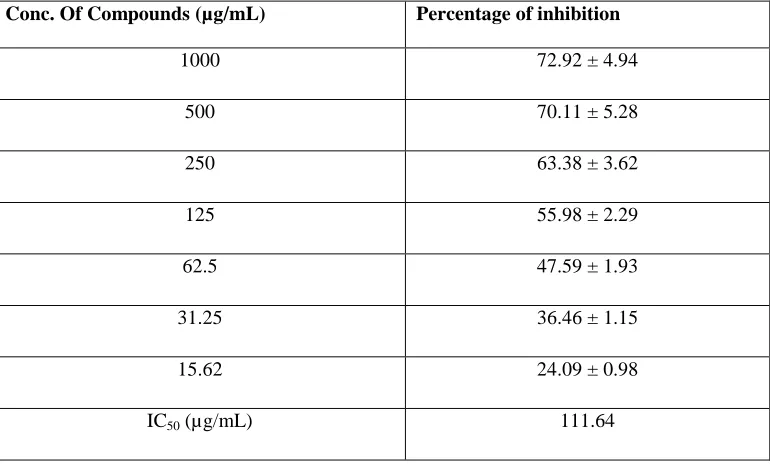
Antioxidant Analysis
Table 3 shows the DPPH scavenging activity of the copolyester PDDS. Antioxidant compounds are used as food
additives and in preventing lifestyle related diseases and ageing [13]. Lower the value of IC50, greater is the
antioxidant property. The IC50 value (111.64) of the synthesized copolyester by DPPH Scavenging Assay shows that
it has good antioxidant property.
Antimicrobial Activity
The antimicrobial activity of Copolyesrer PDDS against two gram negative and gram positive bacteria by well
diffusion method is given in the Table 4 and Figure 6. The copolyester exhibits inhibition range from 10.0-14.0 mm
for the human pathogens which shows that the synthesized copolyester PDDS have excellent antimicrobial activity.
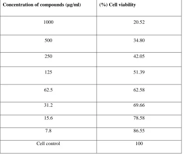
Cytotoxic Activity
The MTT assay of the PDDS against the normal and A549 cell line at various concentrations were carried out and the
results are given in Tables 5 and 6. The concentration required for 50% inhibition of viability IC50 were determined
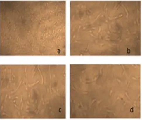
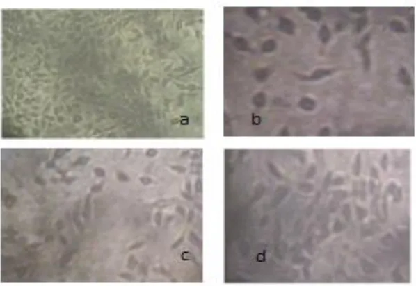
graphically as shown in Figure 7. The affected normal and A549 cells at various concentrations is shown in Figures 8
and 9. The effect of the PDDS on the normal and A549 cell line was expressed in terms of % cell viability. It is
explicit from Tables 5 and 6 that, low concentration of PDDS induced more anticarcinogenic activity on A549 cell
line than normal vero cell line.
CONCLUSION
The Copolyester PDDS was synthesised by direct melt polycondensation method and it showed good solubility in
various organic solvents. The probable structure of the copolyester was confirmed by FT-IR and NMR spectroscopy.
25
copolyester revealed that it is amorphous in nature. The excellent antioxidant, antimicrobial and cytotoxic activity of
[image:4.612.111.493.90.662.2]copolyester indicates that it has versatile application in biomedical field.
Figure 1. FT-IR spectra of PDDS
26
Table 1. 1H-Nmr Spectral data of the PDDS
Chemical Shift in ppm Type of Proton
1.172-1.216 -CH2- protons
1.514-1.564 -C-CH2-CO protons
2.552-2.591 -CH2-CO- protons
3.991 C-CH2-S protons
4.008-4.024 C-CH2-O protons
7.205 CDCl3 Solvent
Figure 3. 13C-NMR spectrum of PDDS
Table 2. 13C-NMR spectral data of PDDS
Chemical Shift in ppm Carbon Assignment
21.79-25.86 -CH2-
28.57-29.42 -CH2-S-
63.03-64.98 -CH-S-
76.73-77.36 -CH2-O-
27
[image:6.612.198.414.83.404.2]Figure 4. DSC Thermogram of PDDS
[image:6.612.115.500.423.655.2]Figure 5. X-Ray Diffractogram of PDDS
Table 3. In vitro Antioxidant Activity of PDDS by DPPH Scavenging Assay
Conc. Of Compounds (µg/mL) Percentage of inhibition
1000 72.92 ± 4.94
500 70.11 ± 5.28
250 63.38 ± 3.62
125 55.98 ± 2.29
62.5 47.59 ± 1.93
31.25 36.46 ± 1.15
15.62 24.09 ± 0.98
IC50 (µg/mL) 111.64
20 40 60 80
0 500 1000 1500
20 40 60 80
28
Figure 6. Antimicrobial activity of Escherichia coli, Klebsiella pneumonia, Bacillus subtilis and Staphylococcus aureus
Table 4. Antimicrobial activity of PDDS by well diffusion method
Human Pathogens Concentration (in
µg/mL)
Zone of inhibition in mm
(Percentage of Inhibition)
Standard Drugs
Tetracycline/(30 µg/mL)
Types of
Organisms
Escherichia coli
1000 12 ± 0.84 (13.33 ± 0.93)
18 (20)
Gram negative
Bacteria
500 11 ± 0.77 (12.22 ± 0.85)
250 -
Klebsiella pneumoniae
1000 14 ± 0.98 (15.55 ± 1.01)
24 (26.66)
500 12 ± 0.84 (13.33 ± 0.93)
250 -
Bacillus subtilis
1000 13 ± 0.91 (14.44 ± 1.01)
32 (35.55)
Gram Positive
Bacteria
500 -
250 -
Staphylococcus aureus
1000 14 ± 0.98 (15.55 ± 1.01)
31 (34.44)
500 10 ± 0.70 ( 11.11 ± 0.77)
[image:7.612.43.576.216.601.2]29
Table 5. Cytotoxic Activity of PDDS on Vero (normal) cell line
Concentration of compounds (µg/ml) (%) Cell viability
1000 51.37
500 58.64
250 63.41
125 69.06
62.5 74.71
31.2 80.49
15.6 88.83
7.8 92.60
Cell control 100
IC 50 value (µg/mL) 1143.90
Table 6. Cytotoxic Activity of PDDS against Lung cancer (A549) cell line
Concentration of compounds (µg/ml) (%) Cell viability
1000 20.52
500 34.80
250 42.05
125 51.39
62.5 62.58
31.2 69.66
15.6 78.58
7.8 86.55
[image:8.612.123.492.406.718.2]30
Figure 7. Graphical representation of activities of PDDS in MTT Assay
Figure 8. In vitro cytotoxic activity of PDDS on Normal vero cell line at different concentrations (a) Normal cell, (b) 7.8 µg/ml, (c) 500
[image:9.612.189.432.347.553.2]31
Figure 9. In vitro cytotoxic activity of PDDS on A549 (lung cancer) cell line at different concentrations (a) Normal cell, (b) 1000 µg/ml, (c)
125 µg/ml and (d) 7.8 µg/ml
ACKNOWLEDGEMENT
The authors would like to thank VIT and CLRI technical persons for their analytical support.
REFERENCES
[1] D Greszta-Franz; HJ Laas; J Krause; D Mager, 2016.
[2] R Xie; D Breed; P Ansems; W Zhou, 2011.
[3] Y Soon Kim; J-C Kim; H H Seo; G J Choi. Ae Ran Park, 2008.
[4] H Hirano; Watase S; Tanaka M. J appl polymer sci. 2004, 91, 1865-1872.
[5] H Hirano; J Kadota; Y Agari; T Harada; M Tanaka; K Hasegawa. Pol Eng Sci, 2007, 47, 3.
[6] T Mamoru; K Shigetoshi; K Seiichi; K Yoshinobu. Polythiol, polymerizable composition, resin and lens,
and process for preparing thiol Compound. EP 1 138 670 B1.
[7] K Okazaki; C Shimakawa; M Tanaka; Y Kanemura; T Nagata; Y Irizato; Kanagawa; Shinichi Umeda.
Polysulfide-Based Resin Composition, Polysulfide-Based Resin, And Optical Material Comprising The
Resin, 1999.
[8] E Didomenico; DL Miller; ME Benz. Polyurethane and polyurea biomaterials for use in Medical Devices.
[9] AJ Coury et al. Adv Urethane Sci Technol. 1984, 9, 130-168. [10]Mosmann T. J Immunol Methods.1983, 65, 55-63.
[11]MS Blois. Nature, 1958, 181, 1199-1200.
[12]C Perez; M Pauli; P Bazerque. Acta Biol Med Exp. 1990, 15, 113-115.