This is an open access journal, and articles are distributed under the terms of the Creative Commons Attribution-Non Commercial-ShareAlike 4.0 License, which allows others to remix, tweak, and build upon the work non-commercially, as long as appropriate credit is given and the new creations are licensed under the identical terms.
© 2018 Journal of Advanced Pharmacy Education & Research | Published by SPER Publication 33
Ameliorative effect of grape seed and ginkgo biloba against
pulmonary damage induced by amiodarone in male albino rats
Sanaa Reda Galaly
*, Manal Abdul-Hamid, Hanaa Mahmoud, Fatma Mostafa
Department of Zoology, Faculty of Science, Beni-Suef University, Beni-Suief, Egypt.
Correspondence: Sanaa Reda Galaly, Department of Zoology, Faculty of Science, Beni-Suef University, Beni-Suief, Egypt. E-mail: [email protected]
ABSTRACT
Amiodarone being an orally effective antiarrhythmic drug widely used throughout the world, had long-term administration side effects such as pulmonary toxicity. Thirty six male albino rats were divided into six equal groups, and doses of different solutions were given to them by gastric tube every day for 8 weeks as follow; the 1st group (G1) which was considered as an untreated control group under the same laboratory conditions, was given distilled water, the 2nd group (G2) was given (100 mg/kg/day) of grape seed , the 3rd group (G3) received (100 mg/kg/day) of ginkgo biloba, the 4th group (G4) amiodarone-treated group that was given (40 mg/kg/day), the 5th group (G5) received amiodarone along with grape seed at the same time, and the 6th group (G6) was given amiodarone parallel with ginkgo biloba at the same time for 8 weeks. The current histological study revealed that amiodarone caused marked changes in the lung including peribronchiolar hyperplasia and inflammatory cells infiltration in addition to thicking of inter-alveolar septa moreover, ultra-structural observations in the lung including vacuolation, degeneration of microvilli and pyknotic nuclei. In addition, histochemical study revealed the depletion of glycogen, and comet assay revealed marked DNA damage. Treatment with the two used antioxidants (grape seed and Ginkgo biloba) reduced the extent of lung damage induced by amiodarone. These antioxidants ameliorated the histopathological structure, increased the contents of glycogen, and improved the ultrastructure alternations of the lung tissue. In conclusion, grape seed is markedly more effective than Ginkgo biloba in protecting rats against amiodarone.
Keywords: Amiodarone, grape seed, ginkgo biloba, comet assay, pulmonary toxicity, histopathology, ultrastructure.
Introduction
Fast rhythms of the heart were suppressed by a group of pharmaceuticals called antiarrhythmic drugs [1]. Amiodarone
was one of the oral effective antiarrhythmic drugs [2], and the
most efficacious anti-dysrhythmic drug [3] which was used all
over the world [4]. The therapeutic dose of human for
amiodarone was recorded to be 600 mg/day [5], and the
calculated therapeutic dose for adult albino rat was (40 mg/kg/day) [6].
Progressive tissue accumulation of amiodarone was the cause of most adverse effects of oral amiodarone such as pulmonary toxicity [7]. Direct cellular toxicity may be due to the inhibition
of lysosomal phospholipase, which results in the accumulation of lysosomal phospholipids leading to cell death [8] or may also
resulted from amiodarone-induced free radical reactions [9].
Amiodarone caused toxicity to lung cells even at therapeutic dose, and was accumulated in lung tissue with 100 - 500 fold of concentration compared with the serum concentration, also the amiodarone clearance in the lung tissue was slow and the compound can last in the lung up to 1 year after cessation of treatment [10]. Pulmonary toxicity by amiodarone involved the
induction of apoptosis in alveolar epithelial cells where the apoptosis was a programmed cell death that played a crucial role in normal development and tissue homeostasis [11].
Approximately 5 % of amiodarone treated patients were affected by pulmonary toxicity of amiodarone, the development of the lung complications appeared to be associated with older age, the duration of the treatment, the cumulative dosage, the high levels of its desethylmetabolite, and probably pre-existing lung diseases as well as co-existing respiratory infections [12].
Antioxidants protectd cells against oxidative damage of amiodarone [13]. The administration of antioxidants (grape seed
or Ginkgo biloba) along with amiodarone reduced lysosomalphospholipidosis [14].
Access this article online
Website:
www.japer.in
E-ISSN: 2249-3379How to cite this article: Sanaa Reda Galaly, Manal Abdul-Hamid, Hanaa Mahmoud, Fatma Mostafa, Ameliorative Effect of Grape Seed and Ginkgo Biloba Against Pulmonary Damage Induced by Amiodarone in Male Albino Rats. J Adv Pharm Edu Res 2018;8(2):33-42.
Source of Support: Nil, Conflict of Interest: None declared.
Grape seed extract had potent antioxidant and anti-inflammatory effects, where grape seed extract significantly eliminated the negative pathological changes of lung [15],
suppressed airway resistance, and reduced the total inflammatory cell and eosinophil counts [16].
Ginkgo biloba extract reversed the oxidative damage induced by mercury in lung with its antioxidant effects [17], also it has
protective effects against lung damage induced by irradiation
[18], and was used for the treatment of cancer thousands of years
ago in China [19].
The aim of this study was to evaluate the possible protective effect of grape seed and Ginkgo biloba against the pulmonary toxicity induced by amiodarone through histological, histochemical, ultrastructural, biochemical studies and comet assay.
Materials and Methods
Experimental animals
In this study, we used thirty-six white male albino rats (Rattusnorvegicus) from breeding unit of Egyptian Organization for Biological Vaccine Production weighing (140 - 180 g), housed at room temperature (25 ± 5 ºC) in stainless steel cages and obtained complete diet pellets and water ad-libitum [20]. Before the beginning of the experiment, we kept
animals under observation for about two weeks to exclude any inter-current infections. In the current study, we followed Directive of European Community (86/ 609/ the 8 th edition
of EEC) and the national rules on animal care that was executed in accordance with the care and use of Laboratory Animals (NIH Guidelines). This was certified by the committee of Zoology Department, Beni-suef University, Egypt.
Chemicals and treatment
Amiodarone (amiodarone hydrochloride) was purchased from the company of Global Napi for pharmaceuticals products under permitting of SanofiSynthel abo. Its brand name is Cordarone tablets, and each tablet contained 200 mg.
Grape seed was purchased from the Arab Company for Pharmceuticals and Medicinal Plants (MEPACO MEDIFOOD). Its common name is Gervital tablets, and each tablet contained 150 mg from grape seed extract.
Ginkgo biloba was purchased from EIMC United Pharmaceuticals for EMA pharm pharmaceuticals. Its common name is ginkgo biloba tablets, and each tablet contained 260 mg from Ginkgo biloba leaf powder extract.
Animal grouping
Each of amiodarone, grape seed or Ginkgo biloba was dissolved in distilled water, and was given by gastric intubation to the rats, daily for eight weeks (time of the experiment). The animals were divided into six groups; the1st group (G1) serving
as the control group under the same laboratory conditions, and was given distilled water. The 2nd group (G2) received (100
mg/kg/day) of grape seed [21]. The 3rd group (G3) received
(100 mg/kg/day) of Ginkgo biloba [22]. The 4th group (G4)
received (40 mg/kg/day) of amiodarone [23]. The 5th group
(G5) received (40 mg/kg/day) of amiodarone parallel with grape seed at the same time. The 6th group (G6) received (40
mg/kg/day) of amiodarone parallel with Ginkgo biloba at the same time.
The animals were sacrificed 8 weeks’ post-treatment. The lungs were freshly separated, washed and used for the histological, histochemical, ultra structural studies and comet assay.
Comet assay:
The alkaline comet assay was performed as described by [24]. To
obtain single cells from liver, the sample must be finely minced using sterile scissors or scalpels, cell dispersion can be achieved by enzymatic digestion of the sample using collagenase. A freshly prepared 10 µL of single liver cells (10,000 - 50,000) in cold Hank’s Balanced Salt Solution, (HBSS) was mixed with 65 µL of 0.7 low melting point agarose (LMA) at 37 °C, and spread onto microscope slide pre -coated with 0.5 % normal melting point agarose (NMA), and the slide was covered with a third layer of LMA, and a cover slip was applied to spread the sample.
The cells then lysed in lysis buffer consisting of 1 % sodium sarcosinate, 2.5 M NaCl, 10 mM Na2 EDTA, 1 % Triton x - 100, 10 % DMSO and 10 mMTris, pH 10 for 1 h at 4 °C. After the lysis, the slides were placed in an electrophoresis unit, the DNA was allowed to unwind for 20 min in the electrophoretic solution consisting of 300 mMNaOH, 1 mM EDTA, pH > 13.
Electrophoresis was conducted at ambient temperature of 4 °C for 20 min at electric field strength of 300 mA. 0.4 M Tris, pH 7.5 were used to neutralize the slides, and 2 µg/ml ethidiumbromide was used to stain them, then they were covered with cover slips.
All the steps mentioned above were applied under dimmed light or in the dark in order to prevent additional DNA damage. A leitzorthoplanepifluorescence microscope (magnification 200 x) equipped with an excitation filter of 515 - 550 nm and a barrier fitter of 590 nm was used to observe the slides. The microscope was connected through a camera to a computer - based image analysis system (Comet Assay IV software).
For each sample, 100 isolated comets (single strand breaks of DNA migrate from nucleus to anode) were randomly selected and measured for comet tail length, % DNA in tail, and tail moment according to the definition.
Tail moment = Tail length X % DNA in tail / 100
Histological preparations
Preparation of paraffin sections
For histological preparations, at the end of the 8th week of the
sectioned into small pieces, each about 3 mm3 in size, and then fixed in 10 % neutral buffered formalin for 24 hours.
Lung specimens were washed to remove the excess of the fixative, and then dehydrated in ascending grades of ethyl alcohol, after that, were cleared in two changes of xylene. Then specimens were impregnated with three changes of paraplast plus at 60 ºC.
Prepared sections of (4 to 5µm) were thickened with a microtome, then stained with haematoxylin and eosin [25] for
histological studies, or with Masson's trichrome staining [26],
and examined under light microscope for histopathological assessment [27].
Histochemical studies
The dehydrated tissue specimens-fixed in carnoy in absolute alcohol two times each for one hour- were cleared in two changes for one hour of xylol, and then impregnated in paraplast at 60 ºC.
Sections of (4 - 5 µm) in thickness were prepared using a rotary microtome, hydrated using a descending ethanolic series after deparafinization, and were stained with periodic acid Schiff’s technique (PAS) for elucidation of polysaccharides [28].
Ultrastructural preparations
The specimens cut from the lung of all animal groups into small pieces, each piece measured about 1 mm3, were fixed
immediately at 4 ºC for 18 - 24 hours in freshly 3 % glutraldehyde-formaldehyde.
The specimens washed in phosphate buffer (pH 7.4), were post-fixed for one hour at 4 ºC in isotonic 1 % osmium tetroxide [29]. Prepared sections for electron microscopic
examination followed the method of [30]. Stained semi-thin
sections with toluidine blue were used for detection of the area of interest, then ultrathin sections were prepared by using the ultra-microtome glass knives.
The specimens were stained by uranyl acetate and lead citrate, and then examined by a Joel CX 100 transmission electron microscope.
Statistical analysis
The Statistical Package was used for the Social Sciences (SPSS for WINDOWS, version 20.0; SPSS Inc, Chicago). The data were analyzed using One-way analysis of variance (ANOVA)
[31] followed by Duncan’s multiple range test post hoc analysis
values. Results for each group were expressed as mean (M) ± standard error (SE). Values of P < 0.05 were considered statistically non-significant, while values of P> 0.05 were considered statistically significant, and values of P> 0.001 were considered statistically highly significant.
Results
Detection of DNA damage by comet assay
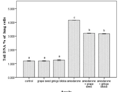
A highly significant (P<0.001) increase in DNA % was revealed in amiodarone treated group when compared with thecontrol group, while the group treated with grape seed plus amiodarone revealed a highly significant (P<0.001) decrease in DNA % when compared with amiodarone group more than the group treated with Ginkgo biloba plus amiodarone (Figure 1). Figure 5 showed DNA in the lung cells by comet assay, where showed normal spots with round shape in control rats (Figure 5a), grape seed treated rats (Figure 5b), and Ginkgo biloba treated rats (Figure 5c). A highly significant (P<0.001) increase in DNA fragmentation (tail length of DNA) in the animals treated with amiodarone compared with the control group appeared as damaged spots where the migrated fragments’ lengths were less than or equal to the basal nuclear DNA diameter; and strongly damaged spots where the comet length was greater than the basal nuclear DNA diameter (tailed) (Figure 5d), while the group treated with the grape seed or Ginkgo biloba plus amiodarone revealed a highly significant (P<0.001) decrease in DNA fragmentation when compared with amiodarone group (Figure 2). Supplementation of the grape seed or Ginkgo biloba respectively with amiodarone for 8 weeks counteracted the effects of amiodarone alone (Figure 5e, 5f), while the animals given grape seed revealed a highly significant (P<0.001) increase in improvement of DNA compared with the control group more than Ginkgo biloba by decreasing the number of damaged spots (tailed % in rat lung cells) and increasing the number of undamaged spots (untailed % in rat lung cells) (Figure 3, 4).
Figure 1: Tail DNA% in rat lung cells of all groups at the end of the experiment
• Number of animals is six for each group.
• Data are expressed as mean ± standard error.
Figure 2: Tail length of DNA in rat lung cells (µm) of all groups at the end of the experiment.
• Number of animals is six for each group.
• Data are expressed as mean ± standard error.
• Data with the same superscript letter are similar (non-significant, P> 0.05), whereas others are significant at P< 0.05, and P< 0.001 is a highly significant effect.
Figure 3: Tailed% in rat lung cellsof all groups at the end of the experiment
• Number of animals is six for each group.
• Data are expressed as mean ± standard error.
• Data with the same superscript letter are similar (non-significant, P> 0.05), whereas others are significant at P< 0.05, and P< 0.001 is a highly significant effect.
Histopathological observations
Microscopic examination of the lung sections of the control, grape seed and Ginkgo biloba treated groups respectively which stained by hematoxylin and eosin (Figure 6a, 6b, 6c) revealed normal histological architecture including blood vessels and numerous clear alveoli with thin interalveolar septa, flattened type I pneumocytes, cuboidal type II pneumocytes. After 8 weeks, amiodarone-treated group showed peribronchiolar hyperplasia (Figure 6d), inflammatory cells infiltration and vacuolation in alveoli (Figure 6e). Supplementation of the grape seed or Ginkgo biloba with amiodarone for 8 weeks counteracted the effects of amiodarone alone, where the group treated with grape seed showed no
aggregation of inflammatory cells or congested blood vessels, and the structure of bronchiole and alveoli appeared normal (Figure 6f), while the group treated with Ginkgo biloba showed less inflammatory cells infiltration; however, the alveoli were normal.
Figure 4: Untailed % in rat lung cells of all groups at the end of the experiment.
• Number of animals is six for each group.
• Data are expressed as mean ± standard error.
• Data with the same superscript letter are similar (non-significant, P> 0.05), whereas others are significant at P< 0.05, and P< 0.001 is a highly significant effect.
Masson's trichrome stain for the control, grape seed and Ginkgo biloba treated group respectively (Figure 7a, 7b, 7c) showed normal distribution of collagen fibers in pulmonary interstitium around bronchiole and alveoli. After 8 weeks, amiodarone-treated group revealed deposition of collagen fibers in pulmonary interstitium around alveolar sacs and in interalveolar septum and severe deposition of collagen fibers around bronchiol (Figure 7d) and the blood vessels (Figure 7e). Supplementation of grape seed or Ginkgo biloba with amiodarone for 8 weeks counteracted the effects of amiodarone alone, where showed mild deposition of collagen fibers in interalveolar septa and the wall of bronchiol and blood vessels (Figure 9f, 9g).
Histochemical observations
The lung sections of control, grape seed and Ginkgo biloba group showed the distribution of PAS-positive material (mainly glycogen) which is represented by the color magenta to red (Figure 8a, 8b, 8c). Amiodarone induced severe depletion of the glycogen content in pulmonary interstitium around alveolar sacs and in interalveolar septum compared to the normal rats (Figure 8d). The administration of grape seed or Ginkgo biloba plus amiodarone resulted in moderate increase in the glycogen content of the lung if compared with amiodarone-treated rats (Figure 8e, 8f).
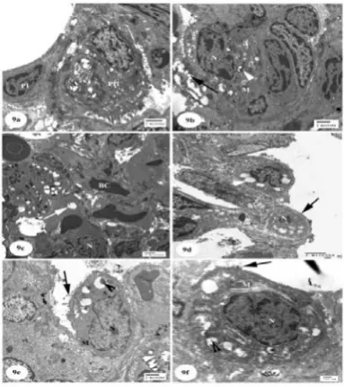
Ultrastructural observations
pneumocyte (Figure 9a), a type II pneumocyte with nucleus, its cytoplasm contained numerous dense mitochondria, lamellar bodies containing electron-dense secretions and short microvilli on the surface of alveoli (Figure 9b).
In rats treated with amiodarone, conspicuous ultrastructure alterations were found in alveolar tissue at the end of the treatment represented by a shrinkage nucleus, congested blood capillaries filled with the blood, vacuolated cytoplasm (Figure 9c), empty lamellar bodies, pyknotic nuclei and degenerated microvilli (Figure 9d).
Ultrastructure study of the rats treated with amiodarone plus grape seed (Figure 9e) or Ginkgo biloba (Figure 9f) revealed a better preservation of such cells compared to those treated with amiodarone alone including short microvilli, partially empty lamellar bodies, mitochondria and irregular nucleus (Figure 9e, 9f). The rats treated with amiodarone plus grape seed revealed a better preservation than the rats treated with amiodarone plus Ginkgo biloba.
Figure 5: Detection of DNA damage by comet assay in lung cells showing (a): control rats with normal spots and round
shape (untailed) (arrows), (b&c): Grape seed group and Ginkgo biloba group, respectively showing normal spots and
round shape, (d): Amiodarone-treated group showing increased the number of damaged spots in which the length of
the comet is greater than the diameter of the basal nuclear DNA (tailed) (arrowheads), (e&f): Amiodarone group treated
with grape seed or Ginkgo biloba, respectively showing decreased the number of damaged spots
Figsure 6:Photomicrographs of lung sections (a): control group showing normal histological architecture including blood
vessels (arrows) and numerous clear alveoli (A) with thin interalveolar septa (arrowhead), (H & E; Scale bar = 100 µm),
(b): grape seed group showing flattened type I pneumocytes (PI), cuboidal type II pneumocytes (PII) (H & E; Scale bar = 200 µm), (c): Ginkgo biloba group (H & E; Scale bar = 100
µm), (d): Amiodarone-treated group showing peribronchiolar hyperplasia (H) (H & E; Scale bar = 100 µm), (e): Amiodarone-treated group showing inflammatory cells infiltration (IF) and vacuolation in alveoli (arrow) (H & E; Scale bar = 100 µm), (f): Amiodarone group treated with grape seed showing recovery of the normal structure of the lung including normal bronchiole (B) and normal alveoli (A) (H & E; Scale bar
= 200 µm), (g): Amiodarone group treated with Ginkgo biloba showing mild recovery of structure of the lung including
Figure 7:Photomicrographs of lung sections (a): control group showing normal distribution of collagen fibers in pulmonary interstitium around bronchiole (B) (Masson's trichrome; Scale
bar = 100 µm), (b): grape seed group showing normal distribution of collagen fibers in pulmonary interstitium around
alveoli (A) (Masson's trichrome; Scale bar = 200 µm), (c): Ginkgo biloba group (Masson's trichrome; Scale bar = 200 µm), (d): Amiodarone-treated group showing deposition of collagen fibers in pulmonary interstitium around alveolar sacs
(A) and in interalveolar septum and severe deposition of collagen fibers around bronchiol (arrow) (Masson's trichrome; Scale bar = 100 µm), (e): Amiodarone-treated group showing severe deposition of collagen fibers around the blood vessels
(arrow) (Masson's trichrome; Scale bar = 100 µm), (f): Amiodarone group treated with grape seed showing nearly normal structure of collagen fibers in interalveolar septa around
alveoli (A) and the wall of bronchiol (B) (Masson's trichrome; Scale bar = 200 µm). (g): Amiodarone group treated with Ginkgo biloba showing mild deposition of collagen fibers in the
wall of blood vessels (arrow) (Masson's trichrome; Scale bar = 100 µm).
Figure 8: Photomicrographs of lung sections (a): control group showing the distribution of PAS-positive material (mainly glycogen) which is represented by magenta to red colour in pulmonary interstitium around alveolar sacs (arrow) (PAS reaction; Scale bar = 200 µm), (b&c): grape seed group
and Ginkgo biloba group, respectively showing increased amounts of glycogen in pulmonary interstitium around alveolar
sacs (arrow) (PAS reaction; Scale bar = 200 µm), (d): Amiodarone-treated group showing depletion of the glycogen content in pulmonary interstitium around alveolar sacs and in interalveolar septum (arrow) (PAS reaction; Scale bar = 200
µm), (e&f): Amiodarone group treated with grape seed or Ginko biloba, respectively showing considerable amount of glycogen (arrows) (PAS reaction; Scale bar = 200 µm).
(PII) (Scale bar = 2 µm), (b): control group showing type II pneumocyte with nucleus (N), cytoplasm contains numerous
dense mitochondria (M), lamellar bodies (arrowhead) containing electron-dense secretions and short microvilli (arrow) on the surface of alveoli (Scale bar = 2 µm), (c): Amiodarone-treated group Showing shrinkage nucleus (N), congested blood capillaries (BC) filled with the blood, vacuoles
in the cytoplasm (arrow) (Scale bar = 2 µm), (d): Amiodarone-treated group Showing empty lamellar bodies (arrowhead), pyknotic nucleus (N) and degenerated microvilli
(arrow) (Scale bar = 2 µm), (e): Amiodarone group treated with grape seed showing short microvilli (arrow), partially empty lamellar bodies (arrowhead), mitochondria (M) and irregular nucleus (N) (Scale bar = 2 µm), (f): Amiodarone group treated with Ginkgo biloba showing short microvilli (arrow), partially empty lamellar bodies (arrowhead), mitochondria (M) and irregular nucleus (N) (Scale bar = 500
nm)
Discussion
Amiodarone was one of the highly effective classes III antiarrhythmic agents used for treating refractory arrhythmias
[32]. The risk for pulmonary and extrapulmonary toxicity was
associated with a high cumulative dose of amiodarone. Amiodarone had a 30-60-day half-life and high lipid solubility. Amiodarone causes the accumulation of phospholipids occuring in inclusion body in alveolar macrophages and type II pneumocytes in the lungs because of possessing potent inhibitory effects on phospholipase, therefore, it led to toxicity after using amiodarone for almost one month [33]. The patient
was diagnosed with amiodarone induced interstitial pneumonitis causing respiratory distress after applying amiodarone infusion, along with taking a 200 mg dosage of amiodarone every day, four months later [34]. Lung damage in
rats can be induced by prolonged administration of amiodarone
[35].
The present study showed that amiodarone treated group revealed a highly significant (P<0.001) increase in DNA % of tail. Amiodarone treatment in albino rats caused a significant increase in the DNA damage by the migration of the DNA from the nucleus towards the anode [36]. Reactive oxygen species
(ROS) can lead to generation of the advanced oxidation molecular products and induce damages to DNA [37].
Accumulation of amiodarone caused DNA damages that could lead to the cell death by apoptosis [38].
In our study, the supplementation of the grape seeds or Ginkgo biloba with amiodarone for 8 weeks counteracted the effects of amiodarone, and decreased the DNA fragmentation or damage. These results agree with [39] who showed that the grape seed
proanthocyanidinpreadministration in cadmium intoxicated rats showed the minimal DNA migration when compared with cadmium alone-treated rats. Grape seed extract prevented DNA oxidative damage in various tissues induced by many
agents [40] and this action may be due to the detoxification of
cytotoxic radicals and presumed contribution to DNA repair [41]
along with its ability to protect against both water- and fat-soluble free radicals providing incredible protection to the cells
[42]. Ginkgo biloba extract had cytoprotective effects and
therapeutic mechanisms against oxidative stress and apoptosis induced by cigarette smoke extract in human pulmonary artery endothelial cells [43]. Prophylactic and therapeutic treatment
with Ginkgo biloba extract significantly reduced N-nitrosodiethylamine induced DNA damage as indicated by reduction in different comet assay parameters (Tail length and % DNA in tail) [44]. Grape seed was more protective than
Ginkgo biloba for DNA in our study.
The present histopathological results of amiodarone-treated group (after 8 weeks) showed peribronchiolar hyperplasia, inflammatory cells’ infiltration and vacuolation in alveoli. These results agree with [45] who reported that after applying
amiodarone, the lung showed injured and collapsed alveoli, mononuclear cell infiltration, thickening of the wall of the bronchioles and pulmonary arterioles with obstruction of some bronchioles. Amiodarone administration caused pulmonology clinic with thickened septa [34].
In our study, the supplementation of the grape seed or Ginkgo biloba with amiodarone for 8 weeks counteracted the effects of amiodarone, where the structure of the alveolar tissue appeared normal. This result was in accordance with Baiomy [46]
who reported that the addition of grape seeds extract improves the hazard of the toxic effects of cadmium chloride in the lung tissue, through its powerful free radical scavenging property, strong antioxidant activity and effective anti-apoptotic potential. The grape seeds’ extract revealed an alleviating effect against AlCl3-induced lesions in various body organs of the rats [47]. Ginkgolides were the major bioactive components of
Ginkgo biloba extracts, ginkgolide B functions as the determinant constituent of ginkgolides in alleviating lipopolysaccharide induced lung injury [48]. The exocarp of
Ginkgo biloba has the effects of anti-cancer, immune promotion, anti-aging [19]. The present study showed mild
deposition of collagen fibers in pulmonary interstitium around alveolar sacs and in interalveolar septum, severe deposition of collagen fibers around the blood vessels and bronchiol. These results agree with [45] who reported that after amiodarone
administration, the lung showed a significant increase in the area % of the inter-alveolar collagen, and the area % of the alveolar wall was detected. These results agree with [49] who
reported that amiodarone significantly enhanced the deposition of lung collagen. Amiodarone administration caused fibrotic changes to the lung [50].
Grape seed extract exerted potent antioxidant properties that could find potential application in the protection against bleomycin-induced lung fibrosis [52]. Ginkgo biloba extract
inhibited bleomycin induced lung injury and fibrosis in rats [53].
Ginkgo biloba has a potent antifibrotic activity against bleomycin-induced lung fibrosis model in rats [54]. Concerning
the present histochemical changes of lung of amiodarone-treated group, it was found that the amount of polysaccharides (mainly glycogen) was markedly decreased at the end of the experiment. This result is similar to the study of [55] who
reported that the treatment of rats with amiodarone showed depletion of glycogen as detected by decreased affinity to Periodic Acid-Schiff’s (PAS) reaction.
The present study showed a marked increase of glycogen level after the administration of grape seed or Ginkgo biloba compared to amiodarone treated rats. These results coincided with that of [56] who showed that the grape seed extract
increased the mRNA levels of glycogen synthase. The grape seed extract increased glycogen concentration [57]. The
percentage of glycogen content was significantly higher in Ginkgo biloba extract group than that in the control group [58].
The effect of Ginkgo biloba extract on glycogen synthesis in cultured SMC was then tested by the incorporation of U14C-glucose into cellular glycogen, the maximal effect increased the above basal activity [59].
The present ultrastructure study of the lung of amiodarone-treated group showed a shrinkage and pyknotic nuclei, congested blood capillaries filled with the blood, vacuoles in the cytoplasm, empty lamellar bodies and degenerated microvilli, [60] revealed increased vacuolations and disrupted
nuclei in amiodarone treated rat lung.
The present ultrastructural study of the lung of rats treated with amiodarone plus grape seed or Ginkgo biloba revealed a better preservation of such cells compared to those treated with amiodarone alone, and showed short microvilli, partially empty lamellar bodies, normal mitochondria and irregular nucleus. This result was in accordance with other researchers who demonstrated that co-administration of grape seed (150 mg/kg) ameliorated oxidative and fibrotic damage in the lung of amiodarone-treated rats [61]. Ginkgo biloba improved the
major ultrastructural signs of cellular injury by reducing the number of cisterns of granular endoplasmic reticule and mitochondria [62].
In conclusion, grape seed was markedly more effective than Ginkgo biloba in protecting rats against amiodarone-induced biochemical, DNA damage, histopathological, histochemical and ultrastructural parameters in lung.
References
1. Wyse, D.G., Waldo, A.L., DiMarco, J.P., Domanski, M.J., Rosenberg, Y., Schron, E.B., Kellen, J.C., Greene, H.L., Mickel, M.C., Dalquist, J.E. andCorley, S.D., 2002. Atrial Fibrillation Follow-up Investigation of Rhythm Management (AFFIRM) Investigators: A
comparison of rate control and rhythm control in patients with atrial fibrillation. N Engl J Med, 347, pp.1825-1833. 2. Kawabata, M., Hirao, K., Hachiya, H., Higuchi, K.,
Tanaka, Y., Yagishita, A., Inaba, O. and Isobe, M.,2011. Role of oral amiodarone in patients with atrial fibrillation and congestive heart failure. Cardiology J, 58, pp.108-115.
3. Lafuente-Lafuente, C., Alvarez, J.C., Leenhardt, A., Mouly, S., Extramiana, F., Caulin, C., Funck-Brentano, C. and Bergmann, J.F., 2009.Amiodarone concentrations in plasma and fat tissue during chronic treatment and related toxicity. Br J ClinPharmacol, 67, pp.511-519. 4. Vassallo, P. and Trohman, R.,2007. Prescribing
Amiodarone: An Evidence-Based Review of Clinical Indications. J AMA, 298, pp.1312-1322.
5. Hardman, J.G., Limbird, L.L., Molinoff, P.B. and Ruddon, R.W.,1996. Antiarrhythmic drugs, in "Good man and Gilman's" "The pharmacological basis of therapeutics". Mc Grow Hill press, New York, St. Vovis, 9th Edn, pp.858-860.
6. El Sayed, O.A., Gawish, S.E. Aweida, G.H. and Auda, E.A., 2007.Histopathological and Biochemical Toxic Effect of Amiodarone on Thyroid Gland in Albino Rat. Egyptian J Hospital Medicine, 29, pp.463-474.
7. Woklove, N. and Baltzan, M., 2009.Amiodarone pulmonary toxicity. Can Resp J, 16, pp.43-48.
8. Somani, P.; Bandyopadhyay, S.; Klaunig, J.E. and Gross, S.A., 1990.Amiodarone and desethylamiodarone induced myelinoid inclusion bodies and toxicity in cultured rat hepatocytes. J Hepatology, 11, pp.81-92.
9. Ruch, R.J., Bandhyopadhyay, S., Somani, P. and Klaunig, J.E.,1991. Evaluation of amiodarone free radical toxicity in rat hepatocytes. ToxicolLett, 56, pp.117-126.
10. Camus, P., Martin, W.J. and Rosenow, E.C., 2004. Amiodarone pulmonary toxicity. Clin Chest Med, 25, pp.65–75.
11. Kapatou, E., Skyrlas, A., Agelaki, M.G., Pantos, C., Kolettis, T.M. and Malamou-Mitsi, V., 2010. Amiodarone attenuates apoptosis, but induces phospholipidosis in rat alveolar epithelial cells. J PhysiolPharmacol, 61, pp.671-677.
12. Papiris, S.A., Triantafillidou, C., Kolilekas, L., Markoulaki, D. and Manali, E.D., 2010. Amiodarone: review of pulmonary effects and toxicity. Drug Saf J, 33, pp.539-558.
13. Halliwell, B., Gutteridge, J.M. and Cross, C.E.,1992. Free radicals, antioxidants, and human disease. J Lab Clin Med, 199, pp.598-620.
14. Honegger, U.E., Scuntaro, I. and Wiesmann, U.N., 1995. Vitamin E reduces accumulation of amiodarone and desethylamiodarone and inhibitsphospholipoidosis in cultured human cells. J BiochemPharmacol, 49, pp.1741-1745.
asthma model: An ultrastructural study. J micron,43, pp.1050-1059.
16. Zhou, D.Y., Du, Q., Li, R.R., Huang, M., Zhang, Q. and Wei, G.Z.,2011. Grape seed proanthocyanidin extract attenuates airway inflammation and hyperresponsiveness in a murine model of asthma by downregulating inducible nitric oxide synthase. J Planta Med, 77, pp.1575-1581.
17. Sener, G., Sehirli, O., Tozan, A., Velioğlu-Ovunç, A., Gedik, N. and Omurtag, G.Z., 2007.Ginkgobiloba extract protects against mercury (II)-induced oxidative tissue damage in rats. J Food ChemToxicol, 45, pp.543-50.
18. Şener, G., Kabasakal, L., Atasoy, B.M., Erzik, C., Velioğlu-Öğünç, A., Çetinel, Ş., Gedik, N. and Yeğen, B.Ç.,2006. Ginkgo biloba extract protects against ionizing radiation-induced oxidative organ damage in rats. J Pharmacological Research,53, pp.241-252.
19. Cao, C., Han, D., Su, Y., Gao, Y., Zhang, M., Chen, H. and Xu, A., 2017.Ginkgobilobaexocarp extracts induces autophagy in Lewis lung cancer cells involving MAPK signaling pathways. J Ethnopharmacol, 23, pp.379-388. 20. Gonder, J.C. and Laber, K.,2007. A renewed look at
laboratory rodent housing and management. ILAR J, 48, pp.29-36.
21. Waggas, AM.,2012. Grape Seed Extract (Vitisvinifera) Alleviate Neurotoxicity and Hepatotoxicity Induced by Lead Acetate in Male Albino Rats. J Behavioral and Brain Science, 2, pp.176¬-184.
22. Dubey, A.K., Devi, A., Kutty, G. and Shankar, R.P., 2005.Hypolipidemic Activity of Ginkgo biloba Extract, EGb 761 in HypercholesterolemicWistar Rats. Iranian J Pharmacology & Therapeutics, 4, pp.9-12.
23. Kannan, R., Sarma, J.S., Guha, M. And Venkataraman, K., 1989. Tissue drug accumulation and ultrastructural changes during amiodarone administration in rats. J ToxicolSci, 13, pp.793-803.
24. Singh, N.P., McCoy, M.T., Tice, R.R. and Schneider, E.L., 1988. A simple technique for quantitation of low levels of DNA damage in individual cells. Exp. Cell Res, 175, pp.184-191.
25. Castro, N.M. and Camargo, J.S.,1951. Coloraçãopolicrômica de corteshistológicos. An. Fac. Farm. Odontol Univ. São Paulo, 9, pp.211-215.
26. Drury, R.A., Wallington, E.A. and Cancerson, R.,1976. Carlton’s Histopathological Techniques: Oxford University press, Oxford, London, New York, 4th Edn. 27. Bancroft, John, D. and Gamble, M., 2002. Theory and
Practice of histological techniques. Churchil-Livingstone, London, New York and Philadelphia, St. Louis, Sydney, Toronto., 5th Edn, pp.377-694.
28. Hotchkiss, R.D.,1948. A microchemical reaction resulting in the staining of polysaccharide structures in fixed tissue preparations. J Arch. Biochem, 16, pp.13-41.
29. Mercer, E.H. and Birbeck, M.S., 1966. Electron microscopy, A Hand book for Biologists. Black well Scientific Publications. Oxford, 2nd Edn.
30. Bozzola, J.J. and Russell, L.D., 1999. Electron Microscopy. Principles and Techniques for Biologists. Jones and Bartlett publishers, Boston., 2nd Edn. 670 p. 31. Rao, P.V., Babu, G.R., Gurappa, K. and Kumar,
A.G.,1985. Larval mosquito control through development of xiphidiocercariae. J Invest Pathol, 46, pp.1-4.
32. Herendael, H.V. and Dorian, P.,2010. Amiodarone for the treatment and prevention of ventricular fibrillation and ventricular tachycardia. JVasc Health Risk Manag, 6, pp.465-472.
33. Kaya, S.B., Deger, S., Hacievliyagil, S.S. and Aytemur, Z. A.,2017. Acute amiodarone toxicity causing respiratory failure. J Rev Assoc Med Bras, 63, pp.210-212.
34. Irfan, O., Gilan, J.A., Irshad, A., Irfan, B. and Khan, J.A., 2017 Pharmacological Threat to Lungs: A Case Series and Literature Review. J Cureus, 9: e1232. 35. Zidan, R.A.,2011. Effect of long-term administration of
amiodarone on rat lung and the possible protective role of vitamin E: A histological and immunohistochemical study. Egypt J Histol, 34, pp.117-128.
36. Sakr, S.A., Zoil, M.E. and El-shafey, S.S.,2013. Ameliorative effect of grape fruit juice on amiodarone-induced cytogenetic and testicular damage in albino rats. Asian Pac J Trop Biomed, 3, pp.573-579.
37. Kinnula, V.L., Fattman, C.L., Tan, R.J. and Oury, T.D., 2005. Oxidative stress in pulmonary fibrosis, a possible role for redox modulatory therapy. Am J RespirCrit Care Med, 172, pp.417-422.
38. Da Silva, C.R., de Andrade Neto, J.B., Sidrim, J.J., Angelo, M.R., Magalhães, H.I., Cavalcanti, B.C., Brilhante, R.S., Macedo, D.S., de Moraes, M.O., Lobo, M.D., Grangeiro, T.B. and NobreJúnior, H.V., 2013. Synergistic effects of amiodarone and fluconazole on Candida tropicalis resistant to fluconazole. J Antimicrob Agents Chemother, 57, pp.1691-700.
39. Miltonprabu, S., bashir, N. and Manoharan, V., 2016 Hepatoprotective effect of grape seed proanthocyanidins on Cadmium-induced hepatic injury in rats: Possible involvement of mitochondrial dysfunction, inflammation and apoptosis. J Tox Rep, 3, pp.63-77.
40. Llopiz, N., Puiggros, F., Cespedes, E., Arola, L., Ardevol, A., Blade, C. and Salvado, M.J., 2004. Antigenotoxic effect of grape seed procyanidin extract in Fao cells submitted to oxidative stress. J Agric Food Chem, 52, pp.1083-1087.
grape seed proanthocyanidin extract. J Res CommunMolPatholPharmacol, 107, pp.137-166.
42. Bagchi, D., Sen, C.K., Ray, S.D., Das, D.K., Patel, D., Bagchi, M., Preuss, H.G. and Vinson, J.A., 2001. Protection against drug and chemical induced multiorgan toxicity by a novel IH636 grape seed proanthocyanidin extract. J Drugs ExpClin Res, 27, pp.3-15.
43. Hsu, C.L., Wu, Y.L., Tang, G.J., Lee, T.S. and Kou, Y.R., 2009. Ginkgo biloba extract confers protection from cigarette smoke extract-induced apoptosis in human lung endothelial cells: Role of heme oxygenase-1. JPulmPharmacolTher, 22, pp. 286-296.
44. El Mesallamy, H.O., Metwally, N.S., Soliman, M.S., Ahmed, K.A. and Abdel Moaty, M.M., 2011. The chemopreventive effect of Ginkgo biloba and Silybummarianum extracts on hepatocarcinogenesis in rats, J Cancer Cell International, 10, pp.11-38.
45. Mahdy, A.A., 2014. The possible ameliorative effect of selenium and vitamins combination against amiodarone induced alveolar damage in albino rat: Histological and Immunohistochemical Study. J Am Sci, 10, pp.61-71. 46. Baiomy, A.A., 2016. Protective Role of Grape Seeds
Extract against Cadmium Toxicity in the Lung of Male Wistar Rats. J CytolHistol, S5, 004.
47. Hasseeb, M.M., Al-Hizab, F.A. and Hussein, Y.A., 2011. A Histopathologic Study of the Protective Effect of Grape Seed Extract Against Experimental AluminumToxicosis in Male Rat. Basic and Applied Sciences, 12, pp.1432. 48. Wu, F., Shi, W., Zhou, G., Yao, H., Xu, C., Xiao, W.,
Wu, J. and Wu, X., 2016.Ginkgolide B functions as a determinant constituent of Ginkgolides in alleviating lipopolysaccharide-induced lung injury. Biomed Pharmacother, 81, pp.71-78.
49. Punithavathi, D., Venkatesan, N. and Babu, M.,2003. Protective effects of curcumin against amiodarone-induced pulmonary fibrosis in rats. Br J Pharmacol, 139, pp.1342-1350.
50. Aboul-Fotouh, G.I., Zickri, M.B., Metwally, H.G., Ibrahim, I.R., Kamar, S.S. and Sakr, W.,2015. Therapeutic Effect of Adipose Derived Stem Cells versus Atorvastatin on Amiodarone Induced Lung Injury in Male Rat.Int J Stem Cells, 8, pp.170-180.
51. Liu, Q., Jiang, J.X., Liu, Y.N., Ge, L.T., Guan, Y., Zhao, W., Jia, Y.L., Dong, X.W., Sun, Y. and Xie, Q.M., 2017. Grape seed extract ameliorates bleomycin-induced mouse pulmonary fibrosis. J ToxicolLett, 273, pp.1-9.
52. Khazri, O., Charradi, K., Limam, F., El May, M.V. and Aouani, E.,2016. Grape seed and skin extract protects against bleomycin-induced oxidative stress in rat lung. J Biomed Pharmacother, 81, pp.242-249.
53. Qi, J., Liu, Y., Li, Q. and Chen, X., 2010. Effect of Ginkgo biloba extract against pulmonary fibrosis and its mechanisms. J ZhongguoZhong Yao ZaZhi., 35, pp.3043-3047.
54. Iraz, M., Erdogan, H., Kotuk, M., Yağmurca, M., Kilic, T., Ermis, H., Fadillioğlu, E. and Yildirim, Z.,2006. Ginkgo biloba inhibits bleomycin-induced lung fibrosis in rats. J Pharmacol Res., 53, pp. 310-316.
55. Sakr, S.A. and El-Gamal, E.M., 2016. Effect of grape fruit juice on amiodarone induced nephrotoxicity in albino rats: J Toxicology Industrial Health, 32, pp.68-75.
56. Aramsri, M., Sirintorn, Y. and Sirichai, A., 2011. Grape seed extract supplementation prevents high-fructose diet-induced insulin resistance in rats by improving insulin and adiponectinsignalling pathways. Nut Brit J, 106, pp.1173-1181.
57. Zhang, H.J., Ji, B.P., Chen, G., Zhou, F., Luo, Y.C., Yu, H.Q., Gao, F.Y., Zhang, Z.P. and Li, H.Y., 2009.A combination of grape seed-derived procyanidins and gypenosides alleviates insulin resistance in mice and HepG2 cells. J Food Sci, 74, pp.1-7.
58. Deng, Y., Li, Z., Shen, T., Yang, S., Yan, X., Zhang, D. and Liu, X., 2002. Clinical study on protective effect of ginaton on ischemia-reperfusion injured myocardium during cardiopulmonary bypass. Chinese J Integrated Traditional Med,8, pp.172-174.
59. Bruel, A., Gardette, J., Berrou, E., Droy-Lefaix, M.T. and Picard, J., 1989. Effects of Ginkgo biloba extract on glucose transport and glycogen synthesis of cultured smooth muscle cells from pig aorta. J Pharmacol Res, 21, pp.421-429.
60. Mehraein, F and Shams, A., 2009. Amiodarone induced morphological changes in rabbit pneumocytes. Yakhteh Med J, 11, pp.13-16.
61. Madkour, N.K. and Ahmed, M., 2013.Amelioration of amiodarone-induced lung fibrosis in rats by grape seed extract. App Sci Res J, 9, pp.3698-3707.