University of Warwick institutional repository: http://go.warwick.ac.uk/wrap
This paper is made available online in accordance with
publisher policies. Please scroll down to view the document
itself. Please refer to the repository record for this item and our
policy information available from the repository home page for
further information.
To see the final version of this paper please visit the publisher’s website.
Access to the published version may require a subscription.
Author(s): Stewart Ranson
Article Title: Longer-range distances by spinning-angle-encoding
solid-state NMR spectroscopy
Year of publication: 2011
Link to published article:
CREATED USING THE RSC ARTICLE TEMPLATE (VER. 3.1) - SEE WWW.RSC.ORG/ELECTRONICFILES FOR DETAILS
ARTICLE TYPE www.rsc.org/xxxxxx | XXXXXXXX
Longer-range distances by spinning-angle-encoding solid-state NMR
spectroscopy
Johanna Becker-Baldus,
a,bThomas F. Kemp,
aJaan Past,
cAndres Reinhold,
cAgo Samoson*
a,cand Steven
P. Brown*
aReceived (in XXX, XXX) Xth XXXXXXXXX 200X, Accepted Xth XXXXXXXXX 200X
5
First published on the web Xth XXXXXXXXX 200X DOI: 10.1039/b000000x
A new spinning-angle-encoding spin-echo solid-state NMR approach is used to accurately determine the dipolar coupling corresponding to a C-C distance over 4 Å in a fully labelled dipeptide. The dipolar coupling dependent spin-echo modulation was recorded off magic angle, switching back to the magic angle for the
10
acquisition of the free-induction decay, so as to obtain optimum sensitivity. The retention of both ideal resolution and long-range distance sensitivity was achieved by redesigning a 600 MHz HX MAS NMR probe to provide fast angle switching during the NMR experiment: For 1.8 mm rotors, angle changes of up to ~5 degrees in ~10 ms were achieved at 12 kHz MAS. A new experimental design that combines a reference and a dipolar-modulated experiment and a master-curve approach to data interpretation is presented.
15
Introduction
Applications of solid-state NMR encompassing biological solids, pharmaceuticals, supramolecular self-assembly and materials science exploit dipolar couplings to measure
20
distances.1-2 In particular, protocols for the determination of three-dimensional structures of biomacromolecules rely upon establishing multiple distance constraints via the inherent inverse cubed dependence on the internuclear distance of a dipolar coupling between two nuclear spins. While utilising
25
dipolar couplings between nuclei with a short through-bond connectivity in the same or neighbouring amino acid residues is important for the assignment of the resonances, it is longer-range distances (> 4 Å) corresponding to inter-residue proximities between nuclei with no through-bond connectivity
30
that are key to determining the secondary and tertiary structure of a protein.3-7
For isolated spin pairs in selectively labelled molecules, homonuclear dipolar couplings can be accurately determined, e.g., by double-quantum magic-angle spinning (MAS) 13C
35
build-up.8 By comparison, distance measurements in multi-spin systems are challenging. Notably, "dipolar truncation", i.e., the dominant effect of the much larger one bond C-C dipolar couplings9-12hinders the measuring of the key longer-range distances (> 4 Å) in fully 13C-labelled
bio-40
macromolecules. Indeed, developing new solid-state NMR approaches to overcome this problem is a current research priority for a number of groups worldwide. Advanced recoupling pulse sequences that seek to counteract dipolar truncation have been presented,13-18while rotational resonance
45
(R2) experiments, i.e., fitting magnetization exchange at a R2 condition19-20 and modifications, e.g., the R2 in the tilted frame (R2TR)21 and R2 width (R2W)22-23 methods enable longer distances to be accurately measured, although typically only for nuclei with large chemical shift differences.
50
In a simple recently presented method, scaled residual dipolar couplings are detected in spin-echo experiments recorded off the magic angle.24-25Specifically, in an approach analogous to that used in selective REDOR and J spin-echo experiments,26-28the selective probing of the dipolar coupling
55
between a specific pair of13C nuclei is achieved by a double-Gaussian spin-echo inversion pulse.25 However, in this previously presented fixed angle experiment, the chosen spinning angle is a compromise that optimises neither dipolar modulation nor resolution. Moreover, it has to date only been
60
shown that distances up to 2.2 Å corresponding to one- and two-bond connectivities could be determined by the off-MAS spin-echo method.25
By using a new switched-angle spinning probe design, this paper presents an uncompromised technical approach to the
65
implementation of the off-MAS spin-echo method that retains both ideal resolution and long-range distance sensitivity. Specifically, a new protocol that combines a reference and a dipolar-modulated experiment as well as a master-curve for data interpretation is shown to allow the dipolar coupling
70
corresponding to an effective C-C distance over 4 Å to be accurately determined in a fully labeled dipeptide.
Experimental details
U-13C,15N L-histidine.HCl.H2O and 1-13C L-alanine were obtained from Cambridge Isotope Laboratory (Andover, MA,
75
USA) and used without further purification. The Ac-VL dipeptide was synthesized by Peptide Synthesis Ltd (Fareham, UK) using U-13C,15N L-valine and U-13C,15N L-leucine as supplied by Cambridge Isotope Laboratory (Andover, MA, USA). The sample was re-crystallized from a 1:1 mixture of
80
water and acetone, with crystals formed by slow evaporation of the solvent.29
recorded off magic angle, switching back to the magic angle for acquisition, to optimise sensitivity. A 600 MHz HX 1.8 mm MAS NMR probe was redesigned to provide fast angle switchingduringthe NMR experiment: angle changes of up to ~5 degrees in ~10 ms were achieved at 12 kHz MAS. Details
5
of the angle-switching mechanism are given in the ESI.
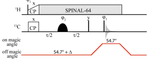
Fig. 1 Switched-angle off-MAS spin-echo pulse sequence, employing SPINAL-64 1H decoupling30and a double-Gaussian inversion pulse to
selectively probe the dipolar coupling due to a specific pair of13C nuclei.
10
Experiments were performed on a Bruker Avance II+ spectrometer operating at 1H and 13C Larmor frequencies of 599.4 and 150.7 MHz, respectively, using a 1.8 mm double-resonance probe at a rotation frequency of 12 kHz. The1H and 13C 90
pulse lengths were 2.5 and 3.0 s, respectively.
15
SPINAL-641H decoupling30 at 100 kHz was used during the spin-echo period, , the z-filter and acquisition. A 1 ms contact time was used for cross polarization from 1H to 13C with a 70 to 100 % ramp on the1H channel.31-32The recycle delay was 3 s. For each spin-echo duration, , 16 transients
20
were co-added. A 16 step phase cycle was used:1= y,y;2 = x, x, y, y;3= x, x, x, x, y, y, y, y,x,x,x,x,y,y, y,y;rec= x,x,x, x, y,y,y, y,x, x, x,x,y, y, y, y(the pulse phases are indicated in Fig. 1).
As for previous (on-angle) MAS spin-echo experiments for
25
determining J couplings,33-36 the spin-echo intensity, s(), is
obtained by integration over the corresponding lineshape (after Fourier transformation with respect to the acquisition time, t2). As discussed further in the ESI, this ensures that only in-phase lineshapes with their cosine spin-echo
30
modulation are considered, i.e., there is no contribution from anti-phase lineshapes which have a sine spin-echo modulation. 1
H decoupling is kept on during thez-filter delay in order to suppress spin diffusion that would otherwise affect an unwanted transfer of magnetisation between the spins. Error
35
bars on fitted parameters are determined by the covariance method, as described in Ref.35
Results and discussion
Fig. 2a and 2b present integrated off-MAS 13C spin-echo (/2/2) intensities, s(), obtained using the pulse
40
sequence in Fig. 1 for a representative range of distances: CO-C, CO-C, CO-C in U-13C,15N L-histidine.H
2O.HCl and C2(V)-C(L) in the dipeptide Ac-U-13C,15N L-valine-L -leucine (AcVL). In each case, two separate experiments were performed, namely a dipolar-modulated (full symbols) and a
45
reference (open symbols) experiment. In the latter reference experiment, an equivalent double Gaussian pulse is used, but
its frequency is positioned such that only one of the two signals is selected, thus allowing the spin-echo dephasing time, T2', to be independently determined. In this work, the
50
offset from the magic angle, , was determined using the sensitivity of the 1-13CL-alanine lineshape to the angle offset (see ESI), though a Hall effect angle sensor37 could also be used.
An extension of the theory of spin-echo MAS modulation
55
for a homonuclear spin pair under aJcoupling38to the case of off MAS is presented in the SI (see eqns E7 & E9) of Ref.24 Specifically, for the case where the through-bond J coupling is zero,s() for the dipolar-modulated experiment is given by
(neglecting here theT20term in Ref.24):
60
2
2
0
( )
exp(
/
')(1/ 2)
cos{
2
(3cos
1)(
s)}sin
s
A
T
D
t
d
, (1)where the integral corresponds to a powder average over the angle, , between the internuclear vector of the dipolar-coupled spins and the rotor axis. The calculation of the time shift, ts, due to the finite length of the refocusing pulse is
65
described in the ESI, using the procedure employed in Ref.25 Note that the offset,, from the magic angle in eqn (1) is in radians, while the dipolar coupling constant,D, is in Hz:
2 0
3
/ 2
4
D
r
, (2)whereris the internuclear distance andis the magnetogyric
70
ratio of the nuclei (here13C).
As well as the use of the angle-switching probe, a novel feature of the experimental protocol described here is the use of the separate reference experiment (see open symbols in Fig. 2). The spin-echo intensities, s(), for the reference
75
experiment are fitted to:
2
( )
exp(
/
')
s
A
T
, (3)thus allowing the independent determination ofT2'. Note that the so-determined T2' for the off-angle spin-echo evolution includes the effect of dipolar modulation due to like spins,
80
i.e., intermolecular 13CO-13CO (U-13C,15N L -histidine.H2O.HCl) and 13C2(V)-13C2(V) (AcVL) dipolar couplings for the experimental data in Fig. 2. The advantage of this procedure is that, for the dipolar-modulated s(), this
leaves onlyDas a free variable for fitting. Best fits in Fig. 2a
85
and 2b are shown as solid and dashed lines for the dipolar-modulated (fit to eqn (1)) and reference (fit to eqn (3)) data, respectively. Table 1 lists the fitted D (together with the corresponding internuclear distance,r) andT2' values as well as the offset from the magic angle,, and the time shift,ts.
90
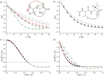
[image:3.595.45.281.145.241.2]root-Fig. 2(a,b) Dipolar-modulated (full symbols) and reference (open symbols) spinning-angle-encoded 13C (150 MHz, 12 kHz MAS) NMR spin-echo
(/2/2) curves recorded using the pulse sequence in Fig. 1 for the (a) CO-C(green squares), CO-C(blue diamonds) and CO-C(red circles) pairs in U-13C,15N-histidine.H
2O.HCl and (b) the C2(V)-C(L) pair in Ac-U-13C,15N-Val-Leu. Best-fits to eqn (1) and eqn (3) are shown as solid and
dashed lines, respectively (see Table 1). The signal intensity was integrated for the CO (His) or C2(V) resonances (after Fourier transformation with 5
respect to the acquisition time,t2), with the experimental noise being less than ± 0.02. (c,d) The analytical expression fors() in eqn (1) defines a master
curve that depends on(in radians),D(in Hz), andts(in s). Using the best-fitDvalues (see Table 1), the green squares, blue diamonds, red circles
and black ellipsoids correspond to the(andandts) values for the experimental data in (a,b). The spin-echo dephasing term, exp(/T2') (see Table 1), is
neglected in (c) and included in (d).
sum-squared dipolar coupling,Drss,39-40that takes into account
10
all (intra- and intermolecular) C-C distances for the same pair of nuclei:
2
rss ij
D
D
. (4)The Drss values determined from the corresponding single-crystal diffraction structures (CSD codes: HISTCM12 and
15
JAYNUS)41-42 are listed in Table 1. Note that while the shortest C-C distances for the CO-C (2.57 Å) and CO-C (3.11 Å) pairs inL-histidine.H2O.HCl are intramolecular, they are intermolecular for the CO-C (4.05 Å) pair in L -histidine.H2O.HCl and the C2(V)-C(L) pair (5.26 Å) in
20
AcVL. Thus, for the C2(V)-C(L) pair in AcVL, the effective distance of 4.36 Å stated in Table 1 corresponds to a sum over multiple distances of 5.26 Å and longer.
A further novel feature of this work is the recognition that the analytical expression for s(), eqn (1), defines a master
25
curve that depends on , D, and ts. This is illustrated by Fig. 2c and 2d for the best-fit D values, where the green squares, blue diamonds, red circles and black ellipsoids
Table 1Spinning-angle-encoded spin-echo fits
spin pair a t
s[ms]b T2' [ms]c distancefittedde distanceeffectivedf
CO-C(His) 2.3° 0.3 41 ± 1 2.59± 0.02 Å (437 ± 9 Hz)
2.55 Å
(460 Hz)
CO-C(His) 2.3° 0.3 33 ± 1 3.25(222 ± 6 Hz)± 0.03 Å (278 Hz)3.01 Å CO-C(His) 2.3° 0.3 30 ± 1 3.72(148 ± 7 Hz)± 0.06 Å (252 Hz)3.43 Å C2(V)-C(L)
(AcVL) 4.2° 1.2 21 ± 1
4.15± 0.06 Å (106 ± 6 Hz)
4.36 Å
(92 Hz)
aDetermined from the sensitivity of the 1-13CL-alanine lineshape to the 30
angle offset (see ESI). Note thatin this Table is specified in degrees, whileis specified in radians in eqn (1) and the master curve representations in Fig. 2c and 2d.bCalculated as described in Ref.25(see
ESI).cDetermined from fitting the reference curve to eqn (3).dThe
corresponding dipolar coupling is given in brackets.eDetermined from 35
fitting the dipolar-modulated curve to eqn (1).fD
rssis given in eqn (4) (all
atoms within 10 Å were considered for the corresponding crystal structures).
correspond to the(andandts) values for the experimental data in Fig. 2a and 2b. Specifically, the master-curve
40
presentation in Fig. 2c shows how the probing of a smallerD
[image:4.595.80.525.60.391.2]using a larger(4.2° as compared to 2.3°).
Fig. 2d shows the effect of the different experimental T2' values (see Table 1); the spin-echo dephasing term is neglected in Fig. 2c. Ideally, the zero crossing should be observed, however spin-echo dephasing which is dependent
5
on 1H decoupling performance43 precludes this for all cases except the shorter CO-C distance (in this case, the determined NMR distance is within 2 % of the effective distance determined from Drss for the crystal structure). Nevertheless, the reference curve approach used here ensures
10
good agreement (within 10 %) between the fitted distance and the effective distance determined from theDrssfor the crystal-structure, for the case of effective distances up to 4.5 Å. In future work, multi-spin simulations will be used to better understand factors that could lead to such small discrepancies.
15
Conclusions
In conclusion, using the simple spin-echo experiment in Fig. 1 and a fast angle switching probe, longer-range C-C distances can be accurately determined in uniformly 13C labeled samples, notably the greater than 4 Å C2(V)-C(L)
20
distance in the dipeptide Ac-VL. The new experimental protocol presented here involves the recording of separate dipolar-modulated and reference spin-echo datasets that correspond to changing the irradiation frequencies of the double Gaussian pulse: this enables the independent
25
determination of the spin-echo dephasing time, T2', such that the dipolar coupling is the only free variable for the fit of the dipolar-modulated data. This method can be extended to larger molecules and other nuclei (e.g., 1H), with the only requirement being the necessity to selectively excite the
30
specific pair of resonances by the cosine-modulated Gaussian pulse, with the further possibility of incorporating off-MAS spin-echo modulation into a higher-dimensional experiment. The master curve presentation in Fig. 2c shows that larger angle offsets would enable the more accurate measurement of
35
longer distances, thus motivating further development of switched-angle probe technology.
Acknowledgements
Funding from EPSRC is acknowledged. We thank M.H. Levitt and G. Pileio for helpful discussions.
40
Notes and references
aDepartment of Physics, University of Warwick, Coventry, UK CV4 7AL.
Fax: +44 24 76150897; Tel: +44 24 76574359; E-mail: [email protected]
bPresent address: Institute of Biophysical Chemistry, Johann Wolfgang
45
Goethe-University Frankfurt, Max-von-Laue-Straße 9, 60438 Frankfurt am Main, Germany.
cMAS Systems and Tallinn University of Technology, Ehitajate 5, Tallinn,
Estonia. E-mail: [email protected]
50
† Electronic Supplementary Information (ESI) available: [Further details: angle switching; angle calibration; the double Gaussian selective pulses]. See DOI: 10.1039/b000000x/
1. A. McDermott,Ann. Rev. Biophys., 2009,38, 385.
55
2. A. Lesage,Phys. Chem. Chem. Phys., 2009,11, 6876.
3. S. O. Smith, D. Song, S. Shekar, M. Groesbeek, M. Ziliox and S. Aimoto,Biochemistry, 2001,40, 6553.
4. F. Castellani, B. van Rossum, A. Diehl, M. Schubert, K. Rehbein and H. Oschkinat,Nature, 2002,420, 98.
60
5. C. Wasmer, A. Lange, H. Van Melckebeke, A. B. Siemer, R. Riek and B. H. Meier,Science, 2008,319, 1523.
6. W. T. Franks, B. J. Wylie, H. L. F. Schmidt, A. J. Nieuwkoop, R. M. Mayrhofer, G. J. Shah, D. T. Graesser and C. M. Rienstra,Proc. Natl. Acad. Sci. U. S. A., 2008,105, 4621.
65
7. I. Bertini, L. Emsley, M. Lelli, C. Luchinat, J. F. Mao and G. Pintacuda,J. Am. Chem. Soc., 2010,132, 5558.
8. M. Carravetta, M. Eden, O. G. Johannessen, H. Luthman, P. J. E. Verdegem, J. Lugtenburg, A. Sebald and M. H. Levitt,J. Am. Chem. Soc., 2001,123, 10628.
70
9. P. Hodgkinson and L. Emsley,J. Magn. Reson., 1999,139, 46. 10. M. Hohwy, C. M. Rienstra and R. G. Griffin,J. Chem. Phys., 2002,
117, 4973.
11. M. J. Bayro, M. Huber, R. Ramachandran, T. C. Davenport, B. H. Meier, M. Ernst and R. G. Griffin, J. Chem. Phys., 2009, 130,
75
114506.
12. V. Ladizhansky,Solid State Nucl. Magn. Reson., 2009,36, 119. 13. I. Marin-Montesinos, G. Mollica, M. Carravetta, A. Gansmuller, G.
Pilelo, M. Bechmann, A. Sebald and M. H. Levitt,Chem. Phys. Lett., 2006,432, 572.
80
14. A. K. Paravastu and R. Tycko,J. Chem. Phys., 2006,124, 194303. 15. R. Tycko,Phys. Rev. Lett., 2007,99, 187601.
16. N. Khaneja and N. C. Nielsen,J. Chem. Phys., 2008,128, 015103. 17. L. A. Straaso, M. Bjerring, N. Khaneja and N. C. Nielsen,J. Chem.
Phys., 2009,130, 225103.
85
18. J. Spano and S. Wi,J. Magn. Reson., 2010,204, 314.
19. P. T. F. Williamson, A. Verhoeven, M. Ernst and B. H. Meier,J. Am. Chem. Soc., 2003,125, 2718.
20. A. Verhoeven, P. T. F. Williamson, H. Zimmermann, M. Ernst and B. H. Meier,J. Magn. Reson., 2004,168, 314.
90
21. K. Nomura, K. Takegoshi, T. Terao, K. Uchida and M. Kainosho,J. Am. Chem. Soc., 1999,121, 4064.
22. R. Ramachandran, V. Ladizhansky, V. S. Bajaj and R. G. Griffin,J. Am. Chem. Soc., 2003,125, 15623.
23. X. H. Peng, D. Libich, R. Janik, G. Harauz and V. Ladizhansky,J.
95
Am. Chem. Soc., 2008,130, 359.
24. G. Pileio, Y. Guo, T. N. Pham, J. M. Griffin, M. H. Levitt and S. P. Brown,J. Am. Chem. Soc., 2007,129, 10972.
25. G. Pileio, S. Mamone, G. Mollica, I. M. Montesinos, A. Gansmuller, M. Carravetta, S. P. Brown and M. H. Levitt, Chem. Phys. Lett.,
100
2008,456, 116.
26. C. P. Jaroniec, B. A. Tounge, J. Herzfeld and R. G. Griffin,J. Am. Chem. Soc., 2001,123, 3507.
27. J. Trebosc, J. P. Amoureux, L. Delevoye, J. W. Wiench and M. Pruski,Solid State Sci., 2004,6, 1089.
105
28. S. Cadars, A. Lesage, N. Hedin, B. F. Chmelka and L. Emsley, J. Phys. Chem. B, 2006,110, 16982.
29. P. L. Stewart, R. Tycko and S. J. Opella,J. Chem. Soc., Faraday Trans., 1988,84, 3803.
30. B. M. Fung, A. M. Khitrin and K. Ermolaev,J. Magn. Reson., 2000,
110
31. G. Metz, X. L. Wu and S. O. Smith,J. Magn. Reson. Ser. A, 1994,
110, 219.
32. S. Hediger, B. H. Meier, N. D. Kurur, G. Bodenhausen and R. R. Ernst,Chem. Phys. Lett., 1994,223, 283.
33. S. P. Brown, M. Perez-Torralba, D. Sanz, R. M. Claramunt and L.
5
Emsley,Chem. Commun., 2002, 1852.
34. S. P. Brown and L. Emsley,J. Magn. Reson., 2004,171, 43. 35. T. N. Pham, J. M. Griffin, S. Masiero, S. Lena, G. Gottarelli, P.
Hodgkinson, C. Filip and S. P. Brown,Phys. Chem. Chem. Phys., 2007,9, 3416.
10
36. I. Hung, A. C. Uldry, J. Becker-Baldus, A. L. Webber, A. Wong, M. E. Smith, S. A. Joyce, J. R. Yates, C. J. Pickard, R. Dupree and S. P. Brown,J. Am. Chem. Soc., 2009,131, 1820.
37. S. Mamone, A. Dorsch, O. G. Johannessen, M. V. Naik, P. K. Madhu and M. H. Levitt,J. Magn. Reson., 2008,190, 135.
15
38. L. Duma, W. C. Lai, M. Carravetta, L. Emsley, S. P. Brown and M. H. Levitt,ChemPhysChem, 2004,5, 815.
39. V. E. Zorin, S. P. Brown and P. Hodgkinson,Mol. Phys., 2006,104, 293.
40. V. E. Zorin, S. P. Brown and P. Hodgkinson,J. Chem. Phys., 2006,
20
125, 144508.
41. J. Donohue and A. Caron,Acta Crystallogr., 1964,17, 1178. 42. P. J. Carroll, P. L. Stewart and S. J. Opella,Acta Crystallogr. C,
1990,46, 243.
43. G. De Paepe, N. Giraud, A. Lesage, P. Hodgkinson, A. Bockmann
25