University of Warwick institutional repository: http://go.warwick.ac.uk/wrap
This paper is made available online in accordance with
publisher policies. Please scroll down to view the document
itself. Please refer to the repository record for this item and our
policy information available from the repository home page for
further information.
To see the final version of this paper please visit the publisher’s website.
Access to the published version may require a subscription.
Author(s): Russell Wallis, Daniel A. Mitchell, Ralf Schmid, Wilhelm J.
Schwaeble and Anthony H. Keeble
Article Title: Paths reunited: Initiation of the classical and lectin
pathways of complement activation
Year of publication: 2009
Link to published version:
http://dx.doi.org/
10.1016/j.imbio.2009.08.006
Publisher statement: Wallis, R. et al. (2009). Paths reunited: Initiation
of the classical and lectin pathways of complement activation.
1
Paths reunited: initiation of the classical and lectin pathways of
complement activation
Russell Wallis*
‡, Daniel A. Mitchell
†, Ralf Schmid
‡, Wilhelm W. Schwaeble* and
Anthony H. Keeble*
*Department of Infection, Immunity and Inflammation and
‡Department of Biochemistry,
University of Leicester, Leicester, United Kingdom and
†Clinical Sciences Research Institute,
Clinical Sciences Building, University of Warwick, Coventry, United Kingdom
Correspondence to:
Dr Russell Wallis
RCUK Academic Fellow
Departments of Infection, Immunity and Inflammation and Biochemistry
Maurice Shock Building
University of Leicester
PO Box 138, Leicester
LE1 9HN UK
2 Abstract
Since its isolation almost fifty years ago, considerable progress has been made towards understanding
the structural organisation and mode of action of C1, the initiating complex of the classical pathway
of complement activation. Nevertheless, knowledge is still incomplete, especially with regard to the
interactions between subcomponents, C1q, C1r and C1s, that trigger activation. Recent studies have
provided new insights into these interactions, and have revealed unexpected parallels with initiating
complexes of the lectin pathway of complement: MBL-MASP and ficolin-MASP. Here, we develop
and expand these concepts and delineate their implications towards the key aspects of complement
activation via the classical and lectin pathways.
Introduction
The classical and lectin pathways of complement activation are central combatants in the fight against
disease. Both are able to lyse pathogens directly via antibody-independent mechanisms, providing
immediate response against invasion (Fujita et al., 2004; Schwaeble et al., 2002). They also stimulate
key cellular and humoral interactions within the innate immune system, including chemotaxis,
phagocytosis and cell adhesion. Crucially, complement activation promotes B-cell differentiation and
maintenance of immune tolerance, building vital bridges with adaptive immunity (Carroll, 2004).
Once established, activation on antibody-antigen complexes further facilitates directed killing (Porter
& Reid, 1978). More recently it has emerged that complement components not only target non-self
but also altered-self structures, and early complement components are key players in tissue
homeostasis through clearance of apoptotic and necrotic cells (Botto, 1998; Stuart et al., 2005; Taylor
et al., 2000).
As expected from their important biological roles, deficiencies in complement components are
associated with increased risk of acute, severe infections (Summerfield, 2003). For example, lack of
C1q increases susceptibility to encapsulated bacteria such as Streptococcus pneumonia (Colten &
Rosen, 1992). It is also linked with defective clearance of immune complexes and cellular debris,
leading to glomerulonephritis (Botto et al., 1998), and increased risk of autoimmune disease,
particularly systemic lupus erythematosus (Lewis & Botto, 2006). Whilst normally protective,
complement can inadvertently contribute to disease pathogenesis. For example, C1q is involved in
the pathology of several amyloid diseases, such as Alzheimer‟s disease and readily binds to prion
proteins to initiate complement activation and exacerbate disease progression (Yasojima et al., 1999).
Reperfusion of tissues (e.g. heart and kidney) following ischemia, triggers lectin pathway-mediated
host damage, therefore specific inhibitors of complement have considerable therapeutic potential
(Møller-Kristensen et al., 2003; Mollnes et al., 2002). Hence, understanding the molecular
interactions triggering complement activation is of great interest from biochemical, immunological
3
Befitting its biological importance, the study of the composition and structure of the C1 complex has a
long history. As early as 1963, the initiating complex of the classical pathway, C1, was shown to
comprise three subcomponent proteins: C1q, C1r and C1s (Lepow et al., 1963). It is now well
established that C1q functions as the recognition subcomponent of the complex by binding to a wide
variety of targets including microorganisms, immune complexes, apoptotic and necrotic cells and
amyloids to initiate the stepwise activation of the associated serine proteases, C1r and C1s. C1q is a
hexamer of subunits, each assembled from three different polypeptide chains, A, B and C (Reid &
Porter, 1976) (Figure 1). N-terminal collagenous tails (subsequently referred to as stalks) splay apart
at an interruption in this domain, called the kink, and terminate in C-terminal globular heads to form
large structures (~530 kDa) that resemble bouquets (Strang et al., 1982). Subcomponents C1r and
C1s are modular proteins, each consisting of two CUB domains separated by an EGF-like domain,
followed by two CCP modules and a serine protease domain (SP) (Schwaeble et al., 2002; Sim &
Tsiftsoglou, 2004). C1r dimerizes through interactions between the SP domain of one polypeptide
and the CCP domains of its partner (Budayova-Spano et al., 2002). In the presence of Ca2+, each chain binds to a C1s polypeptide via the CUB1-EGF domains to form a C1rs tetramer
(C1s-C1r-C1r-C1s; ~340 kDa). The interaction with C1q is also Ca2+-dependent and forms the basis for this review. When C1 binds to a target via its globular heads, C1r activates through auto-cleavage. C1r then
cleaves and activates C1s. To ensure only localised activation, the SP domains stay attached to C1 via
single disulfide bonds to their respective N-terminal fragments. The first substrate of C1s is C4.
Once cleaved it rapidly attaches to the pathogen surface via a reactive thioester bond. C2 then binds
to the C4b fragment and is also cleaved by C1s to form the C3 convertase (C4b2a), the next
component of the pathway. Thus, C1 activation and subsequent steps are limited to the surfaces of
pathogens and other targets, a key regulatory aspect of the pathway.
The lectin pathway of complement was discovered comparatively recently (in 1987) compared to the
classical pathway, but parallels were immediately apparent (Ikeda et al., 1987). MBL and ficolins
resemble C1q in electron micrographs and bind to homologues of C1r and C1s called MBL-associated
serine proteases (MASPs) (Figure 1). Initially upon the discovery of MASP-1 (Ji et al., 1993;
Matsushita & Fujita, 1992) and later MASP-2 (Thiel et al., 1997), it was believed that initiating
complexes would function in the same way as C1, i.e. involving stepwise activation of separate
MASP components. However, subsequent studies revealed that this was not the case; rather MASPs
bind independently to MBLs and ficolins, predominantly as 1:1 complexes (i.e one MASP dimer to
each MBL/ficolin oligomer, (Chen & Wallis, 2001)) and both autoactivate. Differences became even
more apparent, upon discovery of a third MASP, MASP-3, (Dahl et al., 2001) and a non-enzymatic
protein that binds to MBL/ficolins called MAp-19 or sMAP, (Iwaki et al., 2006; Stover et al., 1999).
It is now well accepted that MBL-MASP-2 and ficolin-MASP-2 complexes are able to activate
4
components are still largely undetermined. MASP-2 has a similar catalytic specificity to C1s,
cleaving C2 and C4 (Rossi et al., 2001). The proteolytic activities of C1r/s and the MASPs have been
recently covered in more detail in a recent excellent review (Gal et al., 2009).
Despite the considerable progress that has been made in elucidating the structure of C1 and
MBL-MASP/ficolin-MASP complexes, the mechanism by which subcomponents combine to initiate
complement cascade activation has remained elusive. Until recently, it was generally believed that
subcomponents had evolved distinct strategies to associate with their partner proteins and activate
complement (Gaboriaud et al., 2007). Principally, the C1q binding site on the C1rs tetramer was
proposed to lie at the C1r-C1s interface (Gaboriaud et al., 2004), whilst the MBL binding site was
thought to be localised on the CUB domains of MASPs. However, two recent papers by Phillips et al.
and Bally et al. have challenged this dogma, highlighting the similarities between the classical and
lectin complexes, thus uniting their likely mode of action once more (Bally et al., 2009; Phillips et al.,
2009). In this review, we have focussed on how this recent knowledge helps us to address the key
requirements for a functional model of C1, recently set out by Gal et al. (Gal et al., 2009).
Specifically, any model should answer the following questions: (1) How C1q, C1r and C1s readily
associate to form C1. (2) How the activation signal is transmitted from C1q to the C1r zymogens. (3)
How zymogen C1r autoactivates after receiving the activation signal (and what prevents spontaneous
autoactivation). (4) How C1r activates C1s inside the complex. (5) How C1s is able to cleave C4 and
C2 in the fluid phase. (6) How C1-inhibitor gains access to both C1r and C1s in the complex to block
their activity and disassemble the complex. None of the previous models of C1 have been able to
satisfactorily answer all of these questions. Here we set out how our recent model provides insight
into each question in turn.
Assembly of C1
Recent surface plasmon resonance studies, using purified wild-type and deletion components has
allowed the kinetics of C1 formation to be dissected and has illustrated a number of key points that
impact on structure of C1 (Bally et al., 2009; Phillips et al., 2009): (1) that C1r and C1s can both bind
C1q individually; (2) that the C1rs tetramer binds tighter than C1r or C1s alone indicating that they
both bind within the context of the tetramer; (3) that deletion of the CCP-CCP-SP domains does not
prevent binding of C1r or C1s, implying that they do not interact with C1q significantly; and (4) most
importantly that non-cognate (i.e., MBL-C1rs and C1q-MASP) complexes readily assemble, but with
weaker affinities than cognate ones. This latter striking observation thus shows that the physical basis
for assembly is shared between both complexes and is incompatible with the previous most widely
accepted C1 models (Gaboriaud et al., 2007; Gaboriaud et al., 2004). Instead, the binding data
indicate that C1rs tetramers present six separate binding sites to C1q that are located on the CUB
5
independently proposed that each site binds to a separate collagenous stalk of C1q (Bally et al., 2009;
Phillips et al., 2009). Residues forming the binding sites are conserved in the C1r/C1s/MASP family
and equivalent protease-binding motifs are present on the collagenous stalks of C1qs/MBLs and
ficolins (Figure 2). These similarities explain the early observation that purified MBL oligomers of
the lectin pathway are able to bind and activate C1rs tetramers of the classical pathway (Lu et al.,
1990). The main difference between the C1 and MBL-MASP complexes is in their stoichiometries:
in the lectin pathway, each recognition subcomponent binds to a single MASP dimer, whereas C1q
binds to a C1rs tetramer (the equivalent of two MASP dimers; Figure 2) (Chen & Wallis, 2001). This
distinction accounts for differences in the oligomeric structures of MBLs (dimers to hexamers of
stalks in human MBL, but predominantly tetramers (Teillet et al., 2005)) and ficolins (mainly
tetramers), which are typically smaller that C1q (hexamers). It also explains why the pentameric and
hexameric forms of human MBL (relatively minor components) are most efficient at activating C1rs
tetramers (Lu et al., 1990): these oligomers most resemble C1q so are better able to bind and active
C1rs.
A notable feature of C1, MBL-MASP and ficolin-MASP complexes is their Ca2+ dependence. Ca2+ mediates interactions between C1r and C1s, C1rs and C1q as well as between MBL and MASP and
ficolin and MASP. The Ca2+ binding sites are conserved in each of the CUB domains as well as in the EGF domains of the protease subcomponents (Figure 1). Thus, each C1rs tetramer probably binds 12
Ca2+ and each MASP dimer 6 Ca2+. The interfaces of the MASP homodimers and the two C1r-C1s heterodimers are created by a set of conserved contacts between the CUB1-EGF domains (Feinberg et
al., 2003; Gregory et al., 2003; Gregory et al., 2004). Several residues, which lie in or near the Ca2+ site in the EGF domain, stabilise the dimer by interacting with the CUB1 domain of the adjacent
protomer. The CUB domains co-ordinate the Ca2+ through a group of acidic residues found on surface exposed loops of the CUB domains. Mutation of these residues weakens binding to the
collagenous domains of the respective recognition component implying they play direct roles in the
protein-protein interaction (Teillet et al., 2008). However, mutations that disrupt Ca2+ binding probably also perturb protease binding indirectly, by disrupting the conformation of the surface loops.
Thus the assignment of the roles of the residues in binding remains ambiguous.
C1 subcomponents associate spontaneously in solution and do so even when C1q is already attached
to an activating surface to yield functional complexes (Gigli et al., 1976). This would not be possible
without considerable conformational flexibility of the protease subcomponents. C1rs tetramers adopt
bent but relatively extended structures in solution and in electron micrographs (Perkins & Nealis,
1989), so presumably are able to pass through gaps between C1q stalks, where they adopt a more
constrained bound conformation. The dynamic nature of interactions of individual C1q globular
6
C1q. Such “breathing” of the globular heads would seem likely because each interaction is relatively
weak and high-affinity target binding is instead achieved through multiple head-surface interactions.
Transmission of the activation signal from C1q to C1r
A critical facet of the complement cascade is that activation is only triggered when C1 complexes
bind to an activating surface. There are several factors intrinsic to C1 that combine to prevent
inappropriate activation: (1) the C1r polypeptides are located inside the complex (i.e. within the cone
created by the C1q stalks) so are relatively inaccessible to other serum proteins and proteases; (2) they
circulate as zymogens; and (3) reciprocal interactions between the SP domain of one C1r polypeptide
and the CCP domains of its partner prevent enzyme-substrate interactions that could lead to
autoactivation. Remarkably, each polypeptide must move by some 46 Å to allow activation
(Budayova-Spano et al., 2002). C1q is the only subcomponent that contacts the activating surface
directly, so upon binding it must undergoe considerable conformational changes to trigger
autocatalysis of C1r. Two alternative mechanisms can be considered: “relaxed-to-strained”, in which
binding to the surface distorts C1q thereby driving conversion of the zymogen complex (relaxed) to
the active conformation (strained); or “strained-to-relaxed”, in which C1 converts from a strained
zymogen to a relaxed active conformation. In the former mechanism, the energy for activation comes
from binding to the activating surface, whereas in the latter, some of the energy gained from assembly
of subcomponents primes the complex for activation. Although neither mechanism can be completely
ruled out, the available solution and structural data favour a strained-to-relaxed mechanism (Figure 3).
In particular, both subcomponents are more constrained in the complex compared to their unbound
states. Thus, unliganded C1q is highly flexible in solution, with large differences in the angles
between collagenous stalks, but becomes fixed upon binding to C1rs tetramers (Perkins, 1985).
Severe constraints are also imposed upon C1r and C1s, which adopt more compact configurations on
association with each other and upon binding to C1q (Strang et al., 1982). Importantly, a
strained-to-relaxed mechanism would require relatively little energy input from target binding to trigger
activation of the “primed” complex, hence providing an explanation for the low level of spontaneous
C1 autoactivation observed in vitro, even in the absence of immune complexes or other activators
(Ziccardi, 1982). It would also explain how activation is independent of the nature of the activating
surface, thereby accounting for the diverse targets that trigger activation and the different ways in
which the globular heads of C1q are believed to bind to these targets (Gaboriaud et al., 2003). On the
other hand, a relaxed-to-strained model would seem much less likely, because different activating
targets would somehow have to induce comparable, ordered conformational changes in C1q to trigger
the steps necessary for activation. Such changes would seem particularly unlikely given the innate
flexibility of the C1q stalks.
7
We propose that the inter-protomer C1r CCP-SP interactions not only prevent autoactivation of the
zymogen complex, but also induce strain into circulating C1 complexes (Figure 3). Compatible with
this hypothesis, modelling studies have shown that zymogen C1rs tetramers cannot be docked onto
C1q without distorting the subcomponents, such as through changes at the junction between theEGF
and CUB-2 domains of C1r (which is likely to be flexible), the relativepositions of C1r and C1s and
consequently the relative positions of the C1q stalks (Phillips et al., 2009). For example, in the model
of the zymogen C1 complex proposed by Bally et al. (Bally et al., 2009), the six binding sites on C1rs
tetramers are arranged about a 2 x 3 rectangle when viewed from above, rather than a hexagon as in
the active conformation (Phillips et al., 2009). The overall effect of these changes would be to pull
some (or all) of the C1q stalks closer together, and thus distort the natural 6-fold symmetry of C1q
observed in solution (Perkins, 1985). Complement activation would then be initiated by splaying
apart of the C1q bouquets when they bind to a target, breaking the reciprocal interactions between C1r
polypeptides and releasing the imparted strain. The SP domains would then naturally separate to
allow mutual autocatalysis (Budayova-Spano et al., 2002). This mechanism would explain why
complexes assembled from truncated forms of C1rs, lacking the CCP and SP domains, have
properties compatible with the active (relaxed) rather than the zymogen (strained) conformation
(Phillips et al., 2009).
A strained-to-relaxed activation mechanism shares a number of similarities with the
“split-and-reassembly” model proposed by Gal and colleagues (Kardos et al., 2008). Here, binding of C1q heads
to the target results in a change in the relative position of the C1q arms, which in turn shifts the
equilibrium to the dissociated form of the C1r dimer. However, a major difference concerns the mode
of separation and realignment of C1r. In the split-and-reassembly model, the polypeptides reassemble
spontaneously into an enzyme-substrate complex. Whereas in the strained-to-relaxed model they
would spring apart as the strain is released. In this respect, the motion resembles that proposed in
earlier models (e.g. (Gaboriaud et al., 2007; Gaboriaud et al., 2004)), with the key innovation being
that separation of polypeptides is driven by relaxation, rather than induced by distortion. Irrespective
of the mode of separation, autocatalysis is probably facilitated by stabilizing intermolecular CCP2–SP
interactions, and occurs via a substrate-induced mechanism (discussed in more detail in (Gal et al.,
2009)), followed by cleavage of the remaining zymogen polypeptide by its active partner.
Importantly, by the nature of the strained-to-relaxed mechanism, once activated, the C1r polypeptides
would not tend to reassociate with each other as has been suggested in other models, but rather would
be free to activate C1s.
Activation of C1s by C1r
The next step in C1 activation is the cleavage of C1s by activated C1r. In addition to blocking the
8
intermolecular CCP-SP contacts, the CCP domains of C1r play an additional, more structural role by
acting as spacers to separate the C1q binding domains (CUB1-EGF-CUB2) from the enzymatic SP
domain. Together with inter-domain flexibility, most likely between CUB2 and CCP1 of both C1r
and C1s, this would allow the required protein dynamics for C1r to contact the C1s polypeptide and
activate it. Modelling reveals that there would be sufficient space available for this process to occur
inside the complex, so that activated C1r is protected from C1-inhibitor until after C1s activation has
occurred (Figure 4).
Cleavage of C4 and C2 by C1s
The junctions between the C1q-binding (CUB1-EGF-CUB2) and proteolytic (CCP1-CCP2-SP)
portions C1r and C1s are located towards the outer perimeterof the complex in gaps between adjacent
C1q stalks (Figures 3 and 4). Flexibility at these junctions would allow the proteolytic portions to
swing out into solution through the gaps, placing the CCP1-CCP2-SP domains of C1s in a position to
cleave the next components of pathway – C4 and C2. C4 cleavage results in two products: the peptide
anaphylatoxin C4a and C4b which contains a reactive thioester that anchors C4b covalently to the
target surface. Next, C2 binds C4b (it is unable to bind full-length C4) and is itself cleaved by C1s,
yielding the C4b2a complex. Also known as the C3 convertase, this component proteolytically
activates C3 to form C3a (another peptide analphylatoxin) and C3b, which joins C4b2a to make the
C5 convertase that furthers the complement cascade. MASP-2, within the MBL-MASP complex, has
also been shown to act in a similar manner to produce the C3 convertase (Chen & Wallis, 2004).
Here, accessory binding sites for C4 on the CCP domains of the MASP play key roles in regulating
C3 convertase formation. These additional interactions not only facilitate specific catalysis of C4 by
increasing the affinity of enzyme-substrate complex (Gal et al., 2005), but also reduce the rate of
dissociation of C4b from the MASP. As a consequence, the reaction of the C4b thioester with the cell
surface occurs at least an order of magnitude faster than dissociation of the C4b-MASP-2 complex
(Chen & Wallis, 2004). The overall effect is to ensure that C4b deposits onto the target surface before
it dissociates from the MBL-MASP complex, so that it is close enough for the same MBL-MASP
complex to rapidly activate C2 upon its binding to C4b.
A similar mechanism might regulate C3 convertase formation by the classical pathway, and kinetic
data show that interactions with the CCP modules also facilitate cleavage of C4 by C1s (Rossi et al.,
1998). However, the CCP modules of MASP-2 appear to contribute more towards recognition of C4
than for C1s (Rossi et al., 2005), so this mechanism might be less important in the classical pathway,
perhaps reflecting the greater abundance of components in the serum. This conclusion is supported
by studies comparing C3 convertase formation on different surfaces, which showed that almost every
C4b molecule deposited via the lectin pathway was capableof forming a convertase, compared to only
9
potential for increased amplification in the lectin pathway and the subsequent production of
pro-inflammatory products, such as C3a, C4a,and C5a, provides a possible mechanism to explain why
complement activation via the lectinpathway is a more prominent contributor to the pathologyof
inflammatory reactions.
Binding of C1 inhibitor
C1 inhibitor (C1-INH/SERPING1) has a number of different biological roles including regulation of
vascular permeability and acting as an anti-inflammatory agent (Davis et al., 2008). It functions
primarily by controlling the activities of range of blood-borne proteases including those of the contact
system (factor XII, plasma kallikrein), coagulation (factor XI) and fibrinolysis (plasmin, tissue
plasminogen activator) in addition to the complement proteases (C1r, C1s and MASPs) in their
activated forms. Loss of this regulation by C1-INH through genetic or acquired deficiencies results in
disease states such as angioedema (Cugno et al., 2009). In common with other members of the serpin
family of protease inhibitors, C1-INH functions by acting as a suicide substrate. A reactive centre
loop inserts into the active site and cleavage triggers a startling conformational change. This is
perhaps best illustrated in inhibition of trypsin by α1-antitrypsin, where once cleaved, the reactive
centre loop and the covalently tethered protease move 71 Å to the other end of the serpin, „crushing‟
the protease in the process (Huntington et al., 2000). This remarkable process, sometimes compared
to the mechanism of a „mouse-trap‟, is thermodynamically driven by the cleaved serpin being
hyperstable compared to the uncleaved form (an extreme example of a strained-to-relaxed
mechanism). C1-INH also displays this characteristic property; for example, melting temperatures of
the uncleaved active and cleaved forms are ~60 °C and ~116 °C, respectively (Beinrohr et al., 2007).
Thus, C1-INH probably inhibits C1r and C1s through similar distortion of the SP domains. Notably,
post-C1r activation, the two C1rs hetero-dimers have already separated from each other and
subsequently bind to C1q an order of magnitude weaker than the C1rs tetramer (zymogen form); so
the C1 complex is already intrinsically less stable in this form (Phillips et al., 2009). Association with
C1-INH (~100 kDa) presumably further disrupts and weakens their interactions with C1q by
promoting dissociation and blocking reassociation through steric-electronic repulsion/clashes.
Weakening, rather than total disruption of binding is suggested by the observation that C1-INH strips
C1r and C1s from C1 more efficiently at higher salt concentrations (Sim et al., 1980). Nevertheless,
under physiological conditions the protease and recognition subcomponents separate and much of the
C1q remains bound to the activating surface. Another feature conserved between C1-INH-C1r/s and
some other serpin-protease interactions is that heparin appears to potentiate the protein-protein
interaction by accelerating the second-order association rate constant. For C1-INH binding to C1s,
heparin shields unfavourable electrostatic clashes (Beinrohr et al., 2007). In the case of MASP-1, this
10
C1-INH provides a crucial regulatory function by limiting the lifetime of surface-bound activated C1
and thus regulating C3 convertase formation. Given the relatively compact nature of C1 and the
appreciable size of C1-INH, it is likely that once C1 is bound to an activating surface, C1r and C1s are
only targeted once their SP domains have moved outside of the C1q stalks. Protease-C1-INH
complexes are released rapidly from C1-Ab-Ag complexes, so active C1 exists only transiently on the
activating surface (Sim et al., 1980). Consequently, there would be competition between C4 cleavage
and C1-INH-mediated inhibition. The C1q separated from C1rs but still attached to cell surfaces
would be free to bind to receptors (such as gC1qR/collectin receptor/calreticulin) to further modulate
immune function (Gasque, 2004). Another crucial function of C1-INH is to neutralise spontaneous
C1 activation, which occurs at low-level in serum (Ziccardi, 1982). Such neutralisation is key to
preventing activation in solution, which would lead to depletion of complement components in
plasma. Based on their more exposed positions in unbound C1, C1-INH would be able to readily
access the SP domains of C1r and C1s once activated. The efficiency of this neutralization is likely to
be greatly enhanced by reversible interactions between C1 and C1-INH, as components circulate in
plasma (Ziccardi, 1985).
Parallels between C1 complex and MBL-MASP formation
Given the similarities between the initiating complexes of the classical and lectin pathways and their
abilities to cross-bind and cross-activate in vitro, it is likely that MBL-MASP and ficolin-MASP
complexes undergo similar changes to C1 during activation. Post-activation, MASP-2 is largely
functionally analogous to C1s, although intriguingly, their common catalytic specificities are achieved
through different mechanisms (Harmat et al., 2004). Where the two complexes differ is that two
activation steps (C1r and C1s) are required for C1 activation, but only one step (autoactivation of
MASP-2) is required for MBL-MASP activation. MASP dimers have four separate binding sites for
MBL stalks (Teillet et al., 2008): one for each stalk of a MBL tetramer. Nevertheless, MBL dimers
are able to activate complement (although with reduce activity compared to trimers and tetramers), so
only two of these sites must be occupied for activation to occur (Wallis & Drickamer, 1999). Unlike
C1r, the CCP1-CCP2-SP fragments of each zymogen MASP polypeptide do not interact with each
other significantly (Gal et al., 2005). Nevertheless, MASP binding might still distort the MBL/ficolin,
leading to a similar strained-to-relaxed activation mechanism. Interestingly, in a recent study of MBL
by atomic force microscopy, stalks were not symmetrically arranged in individual oligomers (e.g. 90°
for a MBL tetramers), but rather with only 35–40° between neighbouring stalks. Consequently
significant conformational changes would be required to bind readily to MASPs (Jensenius et al.,
2009). It will be interesting to determine the solution properties of MBL and MBL-MASP complexes
11 Figure legends
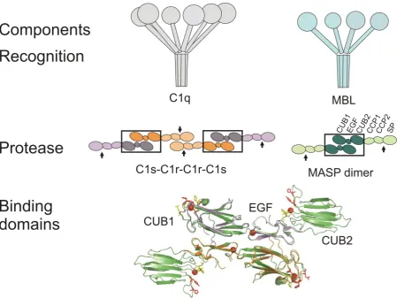
Figure 1: Schematic representations of the individual components of the initiating complexes of lectin and classical complement pathways. Top, C1q, MBL and ficolins (not shown) resemble bouquets, comprising N-terminal collagen-like domains linked to C-terminal target-recognition domains (carbohydrate-recognition, antibody-binding and fibrinogen-like, respectively). Middle, domain organisations of C1rs heterotetramers and MASP homodimers. C1r-C1s and MASP-MASP interactions are conserved (black boxes), so the C1rs tetramer is effectively equivalent to two MASP dimers linked through the CCP-SP domains of C1r. Bottom, crystal structures of MASP-1 CUB1-EGF-CUB2 (green; PDB: 3DEM (Teillet et al., 2008)) and C1s CUB1-EGF (orange and purple; PDB: 1NZI, (Gregory et al., 2003)) fragments. Each EGF domain binds a Ca2+ ion (red sphere) aiding binding to the CUB1 domain of its partner. Additional Ca2+ ions (red spheres) are co-ordinated within the CUB domains and organize the loops that form the binding sites for the MBL/C1q stalks. Putative binding residues are coloured red and yellow for large and lesser effects on binding, respectively. In vivo, C1s does not form homodimers, but each chain instead binds to a C1r polypeptide through homologous interactions.
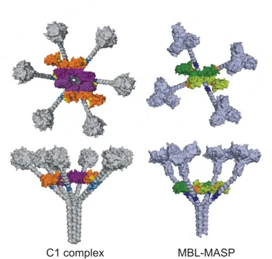
Figure 2: C1 and MBL-MASP complexes. Top (top) and side (bottom) views of C1 and MBL-MASP complexes derived from modelling and binding studies. Only the CUB1-EGF-CUB2 domains of C1r and C1s (purple and orange) and MASP (green) are shown. In both cases the CCP1-CCP2-SP domains are located inside the complex, on top of the CUB1-EGF-CUB2 domains. Binding sites are: C1q (blue), MBL (dark blue), C1rs (yellow) and MASP (orange). Notably, interactions of C1q with C1rs and MBL with MASP are analogous, with four binding sites on each MASP dimer and three on each C1r-C1s (CUB2 of C1s lacks a binding site), making a total of six sites on a C1rs heterotetramer.
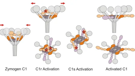
Figure 3: Strained-to-relaxed model of C1 activation. Phase 1: Zymogen C1. The active sites of C1r are kept apart through the interactions of one SP domain with the CCP domains of its partner. Binding to C1q induces strain, pulling the C1q stalks closer together (as indicated by the arrows). Although C1r and C1s are zymogens, their SP domains are relatively exposed in circulating complexes, so are accessible to reversible binding by C1-INH (Ziccardi, 1985). However, irreversible binding and inhibition by C1-INH only occurs upon activation of C1r and/or C1s and serves to neutralise the low level of spontaneous activation that occurs in solution (Ziccardi, 1982). The CCP-SP domains of C1s are not shown for clarity. Phase 2: target binding induced C1r autoactivation. Multiple simultaneous interactions of the C1q heads with an activating surface (e.g., IgG bound to bacterial surfaces) enable the stalks to move apart again (see arrows), releasing the induced strain within the complex. The CCP-SP domains of C1r spring apart allowing autocatalysis of one polypeptide by its partner. Side and top views are shown. Phase 3: C1s activation. Flexibility at the inter-domain CUB2-CCP junctions allows the C1r SP domain to cleave C1s, converting it to the active form. Phase 4: Activated C1. The catalytic domains of C1s, and possibly C1r, move out into solution, where the former activate downstream substrates C4 and C2. Once C1 is bound to a surface, C1-INH can only access the SP domains of C1s and/or C1r after they have moved outside of the complex.
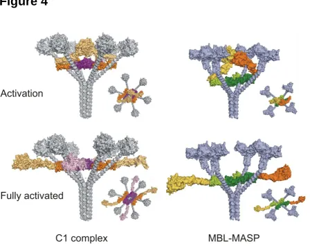
Figure 4: Conformational changes in C1 and MBL-MASP complexes. Side and top views of complexes during activation and fully activated. Autoactivation of C1r and MASPs occurs inside the complexes. Flexibility at the interdomain CUB2-CCP1 junctions would then allow the CCP-protease domains to move outside the cones created by the C1q/MBL stalks and into solution where they would be able to activate downstream substrates C4 and C4b2.
References
12
Beinrohr L., Harmat V., Dobo J., Lorincz Z., Gal P., Zavodszky P. (2007) C1 inhibitor serpin domain structure reveals the likely mechanism of heparin potentiation and conformational disease. J Biol Chem282(29): 21100-21109
Botto M. (1998) C1q knock-out mice for the study of complement deficiency in autoimmune disease. Exp Clin Immunogenet15(4): 231-234
Botto M., Dell'Agnola C., Bygrave A.E., Thompson E.M., Cook H.T., Petry F., Loos M., Pandolfi P.P., Walport M.J. (1998) Homozygous C1q deficiency causes glomerulonephritis associated with multiple apoptotic bodies. Nat Genet19(1): 56-59
Budayova-Spano M., Lacroix M., Thielens N.M., Arlaud G.J., Fontecilla-Camps J.C., Gaboriaud C. (2002) The crystal structure of the zymogen catalytic domain of complement protease C1r reveals that a disruptive mechanical stress is required to trigger activation of the C1 complex. EMBO J 21(3): 231-239
Carroll M.C. (2004) The complement system in regulation of adaptive immunity. Nat Immunol5(10): 981-986
Chen C.B., Wallis R. (2001) Stoichiometry of complexes between mannose-binding protein and its associated serine proteases. Defining functional units for complement activation. J Biol Chem 276(28): 25894-25902
Chen C.B., Wallis R. (2004) Two mechanisms for mannose-binding protein modulation of the activity of its associated serine proteases. J Biol Chem279(25): 26058-26065
Colten H.R., Rosen F.S. (1992) Complement deficiencies. Annu Rev Immunol10: 809-834
Cugno M., Zanichelli A., Foieni F., Caccia S., Cicardi M. (2009) C1-inhibitor deficiency and angioedema: molecular mechanisms and clinical progress. Trends Mol Med15(2): 69-78
Dahl M.D., Thiel S., Matsushita M., Fujita T., Willis A.C., Christensen T., Vorup-Jensen T., Jensenius J.C. (2001) MASP-3 and its association with distinct complexes of the mannan-binding lectin complement activation pathway. Immunity15: 127-135
Davis A.E., 3rd, Mejia P., Lu F. (2008) Biological activities of C1 inhibitor. Mol Immunol 45(16): 4057-4063
Dobo J., Harmat V., Beinrohr L., Sebestyen E., Zavodszky P., Gal P. (2009) MASP-1, a promiscuous complement protease: structure of its catalytic region reveals the basis of its broad specificity. J Immunol183(2): 1207-1214
Feinberg H., Uitdehaag J.C., Davies J.M., Wallis R., Drickamer K., Weis W.I. (2003) Crystal structure of the CUB1-EGF-CUB2 region of mannose-binding protein associated serine protease-2. Embo J22(10): 2348-2359
Fujita T., Matsushita M., Endo Y. (2004) The lectin-complement pathway - its role in innate immunity and evolution. Immunol Rev198(1): 185-202
Gaboriaud C., Juanhuix J., Gruez A., Lacroix M., Darnault C., Pignol D., Verger D., Fontecilla-Camps J.C., Arlaud G.J. (2003) The crystal structure of the globular head of complement protein C1q provides a basis for its versatile recognition properties. J Biol Chem278(47): 46974-46982
13
Gaboriaud C., Thielens N.M., Gregory L.A., Rossi V., Fontecilla-Camps J.C., Arlaud G.J. (2004) Structure and activation of the C1 complex of complement: unraveling the puzzle. Trends Immunol 25(7): 368-373
Gal P., Dobo J., Zavodszky P., Sim R.B. (2009) Early complement proteases: C1r, C1s and MASPs. A structural insight into activation and functions. Mol Immunol
Gal P., Harmat V., Kocsis A., Bian T., Barna L., Ambrus G., Vegh B., Balczer J., Sim R.B., Naray-Szabo G., Zavodszky P. (2005) A true autoactivating enzyme. Structural insight into mannose-binding lectin-associated serine protease-2 activations. J Biol Chem280(39): 33435-33444
Gasque P. (2004) Complement: a unique innate immune sensor for danger signals. Mol Immunol 41(11): 1089-1098
Gigli I., Porter R.R., Sim R.B. (1976) The unactivated form of the first component of human complement, C1. Biochem J157(3): 541-548
Gregory L.A., Thielens N.M., Arlaud G.J., Fontecilla-Camps J.C., Gaboriaud C. (2003) X-ray structure of the Ca2+-binding interaction domain of C1s. Insights into the assembly of the C1 complex of complement. J Biol Chem278(34): 32157-32164
Gregory L.A., Thielens N.M., Matsushita M., Sorensen R., Arlaud G.J., Fontecilla-Camps J.C., Gaboriaud C. (2004) The X-ray structure of human mannan-binding lectin-associated protein 19 (MAp19) and its interaction site with mannan-binding lectin and L-ficolin. J Biol Chem 279(28): 29391-29397
Harmat V., Gal P., Kardos J., Szilagyi K., Ambrus G., Vegh B., Naray-Szabo G., Zavodszky P. (2004) The structure of MBL-associated serine protease-2 reveals that identical substrate specificities of C1s and MASP-2 are realized through different sets of enzyme-substrate interactions. J Mol Biol 342(5): 1533-1546
Huntington J.A., Read R.J., Carrell R.W. (2000) Structure of a serpin-protease complex shows inhibition by deformation. Nature407(6806): 923-926
Ikeda K., Sannoh T., Kawasaki N., Kawasaki T., Yamashina I. (1987) Serum lectin with known structure activates complement through the classical pathway. Journal of Biological Chemistry 262: 7451-7454
Iwaki D., Kanno K., Takahashi M., Endo Y., Lynch N.J., Schwaeble W.J., Matsushita M., Okabe M., Fujita T. (2006) Small mannose-binding lectin-associated protein plays a regulatory role in the lectin complement pathway. J Immunol177(12): 8626-8632
Jensenius H., Klein D.C., van Hecke M., Oosterkamp T.H., Schmidt T., Jensenius J.C. (2009) Mannan-Binding Lectin: Structure, Oligomerization, and Flexibility Studied by Atomic Force Microscopy. J Mol Biol
Ji Y.H., Fujita T., Hatsuse H., Takahashi A., Matsushita M., Kawakami M. (1993) Activation of the C4 and C2 components of complement by a proteinase in serum bactericidal factor, Ra reactive factor. Journal of Immunology150: 571-578
14
Lepow I.H., Naff G.B., Todd E.W., Pensky J., Hinz C.F. (1963) Chromatographic resolution of the first component of human complement into three activities. J Exp Med117: 983-1008
Lewis M.J., Botto M. (2006) Complement deficiencies in humans and animals: links to autoimmunity. Autoimmunity39(5): 367-378
Lu J., Thiel S., Wiedemann H., Timpl R., Reid K.B.M. (1990) Binding of the pentamer/hexamer forms of a mannan-binding protein to zymosan activates the proenzyme C1r2C1s2 complex of the
classical pathway of complement, without involvement of C1q. Journal of Immunology144: 2287-2294
Matsushita M., Fujita T. (1992) Activation of the classical complement pathway by mannose-binding protein in association with a novel C1s-like serine protease. Journal of Experimental Medicine 176: 1497-1502
Møller-Kristensen M., Thiel S., Hansen A.G., Jensenius J.C. (2003) On the Site of C4 Deposition upon Complement Activation via the Mannan-Binding Lectin Pathway or the Classical Pathway. Scandinavian Journal of Immunology 57: 556-561
Mollnes T.E., Song W.C., Lambris J.D. (2002) Complement in inflammatory tissue damage and disease. Trends Immunol23(2): 61-64
Perkins S.J. (1985) Molecular modelling of human complement subcomponent C1q and its complex with C1r2C1s2 derived from neutron-scattering curves and hydrodynamic properties. Biochem J 228(1): 13-26
Perkins S.J., Nealis A.S. (1989) The quaternary structure in solution of human complement subcomponent C1r2C1s2. Biochem J263(2): 463-469
Phillips A.E., Toth J., Dodds A.W., Girija U.V., Furze C.M., Pala E., Sim R.B., Reid K.B., Schwaeble W.J., Schmid R., Keeble A.H., Wallis R. (2009) Analogous interactions in initiating complexes of the classical and lectin pathways of complement. J Immunol182(12): 7708-7717
Porter R.R., Reid K.B.M. (1978) The biochemistry of complement. Nature275: 699-704
Rawal N., Rajagopalan R., Salvi V.P. (2008) Activation of complement component C5: comparison of C5 convertases of the lectin pathway and the classical pathway of complement. J Biol Chem 283(12): 7853-7863
Reid K.B., Porter R.R. (1976) Subunit composition and structure of subcomponent C1q of the first component of human complement. Biochem J155(1): 19-23
Rossi V., Bally I., Thielens N.M., Esser A.F., Arlaud G.J. (1998) Baculovirus-mediated expression of truncated modular fragments from the catalytic region of human complement serine protease C1s. Evidence for the involvement of both complement control protein modules in the recognition of the C4 protein substrate. J Biol Chem273(2): 1232-1239
Rossi V., Cseh S., Bally I., Thielens N.M., Jensenius J.C., Arlaud G.J. (2001) Substrate Specificities of Recombinant Mannan-binding Lectin-associated Serine Proteases-1 and -2. J Biol Chem 276(44): 40880-40887
15
reveals the higher C4 recognition efficacy of the MASP-2 complement control protein modules. J Biol Chem280(51): 41811-41818
Schwaeble W., Dahl M., Thiel S., Stover C., Jensenius J. (2002) The mannan-binding lectin-associated serine proteases (MASPs) and MAp19: four components of the lectin pathway activation complex encoded by two genes. Immunobiology205: 455-466
Sim R.B., Arlaud G.J., Colomb M.G. (1980) Kinetics of reaction of human C1-inhibitor with the human complement system proteases C1r and C1s. Biochim Biophys Acta612(2): 433-449
Sim R.B., Tsiftsoglou S.A. (2004) Proteases of the complement system. Biochem Soc Trans32(Pt 1): 21-27
Stover C.M., Thiel S., Thelen M., Lynch N.J., Vorup-Jensen T., Jensenius J.C., Schwaeble W.J. (1999) Two constituents of the initiation complex of the mannan-binding lectin activation pathway of complement are encoded by a single structural gene. J Immunol162(6): 3481-3490
Strang C.J., Siegel R.C., Phillips M.L., Poon P.H., Schumaker V.N. (1982) Ultrastructure of the first component of human complement: electron microscopy of the crosslinked complex. Proc Natl Acad Sci U S A79(2): 586-590
Stuart L.M., Takahashi K., Shi L., Savill J., Ezekowitz R.A. (2005) Mannose-binding lectin-deficient mice display defective apoptotic cell clearance but no autoimmune phenotype. J Immunol 174(6): 3220-3226
Summerfield J.A. (2003) Clinical potential of mannose-binding lectin-replacement therapy. Biochem Soc Trans31(Pt 4): 770-773
Taylor P.R., Carugati A., Fadok V.A., Cook H.T., Andrews M., Carroll M.C., Savill J.S., Henson P.M., Botto M., Walport M.J. (2000) A hierarchical role for classical pathway complement proteins in the clearance of apoptotic cells in vivo. J Exp Med192(3): 359-366
Teillet F., Dublet B., Andrieu J.P., Gaboriaud C., Arlaud G.J., Thielens N.M. (2005) The two major oligomeric forms of human mannan-binding lectin: chemical characterization, carbohydrate-binding properties, and interaction with MBL-associated serine proteases. J Immunol174(5): 2870-2877
Teillet F., Gaboriaud C., Lacroix M., Martin L., Arlaud G.J., Thielens N.M. (2008) Crystal structure of the CUB1-EGF-CUB2 domain of human MASP-1/3 and identification of its interaction sites with mannan-binding lectin and ficolins. J Biol Chem283(37): 25715-25724
Thiel S., Vorup-Jensen T., Stover C.M., Schwaeble W., Laursen S.B., Poulsen K., Willis A.C., Eggleton P., Hansen S., Holmskov U., Reid K.B., Jensenius J.C. (1997) A second serine protease associated with mannan-binding lectin that activates complement. Nature386(6624): 506-510
Wallis R., Drickamer K. (1999) Molecular determinants of oligomer formation and complement fixation in mannose-binding proteins. J Biol Chem274(6): 3580-3589
Yasojima K., Schwab C., McGeer E.G., McGeer P.L. (1999) Up-regulated production and activation of the complement system in Alzheimer's disease brain. Am J Pathol154(3): 927-936
Ziccardi R.J. (1982) Spontaneous activation of the first component of human complement (C1) by an intramolecular autocatalytic mechanism. J Immunol128(6): 2500-2504
16
17
18
19