The purpose of the following review is to describe the structural adaptations of skeletal muscle tissue in humans in response to temporary or permanent exposure to altitude. Altitude, and in particular the hypoxia that poses its main physiological challenge, can be seen as a stressful environmental condition to which organisms have a capacity to respond by adaptation (Bligh and Johnson, 1973). In the context of this review, we will explore the phenotypic plasticity of skeletal muscle tissue with regard to altitude (acclimatization). Limited evidence is presented that, in some high-altitude populations, some adaptations to altitude may have become genetically fixed. In the context of altitude research, the term acclimation is used to describe phenotypic alterations that are the response to simulated as opposed to real exposure to high altitude (Banchero, 1987). This review will also cover results of acclimation studies in which hypoxia was used during exercise training sessions in humans with the aim of improving athletic performance.
The first report on adaptations of muscle tissue to hypoxia, notably in humans, is the landmark paper of Reynafarje (Reynafarje, 1962), who found oxidative capacity and myoglobin concentration to be elevated in biopsies of sartorius muscle from permanent high-altitude (4400 m) residents compared with sea-level dwellers. Before that, Valdivia (Valdivia, 1958) presented evidence for a significantly increased (by approximately 30 %) capillary supply to skeletal muscle tissue in guinea pigs native to the Andes compared with animals raised at sea level. The data presented in these papers influenced the way in which physiologists thought about the effects of hypoxia on muscle tissue and, as a consequence, also influenced the design of experiments for almost 30 years. Hochachka et al. (Hochachka et al., 1983) condensed the concepts with which high-altitude adaptations in muscle tissue were discussed in what he called an interpretive hypothesis. He defined as the key problem of the organism in hypoxia: ‘to maintain an acceptable high scope for aerobic metabolism in
JEB3288
This review reports on the effects of hypoxia on human skeletal muscle tissue. It was hypothesized in early reports that chronic hypoxia, as the main physiological stress during exposure to altitude, per se might positively affect muscle oxidative capacity and capillarity. However, it is now established that sustained exposure to severe hypoxia has detrimental effects on muscle structure. Short-term effects on skeletal muscle structure can readily be observed after 2 months of acute exposure of lowlanders to severe hypoxia, e.g. during typical mountaineering expeditions to the Himalayas. The full range of phenotypic malleability of muscle tissue is demonstrated in people living permanently at high altitude (e.g. at La Paz, 3600–4000 m). In addition, there is some evidence for genetic adaptations to hypoxia in high-altitude populations such as Tibetans and Quechuas, who have been exposed to altitudes in excess of 3500 m for thousands of generations. The hallmark of muscle adaptation to hypoxia in all these cases is a decrease in muscle oxidative capacity concomitant with a decrease in aerobic work capacity. It is thought that local tissue hypoxia is an important adaptive stress for muscle tissue in exercise training, so these results seem contra-intuitive.
Studies have therefore been conducted in which subjects were exposed to hypoxia only during exercise sessions. In this situation, the potentially negative effects of permanent hypoxic exposure and other confounding variables related to exposure to high altitude could be avoided. Training in hypoxia results, at the molecular level, in an upregulation of the regulatory subunit of hypoxia-inducible factor-1 (HIF-1). Possibly as a consequence of this upregulation of HIF-1, the levels mRNAs for myoglobin, for vascular endothelial growth factor and for glycolytic enzymes, such as phosphofructokinase, together with mitochondrial and capillary densities, increased in a hypoxia-dependent manner. Functional analyses revealed positive effects on V.O∑max (when measured at altitude) on maximal power output and on lean body mass. In addition to the positive effects of hypoxia training on athletic performance, there is some recent indication that hypoxia training has a positive effect on the risk factors for cardiovascular disease.
Key words: skeletal muscle, hypoxia, capillary, mitochondria, human.
Summary
Introduction
Muscle tissue adaptations to hypoxia
Hans Hoppeler* and Michael Vogt
Department of Anatomy, University of Bern, Bühlstrasse 26, CH-3000 Bern 9, Switzerland
*e-mail: [email protected]
the face of the reduced oxygen availability in the atmosphere’. According to this hypothesis, this is achieved by increasing the activities of oxidative enzymes to augment the maximum flux capacity of aerobic metabolism. Supporting a larger oxidative capacity would, in turn, necessitate adaptations of the oxygen-transfer system such as an increased capillarity, shorter diffusion distances and a higher myoglobin concentration in muscle. Hochachka et al. (Hochachka et al., 1983) were able to explain the observations of the classical papers and many subsequent studies within a coherent conceptual framework.
However, doubt was cast on this unifying view of hypoxia adaptations by the review of Banchero (Banchero, 1987). Reporting on animal studies with hypoxia exposures of more than 2 weeks, he came to the conclusion that skeletal muscle capillarity does not respond to normothermic hypoxia even when the muscle is active. He criticized most hypoxia studies on the grounds that they did not control for activity or temperature and that both these factors could be major determinants of muscle adaptive events during hypoxia. It is unclear at present to what extent species differences in responses to hypoxia might be responsible for divergent experimental outcomes when the effects of hypoxia are compared across species.
Muscle structure in lowlanders exposed to acute hypoxia for up to 2 months
A dramatic and consistent consequence of severe altitude exposure, such as during an expedition to the Himalayas (Fig. 1), is a loss of body mass (typically between 5 and 10 %) and a similar loss of muscle volume (see Hoppeler et al., 1990). These decreases in muscle and body mass do not seem to be due to malabsorption (Kayser et al., 1992) and may be circumvented when optimal housing and nutritional conditions
are provided at altitude (Kayser et al., 1993). Concomitant with the decrease in muscle volume, we found a reduction in muscle fibre cross-sectional area in the vastus lateralis muscle of 20 % in 14 mountaineers after 8 weeks at altitudes above 5000 m (Hoppeler et al., 1990). Similar reductions of 25 and 26 % for type II and type I fibres, respectively, were reported for Operation Everest II, an experiment in which subjects were exposed to simulated extreme altitude (MacDougall et al., 1991). However, there is no evidence for fibre type transformations in response to hypoxia exposure in humans (Green et al., 1989).
Capillary density is found to be increased by 9–12 % in human biopsy studies (Green et al., 1989; Hoppeler et al., 1990; MacDougall et al., 1991). The capillary-to-fibre ratio remains unchanged, arguing against capillary neoformation in humans exposed to hypoxia, e.g. during typical expedition conditions. The observed increase in capillary density can therefore be attributed entirely to the reduction in muscle cross-sectional area that is a consequence of muscle fibre atrophy. With regard to oxygen diffusion, however, the situation under given conditions is improved because the same capillary bed (i.e. an identical total capillary length) serves a smaller muscle volume.
Muscle oxidative capacity is found to be moderately reduced by acute exposure to altitude. In seven subjects on a Swiss Himalayan expedition, citrate synthase and cytochrome oxidase activities were reduced by just over 20 % after return to sea level (Howald et al., 1990). Similar decreases in succinate dehydrogenase and hexokinase activities were reported for five subjects on Operation Everest II (Green et al., 1989; MacDougall et al., 1991). Looking at 14 mountaineers after return to sea level, we found a decrease in the volume density of mitochondria of close to 20 % (Hoppeler et al., 1990). In addition, we found that the subsarcolemmal population of mitochondria was reduced significantly more (reduced by 43 %) than the interfibrillar population of mitochondria (reduced by 13 %). The interfibrillar mitochondria make up much the largest fraction of the total mitochondrial population. The significance of this finding is unclear at present. Looking at the individual data for the 14 mountaineers, the subjects with the highest pre-expedition mitochondrial volume densities suffered the greatest decreases in muscle oxidative capacity. The extent to which a reduction in habitual activity from pre-expedition conditions might have contributed to this result remains open.
To appreciate the total extent of the morphological changes in skeletal muscle as a consequence of a ‘typical’ expedition, we have to consider both the reduction in muscle volume and the reduction in oxidative capacity of muscle fibres. The total loss of mitochondria seems to be of the order of 30 %, while the total length of the capillary bed is maintained. From this, we can conclude that the oxygen supply situation for the remaining mitochondria should be improved.
In addition to changes in the structures related to oxygen supply and oxygen utilization, we noted a threefold increase in lipofuscin levels after exposure to high altitude (Martinelli et
Fig. 1. V˙O2max measured before, during the course of and after a typical exposure to altitude for an expedition to Mount Everest base camp (open symbols, sea-level measurements; filled symbols, altitude measurements. Values are means ± S.E.M. for 10 subjects. The asterisk demonstrates that the pre-exposure value is significantly different from all other measurements (P<0.05). From Cerretelli and Hoppeler, 1996, with permission.
V
. O2
max
(l min
-1)
Time (days) 0
1 2 3 4 5
-20 0 20 40 60 80
5050 m 2850 m
0 2 4 6 8
A
lt
itu
de
(
k
m)
[image:2.612.50.287.502.655.2]al., 1990). Lipofuscin is a degradation product formed by peroxidation of lipid and is indicative of muscle fibre damage (Fig. 2). The same study also found evidence for muscle regeneration: the volume density of satellite cells, but not of myonuclei, increased significantly upon return from the expedition. From studies of acute hypoxia exposure of lowlanders, whether in real or simulated ascents to the peaks of the Himalayas, there are some key structural findings that can explain at least some of the functional observations. In particular, the reduction in the maximal rate of oxygen uptake,V.O∑max, after a prolonged exposure to hypoxia can probably be attributed to the combined reduction in muscle cross-sectional area and in muscle oxidative capacity. The qualitative evidence further supports the idea that hypoxia, such as during a typical expedition to the Himalayas, is detrimental to muscle tissue and, hence, to muscle performance capacity. We proposed a direct effect of hypoxia on protein synthesis (Cerretelli and Hoppeler, 1996). In view of the relatively high oxygen costs of protein synthesis, this remains a plausible, but not directly tested, hypothesis (Hochachka et al., 1996).
Muscle structure and aerobic work capacity in permanent high-altitude residents
As discussed in the previous paragraph, acute exposure of lowlanders to high altitude for up to 2 months did not cause the ‘favourable’ adaptations of skeletal muscle tissue, such as a larger capacity for oxygen use and delivery, that had been expected. It was therefore of interest to study the skeletal muscle tissue of high-altitude residents, i.e. of people who could be expected to show complete acclimatization because they had grown up at altitude and were living permanently in hypoxic conditions.
We studied 20 young residents of La Paz of mixed ethnic origin (students at the University of La Paz; altitude 3600–4000 m) before and after 6 weeks of endurance exercise training in local hypoxia or with supplemental oxygen (Desplanches et al., 1996). At the outset of the study, muscle fibre type composition (43 % type I, 34 % type IIA, 4 % type IIAB and 19 % type IIB fibres) was not notably different from the fibre type composition of lowlanders with a similar activity level, as reported in many studies. Fibre cross-sectional area (3500µm2) was found to be normal or slightly reduced compared with that of lowlanders but commensurate with the somewhat lower body mass of these highlanders. Before training, their muscle oxidative capacity, estimated from the total volume density of mitochondria (3.94 %), was smaller by at least 30 % than what would have been expected for untrained
young lowlanders (Hoppeler, 1986). Surprisingly, capillary-to-fibre ratio (1.4) and capillary density (404 mm2) were also considerably lower than in a comparable lowland population. This study therefore indicated a clear reduction in muscle oxidative capacity together with a commensurate reduction in capillarity in untrained permanent high-altitude residents.
After 6 weeks of endurance training, both muscle oxidative capacity and capillarity increased significantly, and the increases were independent of whether training was carried out in hypoxia or normoxia. Moreover, the relative increases in
V.O∑max, in the volume density of muscle mitochondria and in muscle capillarity were similar to those observed in lowland training studies of identical duration and intensity (Hoppeler et al., 1985). These result therefore support the idea that the higher muscle oxidative capacity observed in highlanders than in lowlanders in the classical study of Reynafarje (Reynafarje, 1962) must be attributable to the difference in training status of the two populations studied he studied (B. Saltin, personal communication).
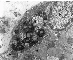
[image:3.612.253.567.70.328.2]A further finding of note is the low content of intracellular lipid droplets (IMCLs, intramyocellular lipids) in muscle biopsies from permanent high-altitude residents. We found on average less than half the IMCL content (0.2 % of muscle fibre volume) of a comparable lowland population (0.5 %). Whereas lowlanders typically double their IMCL content under similar training conditions (Hoppeler et al., 1985), high-altitude residents did not increase the lipid content of their muscle fibres significantly. Large intracellular lipid deposits are observed in endurance athletes, in particular in athletes
competing in events lasting several hours, such as cyclists (Hoppeler, 1986), and may also be induced by consuming a high-fat diet (Hoppeler et al., 1999). These deposits are thought to be of advantage and to be related to the higher reliance of trained muscle on lipids as a substrate for mitochondrial respiration. Low IMCL contents are compatible with the contention that permanent sojourn at high altitude induces a shift in muscle metabolism towards a preferred reliance on carbohydrates as substrate (Hochachka et al., 1996).
In conclusion, we found that permanent high-altitude residents have an unremarkable fibre type composition, slightly reduced fibre cross-sectional area, remarkably low oxidative capacities with a capillary supply reduced in proportion and low intracellular lipid stores. These acclimatory features place high-altitude residents at a distinct disadvantage when exposed to sea-level conditions. Favier et al. (Favier et al., 1995) reported that the V.O∑max of this high-altitude population increased by only 8.2 % when a V.O∑maxtest was carried out in acute hypobaric normoxia (supplementing the inspired air with oxygen). The data compiled by Cerretelli and Hoppeler (Cerretelli and Hoppeler, 1996) indicate that the V.O∑max of a lowlander would increase by 20–25 % when tested under similar experimental conditions.
Muscle structure in Sherpas and Quechuas
The possible effects of phylogenetic adaptation (Hochachka and Somero, 1984) to hypoxia can be studied on populations that have been living at high altitude for thousands of generations, such as Tibetans and Quechas. It has been proposed from a phylogenetic analysis, assuming a species life of 100 000 years for humans, that for a third of that time the Himalayan highlanders and the Andean highlanders did not share common ancestors (Hochachka et al., 1998). Thus, the hypoxia defence mechanisms observed in these two populations arose independently and by positive selection. These authors mention five response systems in which similar traits have evolved in both populations; a blunted hypoxic ventilatory response, a blunted pulmonary vasoconstrictor response, an upregulation of expression of vascular endothelial growth factor, an upregulation of expression of erythropoietin in the kidney and regulatory adjustments of metabolic pathways in skeletal muscle tissue. In the context of the present review, we will concentrate mainly on the latter.
Tibetans (Kayser et al., 1991; Kayser et al., 1996) and Quechas (Rosser and Hochachka, 1993) have a (small) preponderance of slow type I fibres in vastus lateralis muscle (i.e. close to 60 % in Tibetans and 68 % in Quechuas; N=3) compared with approximately 50 % in typical lowland populations. There is also a tendency for the highland population to have estimates of fibre cross-sectional area at the low end of the normoxic spectrum. Muscle oxidative capacity, measured as mitochondrial volume density, is reduced in Tibetans (3.96 %), but their capillary density is within the normal range (467 mm2; Kayser et al., 1991). Comparing second-generation Tibetans (refugees, born and raised in
Katmandu, Nepal, 1300 m) with lowland Nepalese living in the same city, significantly lower volume densities of mitochondria and similarly reduced citrate synthase activities together with lower intramyocellular lipid concentrations and lower 3-hydroxy-acyl-CoA dehydrogenase activities were noted for Tibetans. Moreover, Tibetans had slightly, but significantly, estimates of reduced fibre cross-sectional area. These Tibetans were never exposed to the altitudes at which their ancestors lived (3000–4500 m), emphasizing a hereditary component to these differences.
Together, these adaptations in Tibetans and Quechuas were interpreted as a downregulation of maximum aerobic and anaerobic exercise capacities with a concomitant upregulation of oxidative compared with glycolytic contributions to energy supply (Hochachka et al., 1998). The preponderance of type I fibres is taken as favouring a tighter coupling between ATP demand and ATP supply, reduced lactate accumulation and improved endurance under submaximal conditions. To this, one might add that the shift away from lipid substrates would further optimize the amount of ATP produced per litre of oxygen consumed.
Muscle structure with training in intermittent hypoxia, acclimation studies
Since the seminal paper on muscle tissue of permanent high-altitude residents (Reynafarje, 1962), it had tacitly or openly been assumed that one of the key factors modulating the response of muscle tissue to exercise was local tissue hypoxia. As the evidence from acute and permanent exposure to hypoxia in humans indicated otherwise, it became necessary to design experiments in which a potential hypoxia stimulus could be dissociated from the (negative) effects of permanent exposure to altitude. One way to achieve such a dissociation is to make subjects exercise in hypoxia but to keep them in normoxia for the remainder of the day (intermittent hypoxia exposure). Studies using these protocols encounter the problem of standardization of absolute versus relative exercise intensities at different levels of hypoxia.
than in the normobaric-trained leg. Moreover, the hypobaric-trained leg showed a significantly larger increase in citrate synthase activity and an increase in myoglobin concentration. Another study using single-leg training at a simulated altitude of 3300 m also showed larger increases in citrate synthase activity in the hypoxia-trained than in the normoxia-trained leg, but no significant differences in the improvements in muscle function (Melissa et al., 1997). Desplanches et al. (Desplanches et al., 1993) trained subjects for 3 weeks, (2 h per day, 6 days per week) at simulated altitudes up to 6000 m or under sea-level conditions and found that V.O∑maximproved in hypoxia-trained subjects, but only when measured in hypoxia. The muscle structural adaptations were similar in hypoxia and normoxia training except for an increase in muscle volume and muscle fibre cross-sectional area, which were observed only with hypoxia training. Taken together, these studies are compatible with the generally held idea that altitude training is of advantage for competition at altitude (Cerretelli and Hoppeler, 1996). In addition, they suggest that training in hypoxia could have specific effects on muscle tissue not seen with training of similar intensity in normoxia. However, the data currently available do not indicate which altitude or which training protocol is optimal for improving athletic performance capacity.
To test the hypothesis of a specific response of skeletal muscle to hypoxia training, in particular a possible involvement of hypoxia-inducible factor 1 (HIF-1), we used training programmes in which hypoxia was present only during
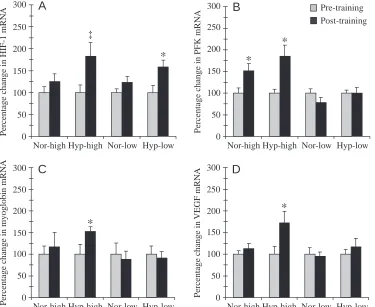
the training sessions (Vogt et al., 2001; Geiser et al., 2001). Four groups of subjects were set up, two of these trained under normoxic and two under hypoxic conditions (corresponding to an altitude of 3850 m) for 30 min, five times a week for a total of 6 weeks on a bicycle ergometer. From each of the two oxotensic groups, one trained at a high intensity, corresponding to the anaerobic threshold, and the other some 25 % below this level. Muscle biopsies were taken from the vastus lateralis muscle before and after the training period and analyzed morphometrically and for changes in mRNA levels of proteins potentially implicated in the response to hypoxia. In vitro experiments had revealed that HIF-1, which is involved in oxygen sensing in mammalian cells, including skeletal muscle, is specifically activated by hypoxia (for reviews, see Wenger, 2000; Semenza, 2000). The transcription factor HIF-1 targets genes coding for proteins involved in oxygen transport (erythropoietin and vascular endothelial growth factor, VEGF) as well as genes coding for glycolytic enzymes and glucose transporters. In our experiments, mRNA levels of the regulatory subunit of HIF-1 increased after training under hypoxic conditions irrespective of training intensity, but not after training in normoxia (Fig. 3A; Hyp-high, +82 %, P<0.10; Hyp-low, +58 %, P<0.05). To us this suggests a specific molecular response to the hypoxic stimulus. Only high-intensity training in hypoxia increased mRNAs coding for myoglobin and VEGF (Fig. 3C,D). The higher level of the VEGF mRNA was reflected by a parallel increase in capillary density (Fig. 4B). Furthermore, we detected increases in levels
Nor-high Hyp-high Nor-low Hyp-low 0
50 100 150 200 250 300
Pe
rcenta
g
e c
h
an
g
e
in
H
IF
-1
mR
N
A
A
‡
*
Nor-high Hyp-high Nor-low Hyp-low 0
50 100 150 200 250 300
Pe
rcenta
g
e c
h
an
g
e
in m
y
og
lo
b
in mR
N
A
C
*
Nor-high Hyp-high Nor-low Hyp-low 0
50 100 150 200 250 300
Pe
rcenta
g
e c
h
an
g
e
in PF
K
mR
N
A
B
*
*
Pre-training Post-training
Nor-high Hyp-high Nor-low Hyp-low 0
50 100 150 200 250 300
Pe
rcenta
g
e c
h
an
g
e
in
VEG
F mR
N
A
D
[image:5.612.198.573.430.737.2]*
Fig. 3. Percentage changes in levels of mRNAs coding for the regulatory subunit of hypoxia-inducible factor-1 (HIF-factor-1) (A), phosphofructokinase (PKF) (B), myoglobin (C) and vascular endothelial growth factor (VEGF) (D) after 6 weeks of endurance training in hypoxia (Hyp) or normoxia (Nor) at individual anaerobic threshold (high) or at a level of training approximately 25 % below individual anaerobic threshold (low) (data from Vogt et al., 2001). Pre-training values are normalized to 100 %. Values are means + S.E.M. (N=8 for both normoxia groups; N=7 for both hypoxia groups). *Significant difference between pre- and post-training values (P<0.05); ‡a
of mRNA coding for phophofructokinase, which is involved in the glycolytic pathway and is an established HIF downstream gene (Fig. 3C), and in mitochondrial volume density (Fig. 4A) after high-intensity training in both hypoxia and normoxia. Both changes in phosphofructokinase mRNA (Fig. 3C) and mitochondrial volume density (Fig. 4) were larger with the hypoxic stimulus (ANOVA).
Taken together, these results support the involvement of HIF-1 in the regulation of adaptation processes in skeletal muscle tissue after training in hypoxia. Our results suggest that high-intensity training in hypoxia leads to adaptations that compensate for the reduced availability of oxygen during training. A high capillarity facilitates the supply of oxygen and substrates to muscle cells. The higher concentration of myoglobin could improve the capacity for storing and transporting oxygen within muscle cells. Finally, by inducing metabolic pathways that favour the use of carbohydrates instead of lipids as substrate (upregulation of glycolytic and oxidative pathways), oxygen would be used more efficiently.
With regard to the increase in muscle myoglobin concentration demonstrated in humans only after high-intensity training in hypoxia, we hypothesize that this could be of advantage to exercise performance at altitude. Endurance training, sprint training and resistance training in normoxia failed to induce changes in muscle myoglobin concentration in humans (Jansson et al., 1982; Svedenhag et al., 1983; Jacobs et al., 1987; Hickson, 1981; Harms and Hickson, 1983; Masuda et al., 1999). In elite cyclists, there is a high correlation between cycling performance and muscle myoglobin content (Faria, 1992). A high myoglobin concentration could facilitate oxygen supply under hypoxic conditions when training or competing at altitude. We have tested the functional consequence of intermittent hypoxia training in several studies with untrained subjects, endurance-trained athletes and elite alpine ski racers (Vogt, 1999; Vogt et al., 1999). Overall, our results indicate an increase in V.O∑max, an increase in maximal power output, an increase in maximal ventilatory response
and an improvement in the rating of perceived exertion, particularly when these variables are measured at altitude.
In addition to the effects of the hypoxic stimuli on exercise performance, there is recent evidence that intermittent hypoxia training might have clinical implications. Bailey et al. (Bailey et al., 2000) trained physically active subjects either in normoxia or in normobaric hypoxia (fractional O2 content 16 %). After training both in normoxia and in hypoxia, concentrations of free fatty acids, total cholesterol, HDL-cholesterol and LDL-HDL-cholesterol were decreased. The concentration of homocysteine, an amino acid implicated in coronary disease, was reduced by 11 % after hypoxia training only. Furthermore, maximal systolic blood pressure was reduced after hypoxia training, indicating a hypotensive effect of hypoxia training, possibly mediated by morphological changes in the endothelium. From these results, the authors concluded that hypoxia training might be beneficial for patients with cardiovascular diseases.
Taken together, intermittent hypoxia training (‘living low – training high’) has been shown to elicit specific molecular responses in skeletal muscle tissue. It is likely that this type of training has the potential to produce a (small) increase in muscle mass not seen in response to normoxia training. It is not presently possible to make specific recommendations as to the best protocols to be used (in terms of intensity, duration and training altitude) to improve performance at altitude. The increase in muscle myoglobin content (initially demonstrated by Reynafarje, 1962) and capillarity may at least partly explain why functional improvements after hypoxia training are more pronounced under hypoxic testing conditions (altitude specificity of training). Intermittent hypoxia training can be considered to be complementary to training schemes in which (mild) hypoxia is applied over hours with the intention of increasing the aerobic performance capacity by increasing haemoglobin concentration (‘living high – training low’). The disadvantage of intermittent hypoxia training is the requirement for technical installations to simulate appropriate
Nor-high Hyp-high Nor-low Hyp-low 0
50 100 150 200
Pe
rcenta
g
e c
h
an
g
e
in Vv
(mt,f)
A
*
Nor-high Hyp-high Nor-low Hyp-low 0
50 100 150 200
Pe
rcenta
g
e c
h
an
g
e
in
Jv (c,f)
B
*
Pre-training Post-training
[image:6.612.113.490.74.231.2]*
*
altitude conditions either by diluting environmental air with nitrogen or by reducing atmospheric pressure.
This work was supported by the Swiss National Science Foundation, Swiss Olympics and the University of Bern. The secretarial help of L. Gfeller-Tüscher is gratefully acknowledged.
References
Bailey, D. M., Davies, B. and Baker, J. (2000). Training in hypoxia: modulation of metabolic and cardiovascular risk factors in men. Med. Sci. Sports Exerc. 32, 1058–1066.
Banchero, N. (1987). Cardiovascular responses to chronic hypoxia. Annu. Rev. Physiol. 49, 465–476.
Bligh, J. and Johnson, K. G. (1973). Glossary of terms for thermal physiology. J. Appl. Physiol. 35, 941–961.
Cerretelli, P. and Hoppeler, H. (1996). Morphologic and metabolic response to chronic hypoxia: the muscle system. In Handbook of Physiology , vol. 2, section 4, Environmental Physiology (ed. M. J. Fregly and C. M. Blatteis), pp. 1155–1181. Oxford: Oxford University Press.
Desplanches, D., Hoppeler, H., Linossier, M. T., Denis, C., Claassen, H., Dormois, D., Lacour, J. R. and Geyssant, A. (1993). Effects of training in normobaric hypoxia on human muscle ultrastructure. Pflügers Arch. 425, 263–267.
Desplanches, D., Hoppeler, H., Tüscher, L., Mayet, M. H., Spielvogel, H., Ferretti, G., Kayser, B., Leuenberger, M., Grünenfelder, A. and Favier, R. (1996). Muscle tissue adaptation of high-altitude natives to training in chronic hypoxia or acute normoxia. J. Appl. Physiol. 81, 1946–1951. Emonson, D. L., Aminuddin, A. H., Wight, R. L., Scroop, G. C. and Gore,
C. J. (1997). Training-induced increases in sea level V.O∑maxand endurance
are not enhanced by acute hypobaric exposure. Eur. J. Appl. Physiol. 76, 8–12.
Faria, I. E. (1992). Energy expenditure, aerodynamics and medical problems in cycling. Sports Med. 14, 43–63.
Favier, R., Spielvogel, H., Desplanches, D., Ferretti, G., Kayser, B., Lindstedt, S. L. and Hoppeler, H. (1995). Maximal exercise performance in chronic hypoxia and acute normoxia in high-altitude natives. J. Appl. Physiol. 78, 1868–1874.
Geiser, J., Vogt, M., Billeter, R., Zuleger, C., Belforti, F. and Hoppeler, H. (2001). Training high – living low: changes of aerobic performance and muscle structure with training at simulated altitude. Int. J. Sports Med. (in press).
Green, H. J., Sutton, J. R., Cymerman, A., Young, P. M. and Houston, C. S. (1989). Operation Everest II: Adaptations in human skeletal muscle. J. Appl. Physiol. 66, 2454–2461.
Harms, S. J. and Hickson, R. C. (1983). Skeletal muscle mitochondria and myoglobin, endurance and intensity of training. J. Appl. Physiol. 54, 798–802. Hickson, R. C. (1981). Skeletal muscle cytochrome c and myoglobin,
endurance and frequence of training. J. Appl. Physiol. 51, 746–749. Hochachka, P. W., Buck, L. T., Doll, C. J. and Land, S. C. (1996). Unifying
theory of hypoxia tolerance: Molecular/metabolic defense and rescue mechanisms for surviving oxygen lack. Proc. Natl. Acad. Sci. USA 93, 9493–9498.
Hochachka, P. W., Gunga, H. C. and Kirsch, K. (1998). Our ancestral physiological phenotype: An adaptation for hypoxia tolerance and for endurance performance? Proc. Natl. Acad. Sci. USA 95, 1915–1920. Hochachka, P. W. and Somero, G. N. (1984). Biochemical Adaptation.
Princeton, NJ: Princeton University Press. 557pp.
Hochachka, P. W., Stanley, C., Merkt, J. and Sumar Kalinowski, J. (1983). Metabolic meaning of elevated levels of oxidative enzymes in high altitude adapted animals: An interpretive hypothesis. Respir. Physiol. 52, 303–313. Hoppeler, H. (1986). Exercise-induced ultrastructural changes in skeletal
muscle. Int. J. Sports Med. 7, 187–204.
Hoppeler, H., Billeter, R., Horvath, P. J., Leddy, J. J. and Pendergast, D. R. (1999). Muscle structure with low- and high-fat diets in well-trained male runners. Int. J. Sports Med. 20, 522–526.
Hoppeler, H., Howald, H., Conley, K., Lindstedt, S. L., Claassen, H., Vock,
P. and Weibel, E. R. (1985). Endurance training in humans: Aerobic capacity and structure of skeletal muscle. J. Appl. Physiol. 59, 320–327. Hoppeler, H., Kleinert, E., Schlegel, C., Claassen, H., Howald, H. and
Cerretelli, P. (1990). Muscular exercise at high altitude. II. Morphological adaptation of skeletal muscle to chronic hypoxia. Int. J. Sports Med. 11, S3–S9.
Howald, H., Pette, D., Simoneau, J. A., Uber, A., Hoppeler, H. and Cerretelli, P. (1990). Muscular exercise at high altitude. III. Effects of chronic hypoxia on muscle enzymes. Int. J. Sports Med. 11, S10–S14. Jacobs, I., Esbjörnsson, M., Sylven, C., Holm, I. and Jansson, E. (1987).
Sprint training effects on muscle myoglobin, enzymes, fiber types and blood lactate. Med. Sci. Sports Exerc. 19, 368–374.
Jansson, E., Sylven, C. and Nordevang, E. (1982). Myoglobin in the quadriceps femoris muscle of competitive cyclists and untrained men. Acta Physiol. Scand. 114, 627–629.
Kayser, B., Acheson, K., Décombaz, J., Fern, E. and Cerretelli, P. (1992). Protein absorption and energy digestibility at high altitude. J. Appl. Physiol. 73, 2425–2431.
Kayser, B., Hoppeler, H., Claassen, H. and Cerretelli, P. (1991). Muscle structure and performance capacity of Himalayan Sherpas. J. Appl. Physiol. 70, 1938–1942.
Kayser, B., Hoppeler, H., Desplanches, D., Marconi, C., Broers, B. and Cerretelli, P. (1996). Muscle ultrastructure and biochemistry of lowland Tibetans. J. Appl. Physiol. 81, 419–425.
Kayser, B., Narici, M., Milesi, S., Grassi, B. and Cerretelli, P. (1993). Body composition and maximum alactic anaerobic performance during a one month stay at high altitude. Int. J. Sports Med. 14, 244–247.
MacDougall, J. D., Green, H. J., Sutton, J. R., Coates, G., Cymerman, A. Y. P. and Houston, C. S. (1991). Operation Everest-II: Structural adaptations in skeletal muscle in response to extreme simulated altitude. Acta Physiol. Scand. 142, 421–427.
Martinelli, M., Winterhalder, R., Cerretelli, P., Howald, H. and Hoppeler, H. (1990). Muscle lipofuscin content and satellite cell volume is increased after high altitude exposure in humans. Experientia 46, 672–676. Masuda, K., Choi, J. Y., Shimojo, H. and Katsuta, S. (1999). Maintenance
of myoglobin concentration in human skeletal muscle after heavy resistance training. Eur. J. Appl. Physiol. 79, 347–352.
Melissa, L., MacDougall, J. D., Tarnopolsky, M. A., Cipriano, N. and Green, H. J. (1997). Skeletal muscle adaptations to training under normobaric hypoxic versus normoxic conditions. Med. Sci. Sports Exerc. 29, 238–243.
Reynafarje, B. (1962). Myoglobin content and enzymatic activity of muscle and altitude adaptation. J. Appl. Physiol. 17, 301–305.
Rosser, B. W. and Hochachka, P. W. (1993). Metabolic capacity of muscle fibers from high-altitude natives. Eur. J. Appl. Physiol. 67, 513–517.
Semenza, G. L. (2000). HIF-1: mediator of physiological and
pathophysiological responses to hypoxia. J. Appl. Physiol. 88, 1474–1480. Svedenhag, K., Henriksson, J. and Sylven, C. (1983). Dissociation of training effects on skeletal muscle mitochondrial enzymes and myoglobin in man. Acta Physiol. Scand. 117, 213–218.
Terrados, N., Jansson, E., Sylven, C. and Kaijser, L. (1990). Is hypoxia a stimulus for synthesis of oxidative enzymes and myoglobin? J. Appl. Physiol. 68, 2369–2372.
Terrados, N., Melichna, J., Sylven, C., Jansson, E. and Kaijser, L. (1988). Effects of training at simulated altitude on performance and muscle metabolic capacity in competitive road cyclists. Eur. J. Appl. Physiol. 57, 203–209.
Valdivia, E. (1958). Total capillary bed in striated muscle of Guinea pigs native to the Peruvian mountains. Am. J. Physiol. 194, 585–589.
Vogt, M. (1999). Hypoxie und Diät als Beeinflussungsvariablen
trainingsbedingter Adaptationsprozesse in der menschlichen Skelettmuskulatur. Dissertation, Philosophisch-Naturwissenschaftliche Fakultät, Universität Bern.
Vogt, M., Puntschart, A., Geiser, J., Zuleger, C., Billeter, R. and Hoppeler, H. (2001). Training high – living low: Molecular adaptations in human skeletal muscle to endurance training under simulated high-altitude conditions. J. Appl. Physiol. 91, 173–182.
Vogt, M., Werlen, L. and Hoppeler, H. (1999). Spielformen des Höhentrainings. Schweiz. Zeitschr. Med. Traumatol. 47, 125–128. Wenger, R. H. (2000). Mammalian oxygen sensing, signalling and gene