Original Article
Expression and contribution of autophagy to the luteal
development and function in the pregnant rats
Zonghao Tang1, Zhenghong Zhang1, Yuxiu Huang2, Yedong Tang1, Jiajie Chen1, Fan Wang1, Zhengchao Wang1
1Provincial Key Laboratory for Developmental Biology and Neurosciences, College of Life Sciences, Fujian Normal
University, Fuzhou 350007, P. R. China; 2Department of Obstetrics and Gynecology, The First Affiliated Hospital of
Fujian Medical University, Fuzhou 350005, P. R. China
Received April 7, 2017; Accepted November 7, 2017; Epub December 15, 2017; Published December 30, 2017
Abstract: Autophagy has been demonstrated as an important regulatory mechanism in the apoptosis of luteal cells during the regression of corpus luteum (CL) in the pseudopregnant rat ovary. However, the role of autophagy during the rat pregnancy still remains unknown. Therefore, the present study was designed to investigate the expression and contribution of autophagy to the luteal development and function in the pregnant rats. Ovaries were obtained from the female rats at the early, middle or late phase of the pregnancy, which correspondingly had three groups, including the early (ELP), middle (MLP) and late luteal phase (LLP). The results of autophagy-associated protein LC-3 showed that autophagy expressed during the pregnant CL and significantly increased during the late luteal phase, which was further confirmed by the mRNA and protein expression changes of the other two autophagy re -lated proteins, Beclin-1 and Atg-5. These results indicated autophagy may play an important role in the luteolysis. Further examinations of cellular apoptosis marker protein cleaved caspse-3 and ovarian prostaglandin2α (PGF2α) levels found that cleaved caspse-3 dramatically increased during the late luteal phase which consistent with the changes of autophagy, while ovarian PGF2α level significantly increased during the middle luteal phase and dramati -cally decreased during the late luteal phase. Together, these results suggested that autophagy may be induced by PGF2α and then contributed to the luteolysis through cellular apoptosis during the late luteal phase of pregnant ovaries. To our knowledge, this will provide a new insight into the important mechanism regulating the luteolysis of the pregnant ovaries in mammals.
Keywords: Autophagy, apoptosis, PGF2α, corpus luteum, pregnant rat
Introduction
Autophagy is an evolutionary conserved cellu-lar bulk degradation system in cells, by which cell may envelop part of cytoplasm in double membrane-bound structures called autophago-somes for the recycle of building blocks [1, 2]. Cell usually maintains a basal autophagy level under physiological conditions, whereas adver- se survival environments such as short of growth factors or nutrients can lead to the up-regulation of cellular autophagy levels so as to bridge the shortage of energy [3, 4]. Accordingly, autophagy was originally thought as a pro-sur-vival mechanism to nutrient deprivation, hypoxia and other types of cellular stress [5, 6]. However, many studies suggested that exces-sive self-digestion or the degradation of es-
sential cellular components also promoted the apoptosis of cell by disrupting the normal regulation of cell physiology and provoke the activation of caspase dependent apoptosis pathways [7-9]. In addition, the morphological changes of steroidogenic cells during corpus luteum (CL) regression, which were character-ized by the vacuolization of cytoplasmic, and lysosomes, the accumulation of
autophago-somes, further confirmed that the apoptosis of
Autophagy in the pregnant corpus luteum
The CL is an important hormone-responsive and hormone-productive reproductive organ in mammals, and the function of which is required for the success of early pregnancy from embryo implantation to development of the conceptus [14]. The main function of CL is to synthesis and secret progesterone [15], which is main-tained during the early phase of pregnancy, and
significantly increased at the middle and late
stages of pregnancy in mammal [14]. However, the absence of pregnancy or the ending of preg-nancy resulted in the removal of CL from ovary, which is mainly caused by the increased level of ovarian prostaglandin F2α (PGF2α) [16]. Therefore, the present study was designed to evaluate the role of autophagy during the devel-opment of corpus luteum in the pregnant rats. The changes of autophagy related protein expressions and related hormones were exam-ined to explore the expression and contribution of autophagy to the luteal development and function in the pregnant rats.
Materials and methods
Animals
Sprague-Dawley rats were purchased from Wushi Experimental Animal Supply Co. Ltd. (Fuzhou, China). The animals were maintained under a 14-h light/10-h dark schedule with continuous supply of chow and water. The experimental protocol was approved by the Institutional Animal Care and Use Committee and the Ethics Committee on Animal Experi- mentation, Fujian Normal University.
Experimental design
The rats were allowed to accommodate for 1~2 week prior to mating with males, which occurred at 2~3 months of age (200-250 g body weight). Previously unmated female rats (three per cage) were mated with a fertile male (one per cage) and were examined every morning for the presence of a vaginal plug. Day 1 of pregnancy
was defined as the day when a vaginal plug was
recovered. The pregnant females were removed and used in the subsequent experiments.
To further confirm the pregnant rat model,
serum levels of progesterone were measured at different phases of pregnancy corresponding to the early, middle and late phase of rat preg-nancy, which correspondingly had three groups
in the present study, including the early luteal phase (ELP, Day 1 to 4), the middle luteal phase (MLP, Day 5 to 16) and the late luteal phase (LLP, Day 17 to 21) [14]. The rats were anesthe-tized with atropine (0.05 mg/kg body weight ip, Sigma-Aldrich, St. Louis, MO, USA) on the days of sample collection prior to opening of the
abdomen, and ≥3.0 ml blood was collected
from the abdominal aorta, and centrifuged at 1,000 x g at 4°C for 10 min. The ovaries were rapidly excised and chilled in ice-cold 0.154 M
NaCl with 14.0 μM indomethacin
(Sigma-Aldrich) immediately following perfusion for measuring the expression levels of autophagy-related proteins.
For histological analysis, one ovary from each
rat was fixed in 4% paraformaldehyde
(Sigma-Aldrich), and the other ovary was snap-frozen and used for the remaining experiments.
Ovarian perfusion
To avoid the effects of the vascular system, ovarian perfusion was performed before the collection of the ovaries. The female rats were perfused in vivo through the abdominal aorta with 0.154 M NaCl. After euthanasia, the abdo-men was opened via a mid-ventral incision and an intravenous cannula was inserted via the
aortic bifurcation. About 40 μl of the perfusion
solution was perfused at ambient temperature through the lower abdominal vascular system for ~5 min at constant pressure using a hand-held syringe. Perfusion was suspended when the corpora lutea become completely pale. The ovaries were then rapidly removed for subse-quent analysis of gene expression.
Immunohistochemistry of LC-3
Generally, the methods for immunohistochemi-cal staining of LC-3 were done according to the manufacturer’s recommendations and
report-ed studies [13]. In briefly, paraffin-embreport-eddreport-ed
tissue sections were dewaxed and rehydarated regularly. Hereafter, the sections were subject-ed to antigen microwave antigen retrieval by 0.01 M citric acid buffer for 10 min. Endogenous peroxide was inhibited by incubation of the
sec-tions in 3% H2O2 for 30 min. The sections were
antibodies used were anti-LC-3 antibody (dilut-ed 1:500, Abcam, Cambridge, MA, USA). After washing with PBS, slides were incubated with the secondary antibodies at room temperature for 20 min. For visualizing, Diaminobenzidine tetrahydrochloride chromogen staining was applied. All section were counterstained with hematoxylin, dehydrated and mounted lastly.
Western blot analysis of LC-3, Beclin-1 and Atg-5 expression levels
Ovarian tissues from each group were homog-enized in ice cold RIPA lysate buffer (Beyotime Institute of Biotechnology, Haimen, China) and centrifuged at 15000 g for 15 min at 4°C, and then the supernatant was collected. Protein concentrations were thereafter determined using BCA Protein Assay Kit (Beyotime Institute of Biotechnology, Haimen, China). Protein
sam-ples were diluted into the equal concentra-
tion and then 20 μg of the protein samples were subjected to 10% SDS-PAGE gel electro -phoresis and then electrophoretically
trans-ferred onto a polyvinylidene difluoride mem -brane (Pall Life Sciences, Port Washington, NY, USA). The membrane was washed with TBS
with 0.2% Tween 20 (TBST; Sigma-Aldrich). Nonspecific binding to the membrane was blocked with 5% nonfat milk in Tris buffered
saline-Tween 20 (TBST, pH 7.4) for 1 h at room temperature. After that, the membranes were incubated overnight at 4°C with LC-3 anti-body (1:1000 dilution, Abcam, Cambridge, MA, USA), anti-Beclin-1 antibody (1:2000 dilution, Protein Tech Group, Wuhan, China), anti-Atg5 antibody (1:1000 dilution, Protein Tech Group,
Wuhan, China) and anti-β-actin antibody
[image:3.612.91.522.69.404.2](1:5000 dilution, Protein Tech Group, Wuhan,
Autophagy in the pregnant corpus luteum
China). After washing with TBST for three times, the membrane were incubated in ho- rseradish peroxidase-conjugated goat anti-rab-bit or mouse IgG (1:5000 dilution, Beyotime Institute of Biotechnology, Haimen, China) for 1 h at room temperature. After that, bands were visualized by using the enhanced chemi- luminescence star (ECL, Beyotime Institute of Biotechnology, Haimen, China). The blots
were quantified using ImageJ 1.49 software
(National Institutes of Health, Bethesda, MD, USA).
RNA extraction and reverse transcription- quan-titative polymerase chain reaction analysis of Beclin-1 and Atg-5 mRNA
Total RNA was extracted from the ovaries using TRIzol solution (Invitrogen Life Technologies, Carlsbad, CA, USA) and then reverse-tran-scribed using a cDNA Synthesis kit (Promega, Biotech Co., Ltd). The reverse-transcribed
prod-ucts were amplified using a FasQuant RT Kit
(TIANGEN BIOTECH CO., LTD, Beijing, China), with Go Taq qPCR Master Mix (Promega corpo-ration, Lot 0000209928), Beclin1 primer (for-ward primer 5-ATG CTG TCC TTT CCC TCT TCC-3’, reverse primer 5’-ACC TTT ACC TCT TGT CCC TTC C-3’) and Atg-5 primer (forward primer 5’-AGA AGA AGA GCC AGG TGA TGA-3’, reverse primer 5’-AAT GCT GAT GTG AAG GAA GTT GT-3’). A kit for detecting the levels of 18S ribo-somal RNA (Hs99999901_s1) was used as an
endogenous control. The 20 μl PCR reaction mix contained 10.0 μl 2X Go Taq qPCR Master Mix (Promega corporation), 0.2 ul CXR refer
-ence Dye, 2.0 μl cDNA template, 7.0 μl
RNase-free water and 0.8 primer (containing 0.4 ul forward and 0.4 ul reverse). The PCR conditions of the RT-qPCR system (Applied Biosystems Life Technologies), were as follows: 50°C for 2 min, 95°C for 10 min, followed by 40 cycles at 95°C for 15 s, and 60°C for 1 min. The relative gene expression levels were calculated in
accordance with the ΔΔCt method, and relative
mRNA levels were expressed as 2-ΔΔCt values
[17, 18].
Radioimmunoassay of progesterone and pros-taglandin F2α
The levels of serum progesterone and ovarian prostaglandin F2α were determined using
spe-cific radioimmunoassay kits, according to the
manufacturer’s instructions. The progesterone
RIA kit [intra-assay coefficient of variation (CV) < 4.3%; inter-assay CV < 7.1%] and the prosta -glandin F2a radioimmunoassay kit (intra-assay
CV < 10.0% and interassay CV < 15.0%) were
from Atomic Gaoke Co., Ltd., Department of Isotope, China Institute of Atomic Energy (Beijing, China). Protein concentrations were determined using a BCA kit (Beyotime Institute of Biotechnology, Haimen, China), with bovine serum albumin standards.
Statistics
Data are presented as means ± SE. The signifi -cance of differences in mean values within and between multiple groups was evaluated using a
one-way ANOVA, followed by a Tukey’s multiple
range test. P < 0.05 was considered
statisti-cally significance. #: P < 0.05, vs. the early
lutral phase (ELP), and &: P < 0.05, vs. the mid-dle lutral phase (MLP).
Results
Expression and localization of LC-3 during the luteal development in the ovaries of pregnant rats
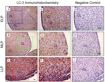
During the pregnancy, corpus luteum is the main part in the ovary. The present results showed LC-3 expressed during this period and mainly localized in steroidogenic cells (Figure 1). Notably, the immunoreactivity of LC-3 main-tained a weak level at ELP (Figure 1A and 1D) and MLP (Figure 1B and 1E) compared with the negative control (Figure 1G-I), LC-3
expres-sions significantly increased at LLP (Figure 1C
and 1F). These results indicated that LC-3 mediated-autophagy expressed during the lute-al development of pregnant rats, especilute-ally luteolysis at LLP.
LC-3II expression increased during the luteal development in the ovaries of pregnant rats
For further confirming the results of LC-3 immu -nohistochemistry, the present study also detected LC-3 protein expression by western
Autophagy-related proteins Beclin-1 and Atg-5 increased during the luteal development in the ovaries of pregnant rats
Beclin1 and Atg-5 are two essential factors that involved in the initiation of autophagy in mam-malian cells though autophagy could also be induced via Beclin-1 independent pathways
[19]. The present study firstly examined the
[image:5.612.327.520.74.354.2]mRNA expression levels of Beclin1 and Atg-5 and found their expressions dramatically increased at LLP compared with ELP and MLP (Figure 3) and then detected the protein expres-sion levels of these two proteins and found the similar results with mRNA expressions (Figure 4). These results demonstrated the expression levels of both proteins were upregulated and the induction of autophagy was occurred in a
Figure 2. Blotting analysis of LC-3I and LC-3II during the luteal development in the ovaries of pregnant rats. A: Representative ECL gel documents of West-ern blot analyses depicting the protein levels of LC-3I and LC-3II. B: Summarized intensities of LC-3II blots normalized to the control. Each value represents the mean ± SE, n=6. One-way analysis of variance (ANOVA) was used to analyze the data. ELP: the early luteal phase, MLP: the middle phase, LLP: the late luteal phase, #: P < 0.05, vs. ELP group, &: P < 0.05, vs. MLP group.
[image:5.612.91.288.74.239.2]Figure 3. Expression changes of Beclin-1 and Atg-5 mRNA during the luteal development in the ova-ries of pregnant rats. A: The relative mRNA levels of Beclin-1 by real-time RT-PCR analysis. B: The relative mRNA levels of Atg-5 by real-time RT-PCR analysis. Each value represents the mean ± SE. n=6, One-way analysis of variance (ANOVA) was used to analyze the data. ELP: the early luteal phase, MLP: the middle phase, LLP: the late luteal phase, #: P < 0.05, vs. ELP group, &: P < 0.05, vs. MLP group.
[image:5.612.94.284.386.591.2]Autophagy in the pregnant corpus luteum
Beclin-1 dependent manner during the late luteal phase of rat pregnancy.
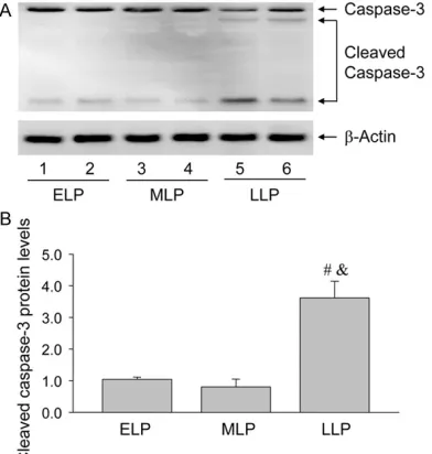
Activation of luteal cell apoptosis during the luteal development in the ovaries of pregnant rats
Given the important role of cell apoptosis in the luteolysis at the late luteal phase of pregnancy, the present study detected the expression of caspase-3 and found the expression of cleaved
caspase-3 significantly increased at LLP com -pared with ELP and MLP (Figure 5), indicating the apoptosis of luteal cells via the activation of caspase-3 may be related with the induction of autophagy during the luteolysis of pregnancy.
Serum progesterone and ovarian prostaglan-din F2a during the luteal development in the ovaries of pregnant rats
It is well-known that serum progesterone levels keep increasing during the pregnancy and ovar-ian prostaglandin F2a contributes to the luteoly-sis of the ovary. Therefore, the present study
measured these two hormone levels and found serum progesterone level was the highest at LLP (Figure 6A) and the lowest at ELP (Figure 6A) as reported previously. Interestingly, ovari-an prostaglovari-andin F2a increased at MLP (Figure 6B) and then decreased at LLP (Figure 6B), implying the changes of prostaglandin F2a were also involved in the role of autophagy during the luteolysis through cell apoptosis in the ova-ries of pregnant rats.
Discussion
The results of the present study clearly de-
monstrated that autophagy was significantly
[image:6.612.326.519.74.288.2]induced and activated at LLP of pregnant rats, while the expression of cleaved caspase-3 in a similar manner. These results suggested that autophagy may play an important role in the luteal regression of pregnant ovaries in vivo in mammals.
Figure 5. Blotting analysis of caspase-3 and cleaved caspase-3 during the luteal development in the ova-ries of pregnant rats. A: Representative ECL gel doc-uments of Western blot analyses depicting the pro-tein levels of caspase-3 and cleaved caspase-3. B: Summarized intensities of cleaved caspase-3 blots normalized to the control. Each value represents the mean ± SE, n=6. One-way analysis of variance (ANOVA) was used to analyze the data. ELP: the early luteal phase, MLP: the middle phase, LLP: the late luteal phase, #: P < 0.05, vs. ELP group, &: P < 0.05, vs. MLP group.
[image:6.612.92.288.75.281.2]In mammals, corpus luteum is an ephemeral
endocrine structure formed from a ruptured and ovulated follicle, the function of which is important for the female reproductive cycle [20-23]. At the end of pregnancy, the CL under-goes a process of regression leading to its dis-appearance from the ovary and allowing the initiation of a new cycle [14]. Previous studies have revealed that autophagy is involved in the regression of mammalian CL, such as the induction of autophagy in the regressing CL of marmoset monkeys [24, 25] and humans [26]. In the pseudopregnant rat, the induction of autophagy was also revealed at the late stage of rat pseudopregnancy [13]. However, there are no studies of the induction and involvement of autophagy in the luteal regression of preg-nancy, since the mechanisms regulating the luteal development and function were different physiologically between pseudopregnancy and pregnancy. In the present study, the results of LC-3 immunohistochemistry indicated LC-3 mediated autophagy expressed during the
lute-al development and significantly increased at
LLP of pregnant ovaries. During the induction of autophagy, LC-3 is converted from LC-3I to LC-3II; LC-3II and then localized to isolated membranes and autophagosomes [27, 28]. Accordingly, the amount of LC-3II expressed is positively correlated with the number of autophagosomes [29]. In present study, the expression of LC-3II was measured and showed the same pattern with LC-3 staining intensity in the luteal cells, which implied that luteal cells might also be the primary site for autophago-some formation at LLP of rat pregnancy. Since Atg-5 is an essential protein that impli-cated in the initiation of autophagy [30] and Beclin1 is another autophagy related protein that involved in the formation of autophago-some in mammalian cells [31, 32], the present study also examined the expressions of anoth-er two autophagy-related proteins, Beclin-1 and Atg-5, during the luteal development of the pregnant ovaries. Consistently, the result show- ed that the expression of Atg5 was simultane-ously increased at LLP of rat pregnancy. However, autophagy could also be induced via a Beclin-1 independent pathway [33, 34]. Previous studies have revealed that Beclin-1 is crucial for the induction of autophagy in mouse [35], but it is still unknown whether Beclin-1 is also involved in the regulation of autophagy in
pregnant rat model. Therefore, the present study checked the expression of Beclin-1 at dif-ferent stages of rat pregnancy and found the
expression of Beclin-1 significantly increased at
LLP compared with ELP and MLP. These results further demonstrated autophagy may play an important role in the luteal regression of preg-nant ovaries in vivo in mammals.
At the late stage of pregnancy, part of CL func-tions is taken over by placenta and pregnant CL thereafter undergoes a process of regression [16, 36, 37]. Given the contribution of cell apop-tosis to the luteal regression, the present study further detected the expression of cleaved cas-pase-3 during the luteal development and found its expression levels obviously increased at LLP, which is consistent with the expressions of autophagy-related proteins. These results together indicated the induction of autophagy contributed to the luteolysis of the pregnant ovaries. Interestingly, the concentrations of ovarian PGF2a significantly increased at MLP
and then dramatically decreased at LLP. In most mammalian species, PGF2a is recognized as a decisive factor in the apoptosis of luteal cells during the functional regression of corpus luteum [38]. In addition to canonical apoptosis pathway induced by PGF2a emergence, the induction of autophagy is also identified as one
of the main factors that contributed to cell apoptosis. Choi et al studies have also demon-strated that administration of PGF2a is capable of inducing autophagy in luteal cells in vitro
[39], further indicating that autophagy may be induced by prostaglandin F2a and then contrib-ute to the lcontrib-uteolysis through cell apoptosis in the ovaries of pregnant rats.
In conclusion, the present study is the first
Autophagy in the pregnant corpus luteum
Acknowledgements
This study was supported by Fujian Provincial Natural Science Foundation (2016J01145 and 2016J01530) and Fujian Province Science and Technology Project of The Education Depart- ment (JAT160118 and JZ160426). We greatly thank Dr. Yang Zhang in University of Houston to help us improve the writing and the quality of our revised manuscript.
Disclosure of conflict of interest
None.
Address correspondence to: Dr. Zhengchao Wang, Provincial Key Laboratory for Developmental Biology and Neurosciences, College of Life Sciences, Fujian Normal University, 8 Shangsan Road, Fuzhou 350007, P. R. China. Tel: +86-591-22868203; Fax: +86-591-22868203; E-mail: [email protected]
References
[1] Galluzzi L, Pietrocola F, Levine B and Kroemer G. Metabolic control of autophagy. Cell 2014; 159: 1263-1276.
[2] Barth S, Glick D and Macleod KF. Autophagy: assays and artifacts. J Pathol 2010; 221: 117-124.
[3] Lum JJ, Bauer DE, Kong M, Harris MH, Li C, Lindsten T and Thompson CB. Growth factor regulation of autophagy and cell survival in the absence of apoptosis. Cell 2005; 120: 237-248.
[4] Russell RC, Yuan HX and Guan KL. Autophagy regulation by nutrient signaling. Cell Research 2014; 24: 42-57.
[5] Bursch W, Karwan A, Mayer M, Dornetshuber J, Fröhwein U, Schulte-Hermann R, Fazi B, Di Sano F, Piredda L, Piacentini M, Petrovski G, Fésüs L and Gerner C. Cell death and autopha-gy: cytokines, drugs, and nutritional factors. Toxicology 2008; 254: 147-157.
[6] Blagosklonny MV. Hypoxia, MTOR and autoph -agy: converging on senescence or quiescence. Autophagy 2013; 9: 260-262.
[7] Booth LA, Tavallai S, Hamed HA, Cruickshanks N and Dent P. The role of cell signalling in the crosstalk between autophagy and apoptosis. Cellular Signalling 2014; 26: 549-555. [8] Gordy C and He YW. The crosstalk between
au-tophagy and apoptosis: where does this lead? Protein Cell 2012; 3: 17-27.
[9] Li MM, Tan J, Miao YY, Lei P and Zhang Q. The dual role of autophagy under hypoxia-involve-ment of interaction between autophagy and apoptosis. Apoptosis 2015; 20: 769-777.
[10] Sugino N, Suzuki T, Kashida S, Karube A, Taki-guchi S and Kato H. Expression of Bcl-2 and Bax in the human corpus luteum during the menstrual cycle and in early pregnancy: regu-lation by human chorionic gonadotropin. J Clin Endocrinol Metab 2000; 85: 4379-4386. [11] Dickson SE, Bicknell R and Fraser HM.
Mid-lu-teal angiogenesis and function in the primate is dependent on vascular endothelial growth factor. J Endocrinol 2001; 168: 409-416. [12] Morales C, Garcíapardo L, Reymundo C,
Belli-do C, SánchezcriaBelli-do JE and Gaytán F. Different patterns of structural luteolysis in the human corpus luteum of menstruation. Hum Reprod 2000; 15: 2119-2128.
[13] Choi J, Jo M, Lee E and Choi D. The role of au-tophagy in corpus luteum regression in the rat. Biol Reprod 2011; 85: 465-472.
[14] Bowen-Shauver JM and Gibori G. The Corpus Luteum of Pregnancy 2003.
[15] Behrman HR, Endo T, Aten RF and Musicki B. Corpus luteum function and regression. Repro-ductive Medicine Review 2009; 2: 153. [16] Pan XY, Zhang ZH, Wu LX and Wang ZC. Effect
of HIF-1a/VEGF signaling pathway on plasma progesterone and ovarian prostaglandin F(2)a secretion during luteal development of pseu-dopregnant rats. Genet Mol Res 2015; 14: 8796-8809.
[17] Zhang Z, Pang X, Tang Z, Yin D and Wang Z. Overexpression of hypoxia-inducible factor pro-lyl hydoxylase-2 attenuates hypoxia-induced vascular endothelial growth factor expression in luteal cells. Mol Med Rep 2015; 12: 3809-3814.
[18] Wang Z, Tang L, Zhu Q, Yi F, Zhang F, Li PL and Li N. Hypoxia-inducible factor-1α contributes to the profibrotic action of angiotensin II in renal medullary interstitial cells. Kidney Internation-al 2010; 79: 300-310.
[19] Scarlatti F, Maffei R, Beau I, Codogno P and Ghidoni R. Role of non-canonical Beclin 1-inde-pendent autophagy in cell death induced by resveratrol in human breast cancer cells. Cell Death Differ 2008; 15: 1318-1329.
[20] Young FM, Rodger FE, Illingworth PJ and Fraser HM. Cell proliferation and vascular morphology in the marmoset corpus luteum. Hum Reprod 2000; 15: 557-566.
[21] Wulff C, Dickson SE, Duncan WC and Fraser HM. Angiogenesis in the human corpus lute-um: simulated early pregnancy by HCG treat-ment is associated with both angiogenesis and vessel stabilization. Hum Reprod 2001; 16: 2515-2524.
[23] Zhang Z, Yu D, Yin D and Wang Z. Activation of PI3K/mTOR signaling pathway contributes to induction of vascular endothelial growth factor by hCG in bovine developing luteal cells. Anim Reprod Sci 2011; 125: 42-48.
[24] Fraser HM, Lunn SF, Cowen GM and Illingworth PJ. Induced luteal regression in the primate: evidence for apoptosis and changes in c-myc protein. J Endocrinol 1995; 147: 131-137. [25] Fraser HM, Lunn SF, Harrison DJ and Kerr JB.
Luteal regression in the primate: different forms of cell death during naturaland gonado-tropin-releasing hormone antagonist or prosta-glandin analogue-induced luteolysis. Biol Re-prod 1999; 61: 1468-1479.
[26] Del CF, Sierralta W, Kohen P, Muñoz A, Rd SJ and Devoto L. Features of natural and gonado-tropin-releasing hormone antagonist-induced corpus luteum regression and effects of in vivo human chorionic gonadotropin. J Clin Endocri-nol Metab 2007; 92: 4436-4443.
[27] Kabeya Y, Mizushima N, Ueno T, Yamamoto A, Kirisako T, Noda T, Kominami E, Ohsumi Y and Yoshimori T. LC3, a mammalian homologue of yeast Apg8p, is localized in autophagosome membranes after processing. EMBO J 2000; 9: 5720-5728.
[28] Kabeya Y, Mizushima N, Yamamoto A, Oshi-tani-Okamoto S, Ohsumi Y and Yoshimori T. LC3, GABARAP and GATE16 localize to au-tophagosomal membrane depending on form-II formation. J Cell Sci 2004; 117: 2805-2812. [29] Nara A, Mizushima N, Yamamoto A, Kabeya Y,
Ohsumi Y and Yoshimori T. SKD1 AAA ATPase-dependent endosomal transport is involved in autolysosome formation. Cell Struct Funct 2002; 27: 29-37.
[30] Otomo C, Metlagel Z, Takaesu G and Otomo T. Structure of the human ATG12~ATG5 conju-gate required for LC3 lipidation in autophagy. Nat Struct Mol Biol 2013; 20: 59-66.
[31] Kang R, Zeh HJ, Lotze MT and Tang D. The Be-clin 1 network regulates autophagy and apop-tosis. Cell Death Differ 2011; 18: 571-580.
[32] Pattingre S, Tassa A, Qu X, Garuti R, Liang XH, Mizushima N, Packer M, Schneider MD and Levine B. Bcl-2 antiapoptotic proteins inhibit Beclin 1-dependent autophagy. Cell 2005; 122: 927-939.
[33] Grishchuk Y, Ginet V, Truttmann AC, Clarke PGH and Puyal J. Beclin 1-independent au-tophagy contributes to apoptosis in cortical neurons. Autophagy 2011; 7: 1115-1131. [34] Mccoy F, Hurwitz JL, Mctavish N, Paul I, Barnes
C, O’Hagan B, Odrzywol K, Murray J, Longley D, McKerr G and Fennell DA. Obatoclax induces Atg7-dependent autophagy independent of be-clin-1 and BAX/BAK. Cell Death Dis 2010; 1: e108.
[35] Gawriluk TR, Ko C, Hong XM, Christenson LK and Rucker EB. Beclin-1 deficiency in the mu -rine ovary results in the reduction of progester-one production to promote preterm labor. Proc Natl Acad Sci U S A 2014; 111: E4194-E4203. [36] Wu L, Zhang Z, Pan X and Wang Z. Expression
and contribution of the HIF-1alpha/VEGF sig -naling pathway to luteal development and function in pregnant rats. Mol Med Rep 2015; 12: 7153-7159.
[37] Wulff C, Dickson SE, Duncan WC and Fraser HM. Angiogenesis in the human corpus lute-um: simulated early pregnancy by HCG treat-ment is associated with both angiogenesis and vessel stabilization. Hum Reprod 2001; 16: 2515-2524.
[38] Kaneko K and Kawakami S. The roles of PGF2α and PGE2 in regression of the corpus luteum after intrauterine infusion of Arcanobacterium pyogenes in cows. Theriogenology 2009; 71: 858-863.