Original Article
Effect of opisids on the immunologic function of
gastrointestinal cancer patients undergoing laparoscopy
Jian-Feng Zeng1, Li Li1, Xiu-Hong Wang3, Shi-Biao Chen2
1Department of Anesthesiology, Sun Yat-sen Memorial Hospital, Guangzhou, Guangdong, China; 2Department of
Anesthesiology, The First Affiliated Hospital of Nanchang University, Nanchang, Jiangxi, China; 3Department of
Anesthesiology, The People’s Hospital of Jiangxi Province, Nanchang, Jiangxi, China
Received December 21, 2015; Accepted April 3, 2016; Epub June 15, 2016; Published June 30, 2016
Abstract: It is well-known that the perioperative period is characterized by immunosuppression and may predispose already immunosuppressed cancer patients to tumor spread. Cancer patients typically show depression of both cellular and humoral immune functions. This study is aimed to evaluate the effect of traditional general anesthesia or without opioids on the immunologic function of the gastrointestinal cancer patients undergoing laparoscopic sur-gery. A total of 90 gastrointestinal cancer patients were randomly divided to 3 groups to accepted three anesthetic methods. RE and SE of Entropy, mean arterial pressure and heart rate during the entire anesthesia process were detected at nine times and changes in the proportion of CD3+, CD4+, CD8+ and CD16+/56+ were measured by flow cytometry and concentration of IFN-γ and IL-10 were measured by enzyme-linked immunoassay (ELISA) at four phas -es. No significant differences were observed in RE and SE of Entropy, mean arterial pressure and heart rate at the nine points in the three groups (P>0.05). As the same time, the time of ambulation after operation, postoperative anal exhaust time and discharge time also showed no significant differences in the three groups. However, Changes in the proportion of CD3+, CD4+, CD8+, CD16+/56+, IFN-γ and IL-10 displayed significant differences in three groups.
This results indicates that patients in laparoscopic surgery who received the general anesthesia combined with spinal anesthesia without opioids has better protective effect on postoperative immunologic function than those received general anesthesia combined with spinal anesthesia with opioids and general anesthesia alone.
Keywords: Opioid, gastrointestinal cancer, immunologic function, postoperative recovery
Introduction
Gastric cancer is the fourth most common can-cer worldwide and the second leading cause of cancer related deaths in the world [1]. Surgery has been the cornerstone of gastric cancer treatment. The overall survival of patients with gastrointestinal cancer remains poor despite improved diagnostic and treatment strategies. Although surgical resection is still one of the
first priorities, surgery may inevitably induce
profound systemic neuroendocrine, metabolic,
inflammatory and immunological stress [2].
Regional anesthesia, including spinal and epi-dural anesthesia, reduces the stress response caused by surgery, which is believed to be a mediator of postoperative immunosuppression [3]. Regional anesthesia attenuates the surgi-cal stress response by blocking afferent neural transmission. The addition of regional
anesthe-sia to general anestheanesthe-sia results in less overall use of opioids and volatile anesthetics [4]. Spinal anesthesia is known to prevent or atten-uate an excessive stress response during or after surgery, which prevents noxious afferent input from reaching the central nervous system [5]. Both preclinical and clinical studies have suggested that the addition of spinal blockade to general anesthesia attenuates the metasta-sis-promoting effect of surgery in the tumor-bearing host [6].
immune-stimulat-ing cytokines, phagocytic activity, and antibody production. Th cells are sub-groups of lympho-cytes that play a central role in orchestrating host immune responses through their capacity to help other cells in the immune system [9]. In the scenario of cancer, Th1 cells mediate
anti-tumor reactivity, by producing interferon-γ (IFN-γ), resulting in tumor regression [10]. The Th
subsets are known for their altered frequen-cies, distribution and balance in cancer-bearing patients [11]. More importantly, recent research has revealed that the balance of Th subsets determines the direction of anti-tumor immune responses and hence patient clinical outcomes [12]. Proper peri-operative management, inclu- ding selection of suitable anesthetic methods, may help recover the disturbed balances of Th subsets or even maintain the balance of anti-tumor responses. IL-10, mainly secreted by Th2 cells, has immunosuppressive effects. Foreign scholars [13] has studied that the mRNA of IL-10 was detected in basal cell carcinoma by the RT-PCR assay and IL10 was detected in its cancer strain culture supernatant by ELISA and in tissue sections by immunohistochemistry, which prompts cancer cells to secrete IL-10 inhibiting the immune response.
In this study, under the control of the depth of anesthesia, the 24 h postoperative VAS pain score, recovery time, extubation time, postop-erative anal exhaust time, discharge time and the time of ambulation after operation were measured and compared between general an- esthesia with opioids (group A), general anes-thesia combined with spinal anesanes-thesia with opioids (group B) and general anesthesia com-bined with spinal anesthesia without opioids (group C). We investigated Th cell subset chang-es in the peripheral blood of gastrointchang-estinal cancer patients before anesthesia induction (T1), at the end of surgery (T2) and 24 hours (T3) and 72 hours (T4) after operation. In par-ticular, we compared the differences in Th cell subsets changes between group A, group B and C to determine whether anesthetic methods have an impact on immune responses and postoperative recovery.
Patients and methods
Patients
The study was approved by the Ethics Com-
mittee of First Affiliated Hospital of Nanchang
University and patients were given oral written informed consent.
Ninety (ASA) scores I-II patients suffering from gastrointestinal cancers who underwent cura-tive surgery from May to September 2013 were allocated randomly by closed envelopes to receive traditional general anesthesia with opi-oids (group A, n=30); general anesthesia com-bined with spinal anesthesia with opioids (group B, n=30) and general anesthesia com-bined with spinal anesthesia without opioids (group C, n=30). No patient had lymph nodes and metastasis. Patients with severe diseases involved in heart, lung, brain, liver, kidney or taking drugs that affect immune function or immune system disorders before operation were refused to participate in this study. The patients who have intraoperative laparotomy surgery or bleeding occurs or hypotension or death or hemolysis case pathological results showed positive surgical margins and lymph cancer positive biopsy were excluded from the study. Informed consent about the objective and methods of our study was obtained from patients in all cases.
Anesthesia and analgesia
In the anesthetic room, before insertion of the spinal catheter, non-invasive blood pressure (NIBP), heart rate (HR), ECG, pulse oxygen satu-ration (SpO2), pressure of end-tidal carbon diox-ide (PETCO2), reaction entropy (RE) and state entropy (SE) were recorded routinely and continuously.
In group A, anaesthesia was induced with dex-medetomidine 1 ug/kg, propofol 1.5-2.5 mg/ kg, atracurium and fentanyl 2-4 ug/kg for 10 minute. Anaesthesia was maintained with pro-pofol 4-12 mg/kg·h, atracurium 0.6-0.7 mg/ kg·h, remifentanil 4-8 ug/kg·h. Postoperative analgesia was achieved with intravenous infu-sions of sufentanilinitially 1.5-2.5 ug/kg for 2 days. In both group B and C, an epidural cathe-ter was inserted at T9-10 or T12-L1 or L2-3 intervertebral space by a paramedian
tech-nique, which was confirmed by 1% lidocaine with 0.5% ropivacaine 5 ml+5 ml to exclude
while no fentanyl in group C. Maintaince of Anesthesia was intravenous propofol 4-12 mg/ kg·h, atracurium 0.6-0.7 mg/kg·h, remifentanil 4-8 ug/kg·h in group B but no remifentanil in group C. Postoperative Analgesia was
intrave-nous 0.15-0.25% ropivacaine with 200 ml for
two days in both group. Flow cytometry analysis
5 mL blood samples were collected in the 100
μl of EDTA tube, treated immediately with 10 mg/mL of Brefeld in A (BFA; Sigma Chemical, St. Louis, Missouri), kept at ambient tempera-ture, and prepared within 4 hours. Brefeld in A, a relatively nontoxic but potent inhibitor of extracellular transport, was used to block cyto-kine secretion, keeping the products within cells. Cell surfaces were stained with anti-CD3, anti-CD4, anti-CD8, anti-CD16 and anti-CD56 Ab. The red cells were lysed with 13 FACS Lysing Solution (Becton Dickinson) for 10 min-utes at room temperature. After washing with phosphate-buffered saline (PBS) containing
0.5% bovine serum albumin (BSA) and NaN3,
cells were permeabilized with 0.5 mL 1 3 FACS permeabilizing Solution (Becton Dickinson) for
10 minutes at room temperature. After two washes, cells were incubated with optimal
con-centrations of anti-IFN-γ and anti-IL-10 mAb. Stained cells were analyzed on an EPICS/XL
fl-ow cytometer (Coulter Electronics, Inc., Hialeah, Florida).
Enzyme-linked immunosorbent assay analysis
The plasma levels of IFN-γ, IL-10 were mea -sured by enzyme-linked immunosorbent assay (ELISA), following the manufacturer’s instruc-tions (R and D Systems, Minneapolis, MN, Uni-
ted States). Intra-assay and inter-assay coeffi
-cients of variation for all ELISAs were <5% and <10%, respectively. All samples were
measur-ed using three independent experiments, in duplicate.
Statistical analysis
All results are presented as mean ± SD from 3 independent experiments. Statistical differenc-es between groups were determined by the one-way analysis of variance and Student’s t-test. Values of P<0.05 were considered
[image:3.612.89.527.86.180.2]statis-tically significant differences.
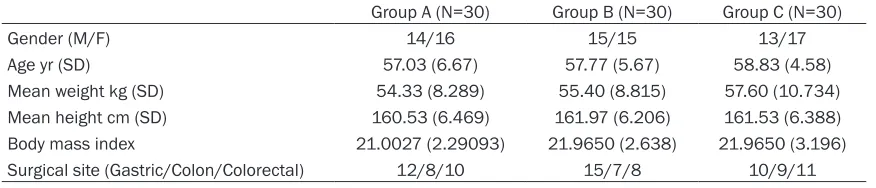
Table 1. Demographic data for groups A, B and C
Group A (N=30) Group B (N=30) Group C (N=30)
Gender (M/F) 14/16 15/15 13/17
Age yr (SD) 57.03 (6.67) 57.77 (5.67) 58.83 (4.58)
Mean weight kg (SD) 54.33 (8.289) 55.40 (8.815) 57.60 (10.734) Mean height cm (SD) 160.53 (6.469) 161.97 (6.206) 161.53 (6.388) Body mass index 21.0027 (2.29093) 21.9650 (2.638) 21.9650 (3.196) Surgical site (Gastric/Colon/Colorectal) 12/8/10 15/7/8 10/9/11
The values in parentheses represent the standard deviation.
[image:3.612.100.523.214.359.2]Results
Patient characteristics
There were no significant differences in gender,
age, weight, height, body mass index between three groups (Table 1).
Depth of anaesthesia
The entropy was declined rapidly to about 40 after induction of anesthesia, and then slowly raised to about 50. RE and SE raised moder-ately during intubation time, skin incision time and other external stimuli time, while slowed down after stimulation and gradually increased
after anesthesia. There was no significant dif -ference (P>0.05) in RE and SE in three groups before induction of anesthesia (t1), before intu-bation (t2), at the time of intuintu-bation (t3), one
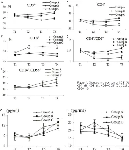
Changes in the proportion of CD3+, CD4+, CD8+, CD4+/CD8+ and CD16+/56+
The results were showed in Figure 4. The pro-portion of CD3+ was no statistically significantly different between three groups (P>0.05) before anesthesia induction (T1) and at the end of sur-gery (T2). The proportion of CD3+ in Group C
was increased more significantly than group A
and B after operative at 24 h (T3) and 72 h (T4),
and there was statistically significant differ -ence (P<0.01) in three groups, while the differ-ences between Group A and B was not
statisti-cally significant (P>0.05). There was no stati-stically significant difference in the proportion
[image:4.612.91.382.74.244.2]of CD4+ between three groups (P>0.05) before anesthesia induction (T1). The proportion of CD4+ was more significantly increased in group B and Group C than Group C and the differenc-es between three groups were statistically sig-Figure 2. Chance of mean arterial pressuren in three groups.
Figure 3. Chance of the HR of three groups.
minute after intubation (t4), three minutes after intub- ation (t5), before incision (t6), skin incision (t7), exp- loratory surgery (t8), at end-ing of administration (t9). Interestingly, the external stimuli entropy change in the both group B and C with epidural anesthesia was smaller than group A, but
there was no significant dif -ference (P>0.05) in three groups (Figure 1). Mean art- erial pressure after induc-tion of anesthesia in three groups decreased transie- ntly, but decreased more greatly in group B and C wi- th epidural anesthesia than in group A, then followed in smoothly low state. There
was no significant differ -ence (P>0.05) among the three groups at nine time points (Figure 2). The heart rate of the patient in three groups was not obviously,
[image:4.612.93.379.284.445.2]nificant (P<0.05) in T2, T3, T4 phase; The pro -portion of CD4+ in group C was increased more
significantly than group B and the difference
between this two groups was statistically
sig-nificant (P<0.05). The proportion of CD8+ between three groups had no statistically dif-ference (P>0.05) in T1 phase, while the propor-tion of CD8+ was significantly more decreased
in group B and group C than Group A in T2, T3,
T4 phase, which was statistically significant
(P<0.05) between three groups. But there was no statistically difference in group B and Group
[image:5.612.92.520.72.571.2]C. There was no statistically significant differ -ence (P>0.05) in the ratio of CD4+/CD8+ between three groups in T1 Phase, but the ratio of CD4+/CD8+ in group C was increased more Figure 4. Changes in proportion of CD3+ (A),
CD4+ (B), CD8+ (C), CD4+/CD8+ (D), CD16+/
CD56+ (E).
significantly than in group A and B in T2, T3, T4
phase, when the difference was statistically
significant between three groups (P<0.01),
while difference between group A and B was
not statistically significant. There was no statis
-more obviously than group A in T2 phase, which was characterized with statistically significant
difference (P<0.005), while there was no
statis-tically significant difference between Group B
[image:6.612.96.386.76.330.2]and C (P>0.05). Figure 6. Dimensional flow cytometry (From lower left to upper right is red blood cell debris, lymphocytes, monocytes, neutrophils, where the aim region in lym-phocytes was T cells, NK cells).
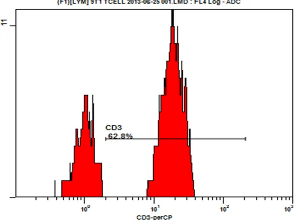
Figure 7. CD3+ cells in the histogram (the proportion of CD3+ cells was 62.8%).
tically significant difference
(P>0.05) in the ratio of CD16+/CD56+ between the three groups in T1 and T2 Phase. The ratio of CD16+/ CD56+ was increased more
significantly in group A and
B than group C and the dif-ference between three gr- oups was statistically
sig-nificant (P<0.05) in T3 and
T4 phase, when there was
statistically significant
diff-erence between three gro- ups, but the ratio of CD16+/ CD56+ in group C was incr-
eased more significantly
than group B and the differ-ence in the two groups was
statistically significant (P<
0.05) (Figures 7-9). Plasma concentrations of IFN-γ and IL-10
The Plasma concentratio-
ns of IFN-γ and IL-10 were
measured with witch ELISA kits (Quantikine, R&D Sys- tems and Minneapolis, MN, USA). The result was sho- wed in Figures 5-9. There
was no statistically signifi -cant difference in the
con-centration of IFN-γ in three
groups in T1, T2 and T3 phase. The concentration
of IFN-γ in group B and C was increased more signifi -cantly than Group A in T4 phase, when there was
sig-nificant difference between
three groups. There was no was significant difference
[image:6.612.93.385.397.615.2]Postoperative recovery
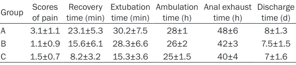
Recovery time is recorded from the end of anesthesia to regaining consciousne- ss; extubation time is mea-sured from the start of anesthesia to full regain consciousness. The result of recovery time and extu-bation time was showed in Table 2. Compared with group A, the recovery time of group B and C was
sig-nificantly reduced, while
the difference in two groups
was statistically significant
(P<0.05); The recovery time of group C decreased more
significantly than group A
and B, while the difference in group B and Group C was
statistically significant (P
<0.05). The extubation ti- me of Group C was reduced
more significantly than
Gr-oup A and B, but the differ-ence in three groups was
statistically significant (P<
0.05); There was no
statis-tically significant difference
between group A and B (P>0.05).
Pain severity was assessed at rest using a visual ana-logue scale (VAS) with val-ues from 0 (no pain) to 10 (worst pain imaginable). Table 2 showed the medi-ans and ranges of pain, sedation in each of three groups 24 hours after sur-gery. The scores of VAS of group B and C was decrease
more significantly than
gr-oup A, and the difference in three groups was
statisti-cally significant (P<0.05),
while there was no
statisti-cally significant difference
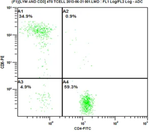
[image:7.612.93.387.76.334.2]between group B and Gr- oup C (P>0.05). There was Figure 8. CD4+ cells and CD8+ cells parameter (The proportion of CD8+ cells was
[image:7.612.95.383.391.669.2]34.9% and CD4+ cells were 59.3%).
no significant difference in the mean time of
Postoperative ambulation, postoperative anal exhaust and discharge from hospital in three groups (Table 2).
Discussion
In this study, we had compared three groups of patients undergoing major or super major oper-ation in immunosuppression, prognosis and the survival rate. Gottschalk et al [15] has shown that anesthesia, surgical procedures and excessive stress response caused by
sys-temic inflammation in patients had effect on
cancer recurrence and prevented immunosup-pression at perioperative stage. Perioperative immune function, postoperative infection and cancer metastasis and recurrence are closely related with each other, which indirectly affect
the final outcome and prognosis of the patient.
Thus, the surgical removal of cancer was fol-lowed by increasing the likelihood that cancer metastasis occurs and further weakens the immune system at the same time, which result-ed to decrease perioperative immunity. Such views were consistent with the results in this study. In this study, it was showed that the
con-centration of IFN-γ, the proportion of CD3+, CD4+, the ratio of CD4+/CD8+ and CD16+/56+ in the patient decreased at the end of surgery and
on the first postoperative day, while the con -centration of IL-10 and the proportion of CD8+
raised at the end of surgery and on the first
postoperative day, which was consistent with the study of Tan Wei [15]. Perioperative
immu-nosuppression may have significant impact on
patient recovery and long-term survival rate; therefore, it is necessary to take measures to investigate how to protect perioperative immu-nity of the cancer patient.
In this study it had been noticed that the RE and SE of entropy, the mean arterial pressure and HR in general anesthesia combined with spinal anesthesia group were more stable than that of general anesthesia, which indicts that
anesthesia can be attributed to at least three different mechanisms [17].First, regional anes-thesia attenuates the immunosuppressive effect of surgery. Neuraxial anesthesia can inhibit the neuroendocrine stress response and paravertebral analgesia in humans having breast surgery has also been shown to inhibit this surgical stress response [18]. Secondly, patients who receive regional analgesia have lower opioid requirements. Paravertebral anal-gesia can reduce opioid requirements after breast surgery [19]. Opioids may themselves inhibit cell-mediated immunity and host anti-tumor defenses. Finally, when regional anes-thesia is used in addition to general anesthe-sia, the amount of general anesthetic required during surgery is reduced. In this study, we also
found that the concentration of IFN-γ, the pro -portion of CD3+, CD4+, the ratio of CD4+/CD8+ and CD16+/56+ in the patient increased at the
end of surgery and on the first postoperative
dayin general anesthesiacombined with spinal anesthesia group, compared to the general anesthesia group, while the proportion of CD4+
was statistically significant different at the end of surgery and on the first postoperative day
and the third postoperative day. The ratio of CD16+/56+ was statistically significant different
on the first day after surgery and the third post
-operative day and the concentration of IFN-γ was statistically significant different on the
third postoperative day. Correspondingly, the concentration of IL-10 and the percentage of CD8+ had decreased at the end of surgery, on
the first postoperative day and the third postop -erative day, while there was no statistically
sig-nificant difference in the percentage of CD8+ at
the end of surgery, the first day after surgery
[image:8.612.87.387.85.151.2]and the third postoperative day, also was the percentage of IL-10 at the end of surgery. These results illustrated that the general anesthesia combined with spinal anesthesia has immune protection of advantages over general anesthe-sia advantage. Zhou D et al [20] conducted a study on general anesthesia and general anes-thesia combined with epidural anesanes-thesia on Table 2. Statistics of recovery in three groups
Group Scores of pain time (min)Recovery Extubation time (min) Ambulation time (h) Anal exhaust time (h) Discharge time (d) A 3.1±1.1 23.1±5.3 30.2±7.5 28±1 48±6 8±1.3 B 1.1±0.9 15.6±6.1 28.3±6.6 26±2 42±3 7.5±1.5 C 1.5±0.7 8.2±3.2 15.3±3.6 25±1.5 40±4 7±1.6
perioperative liver Th cell differentiation, and the results showed that compared with general
anesthesia, anesthesia combined hard film
anesthesia can better protect the anti-tumor immune effect differentiation of Th cells, reduc-ing the proportion of Treg cells after surgery,
which is to be beneficial for patients with hepa -tocellular carcinoma. A study conducted by Chen WK [16] in the effect of anesthetic tech-nique on survival in human cancerssuggested that epidural anesthesia and analgesia may improve overall survival rate in patients with tumors, compared with general anesthesia. For Postoperative recovery in this study, it was demonstrated that the recovery time and
extu-bation time were shortened significantly in two
general anesthesia combined with spinal anes-thesia groups, compared to general
anesthe-sia, but it was shorten more significantly in
anesthesia combined with spinal anesthesia with opioids group than without opioids group.
There was statistically significant difference in
recovery time and extubation time, but no
sta-tistically significant difference in Scores of pain
after 24 hours postoperative, postoperative
ambulation time, postoperative flatus and dis -charge time in two groups, which probably due to the promotion of the idea of fast track surgery.
Several major differences between the two anesthetic methods may be responsible for the distinct patterns in Th1/Th2 balance, as well as Th17 and Treg frequencies between the two groups. First, regional anesthesia substantially attenuates surgery-induced stress responses, including increases in levels of corticosteroid hormone and catecholamine [21]. Second, opi-oids inhibit both cellular and humoral immune function in humans [22]. Animal experiments have shown that morphine suppresses the lym-phocyte proliferative response to mitogens when given systemically, but not when given intrathecally [23]. Similarly, patients receiving an epidural mixture of opioids and local anes-thetics exhibited better preservation of lympho-cyte proliferation and cytokine production than those receiving intravenous opioids alone. Third, intravenous and inhalation anesthetics are associated with elevated serum concentra-tions of catecholamines and cortisol [24]. Glucocorticoids and catecholamines can heav-ily influence immunomodulation, including dec-
reases in Th1/Th2 cytokine production and an increase in FoxP3 mRNA expression [25]. Consequently, it is not surprising that general anesthesia used alone suppressed the surgical stress-induced immune response more pro-foundly than general anesthesia combined with spinal anesthesia.
Of course, other immune mediators such as immune cells, cytokines factually are involved in the various steps of tumor occurrence, including tumor formation, tumor progression and metastasis, when interactions between tumor cells and immune mediators are exten-sive and dynamic. Currently, the mechanisms
of inflammation promoting tumorigenesis have
not yet been fully revealed in molecular level,
and mechanisms of the effect of inflammatory
microenvironment on different caners are var-ied. In this study, we try to put forward an idea of non-opioid anesthesia to preliminarily inves-tigate gastrointestinal cancer with the current high incidence by comparing the effect of three anesthesia methods on the immune function and rehabilitation in patients with gastrointesti-nal cancer.
Disclosure of conflict of interest
None.
Address correspondence to: Dr. Shi-Biao Chen, Department of Anesthesiology, The First Affiliated Hospital of Nanchang University, 17 Yongwai- zheng Avenue, Nanchang 330000, Jiangxi, China. E-mail: [email protected]
References
[1] Price TJ, Shapiro JD, Segelov E, Karapetis CS, Pavlakis N, Van Cutsem E, Tebbutt NC. Man-agement of advanced gastric cancer. Expert Rev Gastroenterol Hepatol 2012; 6: 199-208. [2] Bruix J, Sherman M. Management of
hepato-cellular carcinoma: an update. Hepatology 2011; 53: 1020-1022.
[3] Exadaktylos AK, Buggy DJ, Moriarty DC, Mas-cha E, Sessler DI. Can anesthetic technique for primary breast cancer surgery affect recur-rence or metastasis? Anesthesiology 2006; 105: 660-664.
[4] Snyder GL, Greenberg S. Effect of anesthetic technique and other perioperative factors on cancer recurrence. Br J Anaesth 2010; 105: 106-615.
the cardiovascular, respiratory and gastroin-testinal systems. Minerva Anestesiol 2008; 74: 549-563.
[6] Biki B, Mascha E, Moriarty D, Fitzpatrick J, Ses-sler D, Buggy D. Anesthetic technique for radi-cal prostatectomy surgery affects cancer re-currence: a retrospective analysis. Anesthe- siology 2008; 109: 180-187.
[7] Yeager MP, Colacchio TA, Yu CT, Hildebrandt L, Howell AL, Weiss J, Guyre PM. Morphine inhib-its spontaneous and cytokine-enhanced natu-ral killer cell cytotoxicity in volunteers. Anesthe-siology 1995; 83: 500-508.
[8] Vallejo R, Leon-Casasola O, Benyamin R. Opi-oid therapy and immunosuppression: a review. Am J Ther 2004; 11: 354-65.
[9] Zhu J, Paul WE. CD4 T cells: fates, functions, and faults. Blood 2008; 112: 1557-1569. [10] Zhu J, Paul WE. Heterogeneity and plasticity of
T helper cells. Cell Res 2010; 20: 4-12. [11] Miossec P, Korn T, Kuchroo V. Interleukin-17
and type 17 helper T cells. N Engl J Med 2009; 361: 888-898.
[12] Sakaguchi S, Yamaguchi T, Nomura T, Ono M. Regulatory T cells and immune tolerance. Cell 2008; 133: 775-787.
[13] Borman A, Ciepielewski Z, Wrona D, Stojek W, Glac W, Leszkowicz E, Tokarski J. Small doses of morphine can enhance NK cell cytotoxicity in pigs. Int Immunopharmacol 2009; 9: 277-283.
[14] Gottschalk A, Sharma S, Ford J, Durieux ME, Tiouririne M. The role of the perioperative pe-riod in recurrence after cancer surgery. Anesth Analg 2010; 110: 1636-43.
[15] Tan W, Zhang W, Strasner A, Grivennikov S, Cheng JQ, Hoffman RM, Karin M. Tumour-infil -trating regulatory T cells stimulate mammary cancer metastasis through RANKL-RANK sig-nalling. Nature 2011; 470: 548-553.
[16] Chen W, Miao C. The effect of anesthetic tech-nique on survival in human cancers: a meta-analysis of retrospective and prospective stud-ies. PLoS One 2013; 8: 1-5.
[17] Sessler D. Does regional analgesia reduce the risk of cancerrecurrence? A hypothesis. Eur J Cancer Prev 2008; 17: 269-272.
[18] O’Riain SC, Buggy DJ, Kerin MJ, Watson RW, Moriarty DC. Inhibition of the stress response to breast cancer surgery by regional anesthe-sia and analgeanesthe-sia does not affect vascular en-dothelial growth factor and prostagland in E2. Anesth Analg 2005; 100: 244-249.
[19] Moller JF, Nikolajsen L, Rodt SA, Ronning H, Carlsson PS. Thoracic paravertebral block for breast cancer surgery: a randomized double-blind study. Anesth Analg 2007; 105: 1848-1851.
[20] Zhou D, Gu FM, Gao Q, Li QL, Zhou J, Miao CH. Effects of anesthetic methods on preserving anti-tumor T-helper polarization following hep-atectomy. World J Gastroenterol 2012; 18: 3089-3098.
[21] Clemente A, Carli F. The physiological effects of thoracic epidural anesthesia and analgesia on the cardiovascular, respiratory and gastroin-testinal systems. Minerva Anestesiol 2008; 74: 549-563.
[22] Sacerdote P. Opioid-induced immunosuppres-sion. Curr Opin Support Palliat Care 2008; 2: 14-18.
[23] Hamra J, Yaksh T. Equianalgesic doses of sub-cutaneous but not intrathecal morphine alters phenotypic expression of cell surface markers and mitogen-induced proliferation in rat lym-phocytes. Anesthesiology 1996; 85: 355-365. [24] Inada T, Yamanouchi Y, Jomura S, Sakamoto S,
Takahashi M, Kambara T, Shingu K. Effect of propofol and isoflrane anaesthesia on the im -mune response to surgery. Anaesthesia 2004; 59: 954-959.