Original Article
Value of laparoscopic and fast-track surgery in the
application of sigmoid colon cancer resection
Haixing Fang1, Shijie Shao1, Qunfeng Xia1, Jing He1, Chunliang Wang1, Jianfeng Cai1, Xiujun Cai2
1Department of Hepatobiliary Surgery, The First People’s Hospital of Fuyang District of Hangzhou, Hangzhou, Zhejiang, China; 2Department of General Surgery, Shaoyifu Hospital, Hangzhou, Zhejiang, China
Received May 15, 2018; Accepted June 15, 2018; Epub November 15, 2018; Published November 30, 2018
Abstract: Objective: The aim of this study was to evaluate the value of laparoscopic and fast-track surgery (FTS) in the application of sigmoid colon cancer resection. Methods: Patients with sigmoid colon cancer (n=582) were selected as subjects for retrospective analysis. They were divided into experimental groups A (laparoscopic surgery combined with FTS; 249 cases), B (laparoscopic surgery alone; 174 cases), and C (traditional laparotomy alone; 159 cases). The three groups were compared in terms of patient operation time, volume of intraoperative blood loss, length of hospital stay, postoperative exhaust time, postoperative defecation time, complications, nursing sat-isfaction, and scoring of gastrointestinal recovery. Results: Operation time and volume of intraoperative blood loss in group C were significantly greater than those in groups A and B (P<0.05). Length of hospital stay, postoperative exhaust time, and defecation time were the shortest (P<0.05) in group A, followed by group B. Length of hospital stay, postoperative exhaust time, and defecation time were the longest in group C (P<0.05). Complications were fewer and nursing satisfaction was better in group A than those in the other two groups (P<0.05). Differences in gas-trointestinal function recovery scores among the three groups were statistically significant (P<0.05). Group A scored the highest (P<0.05), followed by group B (P<0.05), with group C scoring the lowest (P<0.05). Conclusion: Use of laparoscopy combined with FTS can effectively reduce incidence of injury and complications in patients undergoing sigmoid colon cancer resection, significantly improving patient prognosis.
Keywords: Laparoscopy, concept of fast-track surgery, sigmoid colon cancer, application value
Introduction
Colon cancer is an extremely common malig-nant tumor of the digestive tract. It mostly aris-es at the junction of the rectum and sigmoid colon. Sigmoid colon cancer is the most com-mon type of colon cancer, accounting for the third highest incidence among all gastrointesti-nal cancers [1]. According to statistics reported by Bertelsen et al. [2], in 2015, there were approximately 4.2 million new cases of sigmoid colon cancer, worldwide, with 2 out of every 3 patients being male. With the development of society, living standards have progressed and incidence of colon cancer has increased. Kim et al. [3] demonstrated that incidence of colon cancer has risen in recent years. Colon cancer is mostly found in middle-aged and elderly people, but increasingly more studies [4-6] have demonstrated that incidence of
post-operative infection [9]. This is a major issue in the treatment of colon cancer.
Clinical research and discussion have been continuously conducted to identify an effective resolution for these problems. In recent years, improvement and popularization of laparosco-py have played important roles in improving resection of various types of cancer diseases [10]. Laparoscopic surgery is characterized by less bleeding, less pain, and quicker recovery, which can effectively relieve patient infections after resection. It has been increasingly studi- ed in combination with fast-track surgery (FTS) for application in all types of surgical patients [11-13]. These studies conjecture that laparo-scopic surgery combined wzith FTS in sigmoid colon resection surgery will have high applica-tion value. Currently, there is little-related re- search. To provide effective references and guidance for future clinical treatment, this stu- dy retrospectively analyzed patients with sig-moid colon cancer undergoing laparoscopic surgery and FTS.
Materials and methods
General information
Five hundred and eighty-two patients with sig-moid colon cancer, from The First People’s Hospital of Fuyang District of Hangzhou, were selected as subjects for retrospective analysis. Patients were 30-60 years old, with a mean age of 42.58±9.74 years. According to different surgical methods, they were divided into experi-mental groups A (laparoscopic surgery com-bined with FTS; 249 cases), B (laparoscopic surgery alone; 174 cases), and C (traditional laparotomy alone; 159 cases).
Inclusion and exclusion criteria
Inclusion criteria were: patients exhibited symp-toms highly consistent with clinical sympsymp-toms of sigmoid colon cancer, were diagnosed with sigmoid colon cancer based on pathological biopsy, underwent surgical treatment after diagnosis, had complete case data, and were willing to cooperate with hospital staff. Exclu- sion criteria were: surgical intolerance, other tumor diseases, other cardiovascular and cere-brovascular diseases, intestinal perforation and infarction, mental illness, previous history of open surgery, history of chemotherapy,
mul-tiple tumor metastases, tumor lesions that could not be completely isolated during sur-gery, and transfer to another hospital. All re- cruited patients provided informed consent.
Method
operative epidural analgesia was continued for 48 hours after surgery. Patients were orally administered anti-inflammatory and analgesic drugs. Catheters were indwelled for 24 hours after surgery and body cavity drainage tubes were removed 48-72 hours after surgery. Tan- gerine peels were administered to mix protein concentrates with water within 24 hours after surgery. Some simple physical activity, with the assistance of a nurse, was conducted 1 day after surgery. The amount of exercise was grad-ually increased to help patients get out of bed as soon as possible.
Observation indicators
Observation indicators were based on clinical data (age, course of illness, and pathological stage). Intraoperative indicators, including pa- tient operative time (from the beginning to the end of surgery) and volume of intraoperative blood loss. Postoperative index included length of hospital stay (from time of admission to dis-charge). Hospital discharge standards strictly followed 2012 guidelines for colon cancer reha-bilitation [15]: oral semiliquid diets could be consumed, intravenous fluids did not need to be added, free exercise could be performed without assistance, and oral analgesic drugs could be administered to effectively control pain. Postoperative index also included postop-erative exhaust time (first exhaust total time after surgery), postoperative defecation time (first defecation total time after surgery), inci-dence of complications (such as incision bleed-ing, pulmonary infections, urinary tract
infec-ed overall gastrointestinal recovery of patients (scores were excellent and good).
Statistical method
Data were analyzed and processed using SP- SS 22.0 statistical software (Asia Analytics, for-merly SPSS China). Measurement data, such as patient age, patient operating time, and intraoperative blood loss, are expressed in terms of mean ± standard deviation. Com- parison of variance analysis was used to pare multiple groups. Pairs of groups were com-pared using t-test. Count data, such as patient gender, nursing satisfaction, and complica-tions, are expressed in the form of rates. Chi-squared test was used for comparison between groups. P<0.05 was considered statistically significant.
Results
Patient clinical data
To demonstrate that experimental results were effective and reliable, age, weight, course of disease, gender, pathological stage, and blood counts were compared among the three gr- oups of patients. There were no significant dif-ferences between the three groups (P>0.05), proving that the three groups of patients were comparable (Table 1).
Intraoperative indicators for patients
[image:3.612.91.390.85.268.2]Operation times in experimental groups A, B, and C were 128.47±11.57 minutes, 125.47±
Table 1. Comparison of clinical data of three groups of patients [n (%)] Experiment A
group (n=249) group (n=174)Experiment B group (n=159)Experiment C F P Age 41.86±8.73 40.96±10.57 41.36±9.85 0.46 0.63 Body weight (KG) 75.84±10.62 73.44±12.57 74.38±12.49 2.23 0.11 Disease course (d) 24.68±13.62 25.94±12.44 24.37±14.37 0.66 0.52 RBC (×1012/L) 1.84±1.04 1.72±1.34 1.62±1.08 1.84 0.16
WBC (×109/L) 2.86±1.48 3.04±2.33 2.74±1.69 1.15 0.32
PLT (×109/L) 86.17±29.39 91.52±30.54 88.93±31.58 1.61 0.20
Gender 0.55 0.62
Male 168 (67.47) 108 (62.07) 108 (67.92) Female 81 (32.53) 66 (37.93) 51 (32.08)
Pathological stage 9.45 0.05
I~II 115 (46.18) 79 (45.40) 74 (46.54) III~IV 134 (53.82) 95 (54.60) 85 (53.46)
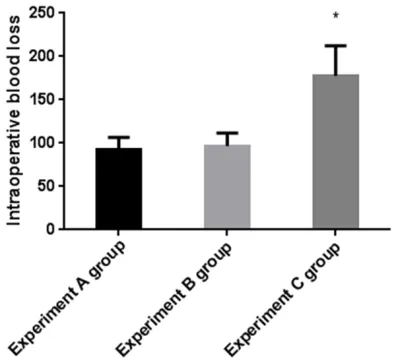
record-and B (P<0.05) (Figure 1). Volumes of intra- operative blood loss in groups A, B, and C were 92.86±13.77 mL, 96.73±14.85 mL, and 177.52±34.96 mL, respectively. Differences in volumes of intraoperative blood loss were sta-tistically significant between the three groups (P<0.05). There were no significant differences in volumes of blood loss between experimental groups A and B (P>0.05). Volumes of intraop-erative blood loss were significantly greater in experimental group C than those in experimen-tal groups A and B (P<0.05) (Figure 2).
Postoperative indicators for patients
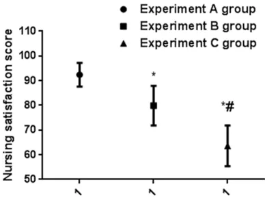
[image:4.612.90.288.71.253.2]Lengths of stay in experimental groups A, B, and C were 6.82±1.63 days, 10.57±2.78 days, and 14.87±3.67 days, respectively. Differences in length of hospital stay between the three groups were statistically significant (P<0.05). Lengths of hospital stay in experimental group C were the longest, followed by experimental group B. Patients in experimental group A had the shortest lengths of hospital stay (all P< 0.05) (Figure 3). Postoperative exhaust times in experimental groups A, B, and C were 41.74±8.75 hours, 56.84±10.27 hours, and 86.77±13.34 hours, respectively. There were statistically significant differences in postoper-ative exhaust time (P<0.05) between the three groups. Postoperative exhaust times in experi-mental group C were the longest, followed by experimental group B. Times were the shortest in experimental group A (all P<0.05) (Figure 4). Postoperative defecation times in experimen- tal groups A, B, and C were 3.27±1.26 days, 4.53±1.17 days, and 5.46±1.07 days, respec-tively. Differences in defecation times were sta-tistically significant between the three groups (P<0.05). Defecation times in experimental gr- oup C were the longest, followed by experimen-tal group B. Times were the shortest in experi-mental group A (all P<0.05) (Figure 5). Nursing satisfaction scores in experimental groups A, B, and C were 92.37±4.82 points, 79.86± 8.04 points, and 63.59±8.24 points, respec-tively. Differences in nursing satisfaction sco- res were statistically significant between the three groups (P<0.05). Nursing satisfaction scores in experimental group C were the low-est, followed by experimental group B. Scores were the highest in experimental group A (all P<0.05) (Figure 6). The incidence of complica-tions (including one and more than one compli-cations) in experimental groups A, B, and C Figure 1. Operating times for the three groups.
*Rep-resents comparison with the experimental time of experimental group A, P<0.05. There were no signifi-cant differences in operation times between group A and group B (P>0.05); Duration of operation in group C was significantly longer than that of group A and B.
Figure 2. Amount of bleeding in the three groups of patients. *Represents intraoperative blood loss com-pared with the experimental group A, P<0.05. There were no significant differences in intraoperative blood loss between group A and group B (P>0.050). Intraoperative blood loss was significantly more in group C than in group A and B.
[image:4.612.90.287.345.524.2]Figure 3. Time of hospitalization for the three groups of patients. *Represents hospitalization time com-pared with experimental group A, P<0.05. #repre-sents hospitalization time compared with experimen-tal group B, P<0.05. Experimenexperimen-tal group C had the longest hospital stay, followed by the experimental group B, and experimental group A had the shortest hospital stay.
Figure 4. Postoperative venting times for the three groups of patients. *Represents postoperative ven-tilatory time compared with experimental group A, P<0.05. #Represents postoperative ventilatory time compared with experimental group B, P<0.05. Among them, group C had the longest postoperative exhalation time, followed by experiment group B, and experiment group A had the shortest postoperative exhalation time.
was 1.61%, 11.49%, and 37.11%, respectively. There was statistically significant differences in incidence of complications between the th- ree groups (P<0.05). The incidence of
[image:5.612.330.521.76.257.2]compli-Figure 5. Postoperative defecation times in three groups of patients. *Represents postoperative def-ecation time compared with experimental group A, P<0.05. #Represents postoperative defecation times compared with experimental group B, P<0.05. Experimental group C had the longest postoperative defecation time, followed by experimental group B. Experimental group A had the shortest postoperative defecation times.
Figure 6. Three groups of patient care satisfaction scores. *Represents a comparison with experimen-tal A group’s nursing satisfaction score, P<0.05. #Represents the score of satisfaction with the ex-perimental group B, P<0.05. Exex-perimental group C had the lowest satisfaction rate of care, followed by experimental group B, and experimental group A had the highest satisfaction rate.
(P<0.05), followed by experimental group B (P<0.05). The incidence was the lowest in experimental group A (P<0.05) (Table 2).
Prognostic indicators for patients
[image:5.612.91.284.372.558.2] [image:5.612.325.519.381.527.2]Table 2. Comparison of complications in three groups of patients [n (%)] Experiment A group
(n=249) Experiment B group (n=174) Experiment C group (n=159) F P Incision bleeding 1 (0.40) 2 (1.15) 12 (7.55) 0.94 0.06
lung infection 2 (0.80) 10 (5.75) 19 (11.95) 0.93 0.07
Nausea and vomiting 5 (2.01) 12 (6.90) 26 (16.35) 0.93 0.08 Intestinal obstruction 0 (0.00) 2 (2.30) 14 (8.81) 0.93 0.07 Urinary system infection 0 (0.00) 1 (0.57) 9 (5.66) 0.94 0.06 Incidence of complications (%) 1.61 11.49* 37.11*,# 8.64 0.02 Note: *Represents complication rate compared with experimental group A, P<0.05. #Represents incidence of complications compared with experimental group B, P<0.05.
these, 1 in experimental group A and 1 in ex- perimental group C were lost to follow up. Recovery scores of gastrointestinal function in experimental groups A, B, and C were 86.57± 7.37 points, 70.54±10.53 points, and 62.53± 8.69 points, respectively. Differences in gas- trointestinal function recovery scores between the three groups were statistically significant (P<0.05). Scores in experimental group A were the highest, followed by experimental group B. Scores were lowest in experimental group C (all P<0.05) (Figure 7).
Discussion
Sigmoid colon cancer is a common malignancy, mostly found in middle-aged and elderly
peo-ple. As human body function weakens with age, patient surgical tolerance naturally decre- ases. Natural resuscitation capabilities are weakened after patients undergo a series of post-traumatic surgeries, including surgical re- section and lymphatic dissection. This is one of the reasons contributing to differences in patient prognosis [16, 17]. With recent improve-ments in laparoscopic techniques, they have been widely used in clinical surgery for the removal of various types of tumors [18]. La- paroscopic invasive surgery is characterized by minimal trauma and rapid recovery. It is also very effective in the investigation of subtle tumor lesions. At present, there are many relat-ed studies [19-21] reporting that clinical and prognostic effects of laparoscopic surgery for tumor diseases are significantly better than tho- se of traditional open surgery. Laparoscopy is the first choice for tumor resection. For some obstinate tumors, such as sigmoid colon can-cer, high local recurrence and metastatic rates after surgery remain barriers to overcome in clinical practice. FTS was first proposed by Danish abdominal surgeons Kehlet and Wile- more. They mentioned that multiple pathophys-iological adjustments before, during, and after abdominal surgery should be carried out to reduce risk of stress and infection caused by surgery [22]. With continuous progress in re- search, FTS has been demonstrated to have extremely high application value in all types of abdominal surgery. However, there are few related studies on sigmoid colon cancer. The- refore, through research and analysis, laparo-scopic surgery combined with FTS may maxi-mize the prognosis of patients with sigmoid colon cancer and provide scientific reference and guidance for clinical treatment of sigmoid colon cancer.
[image:6.612.91.286.234.403.2]Results of this present study demonstrated that laparoscopic surgery combined with FTS (experimental group A) was significantly superi-or to conventional large-open surgery (experi-mental group C) in terms of intraoperative and postoperative indicators. Compared with lapa-roscopic surgery alone (experimental group B), there were no significant differences in intraop-erative indicators, suggesting that laparos- copic surgery can reduce the degree of body damage in patients with sigmoid colon cancer. Compared with traditional open surgery, it has higher application value. Combined with appli-cation of the FTS model, it can effectively improve prognosis and is the best choice for treatment of sigmoid colon cancer among the three methods. Laparoscopic surgery, using the latest technology, can achieve a more pre-cise surgical incision, with less severe surgical trauma, better internal environment stability, and shorter postoperative recovery times. It can not only greatly increase the visual field of small lesions during surgery and enhance the integrity of lesion resection and lymphatic dis-section, but also ensure the stability of pa- tient internal environments through a smaller traumatic incision, reduce stress response caused by surgery, and minimize incidence of postoperative complications and risk of infec-tion [23]. This study also compared incidence of postoperative complications among the th- ree groups of patients. However, laparoscopic surgery requires that surgeons have higher pro-fessional quality knowledge to improve suc- cess rates of surgery. Application of FTS is based on laparoscopic surgery and requires a series of physiological arrangements before surgery to reduce patient insulin resistance and decomposition of metabolic capacity. The- se include strict intraoperative control and adjustment of patient anesthesia and fluid sup-plements to reduce unforeseen accidents dur-ing surgery and postoperative assistance en- abling patients to undergo timely rehabilitation training, promoting healing of muscles and inci-sions, and improving the body’s immune me- tabolism [24]. In the process of patient admis-sion, FTS requires nurses to pay close atten- tion to patient vital signs and increase the time of communication with patients [25]. This not only allows patients to be more fully prepared during surgery but also improves patient psy-chological states, promoting prognosis. Intake of an appropriate diet as soon as possible after
surgery not only plays a protective role in the intestinal mucosa but can also inhibit patient intestinal flora shift phenomenon, further pro-moting the recovery of patients. In this present study, patients with intestinal obstruction in experimental group A demonstrated this fact. Patient physical activity after surgery can also greatly improve occurrence of venous thrombo-sis in the lower extremities and reduce risk of pulmonary infection. Rapid recovery of body function is a key factor determining prognosis. This study compared physiological parameters of patients with sigmoid colon cancer undergo-ing laparoscopic surgery combined with FTS, laparoscopic surgery alone, or traditional large open laparotomy. The aim of this study was to explore the application value of laparoscopic surgery combined with FTS for sigmoid colon cancer. There are limitations to this study. There was a relatively limited number of study subjects with a small age span. The present researchers will conduct a long-term follow up survey for this study’s subjects and continue to improve and perfect experiments in the future to achieve the best experimental results. In summary, the use of laparoscopy combined with FTS can effectively reduce incidence of injury and complications in patients undergoing sigmoid colon resection. It can greatly improve the prognosis of patients and is worthy of pro-motion in clinical practice.
Acknowledgements
Provincial Health Planning Commission Project (2017KY566).
Disclosure of conflict of interest
None.
Address correspondence to: Haixing Fang, De- partment of Hepatobiliary Surgery, The First People’s Hospital of Fuyang District of Hangzhou City, No. 429, Beihuan Road, Fuchun Street, Fuyang District, Hangzhou 311400, Zhejiang, China. Tel: +86-136- 83189288; E-mail: [email protected]
References
sigmoid colon cancer: a propensity-score matching analysis. Ann Surg Oncol 2015; 22: 924-930.
[2] Bertelsen CA, Neuenschwander AU, Jansen JE, Wilhelmsen M, Kirkegaard-Klitbo A, Tenma JR, Bols B, Ingeholm P, Rasmussen LA and Jepsen LV. Disease-free survival after complete meso-colic excision compared with conventional co-lon cancer surgery: a retrospective, popula-tion-based study. Lancet Oncol 2015; 16: 161-168.
[3] Kim CW, Kim WR, Kim HY, Kang J, Hur H, Min BS, Baik SH, Lee KY and Kim NK. Learning curve for single-incision laparoscopic anterior resection for sigmoid colon cancer. J Am Coll Surg 2015; 221: 397-403.
[4] Sinicrope FA, Shi Q, Smyrk TC, Thibodeau SN, Dienstmann R, Guinney J, Bot BM, Tejpar S, Delorenzi M and Goldberg RM. Molecular markers identify subtypes of stage III colon cancer associated with patient outcomes. Gas-troenterology 2015; 148: 88-99.
[5] Tejpar S, Stintzing S, Ciardiello F, Tabernero J, Van Cutsem E, Beier F, Esser R, Lenz HJ and Heinemann V. Prognostic and predictive rele-vance of primary tumor location in patients with RAS wild-type metastatic colorectal can-cer: retrospective analyses of the CRYSTAL and FIRE-3 trials. JAMA Oncol 2017; 3: 194-201. [6] Kogita A, Yoshioka Y, Sakai K, Togashi Y,
So-gabe S, Nakai T, Okuno K and Nishio K. Inter-and intra-tumor profiling of multi-regional co-lon cancer and metastasis. Biochem Biophys Res Commun 2015; 458: 52-56.
[7] Tamas K, Walenkamp A, De Vries E, Van Vugt M, Beets-Tan R, Van Etten B, de Groot D and Hospers G. Rectal and colon cancer: not just a different anatomic site. Cancer Treat Rev 2015; 41: 671-679.
[8] Bae SU, Jeong WK and Baek SK. Robotic ante-rior resection for sigmoid colon cancer using reduced port access. Dis Colon Rectum 2016; 59: 245-246.
[9] Loupakis F, Yang D, Yau L, Feng S, Cremolini C, Zhang W, Maus MK, Antoniotti C, Langer C and Scherer SJ. Primary tumor location as a prog-nostic factor in metastatic colorectal cancer. J Natl Cancer Inst 2015; 107.
[10] Fernández-Hevia M, Delgado S, Castells A, Tasende M, Momblan D, del Gobbo GD, DeLa-cy B, Balust J and LaDeLa-cy AM. Transanal total me-sorectal excision in rectal cancer: short-term outcomes in comparison with laparoscopic surgery. Ann Surg 2015; 261: 221-227. [11] Chen WK, Ren L, Wei Y, Zhu DX, Miao CH and
Xu JM. General anesthesia combined with epi-dural anesthesia ameliorates the effect of fast-track surgery by mitigating immunosuppres-sion and facilitating intestinal functional
recovery in colon cancer patients. Int J Colorec-tal Dis 2015; 30: 475-481.
[12] Chen S, Zou Z, Chen F, Huang Z and Li G. A meta-analysis of fast track surgery for patients with gastric cancer undergoing gastrectomy. Ann R Coll Surg Engl 2015; 97: 3-10.
[13] Bu J, Li N, Huang X, He S, Wen J and Wu X. Feasibility of fast-track surgery in elderly pa-tients with gastric cancer. J Gastrointest Surg 2015; 19: 1391-1398.
[14] West NP, Sutton KM, Ingeholm P, Hagemann-Madsen RH, Hohenberger W and Quirke P. Im-proving the quality of colon cancer surgery through a surgical education program. Dis Co-lon Rectum 2010; 53: 1594-1603.
[15] Alfano CM, Ganz PA, Rowland JH and Hahn EE. Cancer survivorship and cancer rehabilitation: revitalizing the link. J Clin Oncol 2012; 30: 904-906.
[16] Bakker N, Cakir H, Doodeman H and Houdijk A. Eight years of experience with enhanced recov-ery after surgrecov-ery in patients with colon cancer: impact of measures to improve adherence. Surgery 2015; 157: 1130-1136.
[17] Siani L and Pulica C. Laparoscopic complete mesocolic excision with central vascular liga-tion in right colon cancer: long-term oncologic outcome between mesocolic and non-meso-colic planes of surgery. Scand J Surg 2015; 104: 219-226.
[18] Spanjersberg W, Van Sambeeck J, Bremers A, Rosman C and van Laarhoven C. Systematic review and meta-analysis for laparoscopic ver-sus open colon surgery with or without an ERAS programme. Surg Endosc 2015; 29: 3443-3453.
[19] Hiki N, Nunobe S, Matsuda T, Hirasawa T, Ya-mamoto Y and Yamaguchi T. Laparoscopic en-doscopic cooperative surgery. Dig Endosc 2015; 27: 197-204.
[20] Irino T, Nunobe S, Hiki N, Yamamoto Y, Hirasa-wa T, Ohashi M, Fujisaki J, Sano T and Yamagu-chi T. Laparoscopic-endoscopic cooperative surgery for duodenal tumors: a unique proce-dure that helps ensure the safety of endoscop-ic submucosal dissection. Endoscopy 2015; 47: 349-351.
[21] Boni L, David G, Mangano A, Dionigi G, Rausei S, Spampatti S, Cassinotti E and Fingerhut A. Clinical applications of indocyanine green (ICG) enhanced fluorescence in laparoscopic surgery. Surg Endosc 2015; 29: 2046-2055. [22] Shao Z, Jin G, Ji W, Shen L and Hu X. The role
of fast-track surgery in pancreaticoduodenec-tomy: a retrospective cohort study of 635 con-secutive resections. Int J Surg 2015; 15: 129-133.
health care utilization and costs in patients who undergo colectomy. JAMA Surg 2015; 150: 410-415.
[24] Philp S, Carter J, Pather S, Barnett C, D’abrew N and White K. Patients’ satisfaction with fast-track surgery in gynaecological oncology. Eur J Cancer Care (Engl) 2015; 24: 567-573.
![Table 1. Comparison of clinical data of three groups of patients [n (%)]](https://thumb-us.123doks.com/thumbv2/123dok_us/1377805.671554/3.612.91.390.85.268/table-comparison-clinical-data-groups-patients-n.webp)
![Table 2. Comparison of complications in three groups of patients [n (%)]](https://thumb-us.123doks.com/thumbv2/123dok_us/1377805.671554/6.612.91.286.234.403/table-comparison-complications-groups-patients-n.webp)
