Original Article
Influence of renal sympathetic denervation on the
cardiac function of dogs with heart failure
Damin Huang1*, Yong Li2*, Shuxin Hou1, Jinchun Zhang1, Lei Song1, Yingmin Lu1
1Department of Cardiology, Xin Hua (Chongming) Hospital Affiliated to Shanghai Jiao Tong University School of
Medicine, Shanghai 200092, China; 2Department of Emergency Medicine, Wenzhou Central Hospital, Affiliated
Dingli Clinical Institute of Wenzhou Medical University Zhejiang, China. *Equal contributors.
Received August 17, 2015; Accepted November 16, 2015; Epub February 15, 2016; Published February 29, 2016
Abstract: Objective: This study aimed to investigate the influence of renal sympathetic denervation (RSDN) on the cardiac function of dogs with heart failure. Methods: A total of 40 dogs were randomly assigned into RSDN group and control group (n=20 per group). In RSDN group, dogs received radiofrequency ablation of bilateral renal arter
-ies; dogs in control group received femoral artery puncture alone. Pacemaker (VOO module) was implanted in 40 dogs, and rapid right ventricular pacing was introduced to establish heart failure model. The maximum left ven
-tricular end systolic diameter (LVESD), maximum left ven-tricular end-diastolic diameter (LVEDD), cardiac output (CO), cardiac index (CI), left ventricular systolic pressure (LVSP), left ventricular diastolic pressure (LVDP), maximum
systolic blood pressure rise rate (dp/dtmax), maximum diastolic blood pressure drop rate (-dp/dtmax), heart rate
(HR), peripheral renin, norepinephrine, angiotensin II, aldosterone, glomerular filtration rate (GFR) and renal blood flow were measured. Statistical analysis was performed with SPSS version 17.0. Results: The peripheral renin, nor
-epinephrine, angiotensin II, aldosterone, LVDP, LVESD and LVEDD in RSDN group were significantly lower than in control group, but the left ventricular ejection fraction, CI, CO, LVSP, dp/dtmax, and -dp/dtmax in RSDN group were markedly higher than in control group. Conclusion: In heart failure dogs, RSDN may inhibit the renal sympathetic
activity and reduce systemic sympathetic activity, attenuate heart enlargement, mitigate reduced myocardial
con-tractility, suppress the myocardial remodeling due to right ventricular pacing induced heart failure and improve the symptoms of heart failure and cardiac function.
Keywords: Heart failure, renal sympathetic denervation, dog, right ventricular pacing, sympathetic nervous system
Introduction
Heart failure is a clinical syndrome due to the structural or functional abnormality of the heart which may affect the cardiac perfusion or heart beat, and is also the end stage of heart diseases [1]. Heart failure is usually accompa -nied by extensive adrenergic nervous system excitation, parasympathetic nervous system in- hibition, renin-angiotensin-aldosterone system (RAAS) activation, extensive vasoconstriction, and sodium and water retention. In the
pres-ence of heart failure, the density of β-adrenergic receptor reduces, myocardial contractility fails
to increase, sodium and water retention and
vasoconstriction further increase the cardiac preload and afterload and visceral injury, and the renal blood flow reduces, which deterio
-rates heart failure, resulting in a vicious cycle of
disease progression [1].
RAAS is an important humoral regulation sys-tem in humans, and its excitation may promote
the release of neurotransmitters from the adre-nergic nerves, induce the contraction of sys -temic arterioles and veins leading to blood pre- ssure increase, elevate the central sympathetic
vasoconstrictor tone, facilitate the release of
vasopressin and adrenocorticotropic hormone,
increase the synthesis and secretion of aldoste -rone leading to sodium and water retention,
inhibit the left ventricular systolic function and
cause the myocardial remodeling. RASS may
participate in the pathophysiology of different diseases including heart failure, hypertension,
and arrhythmias by above mechanisms. Renal sympathetic nerve may activate RASS via the adrenergic nerves [1-3].
In 2009, Krum et al. for the first time used renal
4325 Int J Clin Exp Med 2016;9(2):4324-4331
of refractory hypertension and confirmed the safety of renal sympathetic nerve ablation with simplicity catheter and its effectiveness in the therapy of refractory hypertension [4]. Recently,
clinical trials reveal that RSDN may not only reduce the renal sympathetic activity, but
inhib-it the activinhib-ities of other sympathetic nerves, and the heart volume and cardiac function are improved after RSDN in patients with refractory
hypertension [5-7]. Animal experiments also indicate that RSDN may delay the progression
of myocardial hypertrophy [8]. However, evi
-dence on the therapy of heart failure with RSDN is still insufficient. In the present study, right
ventricular pacing was introduced in adult dogs
to establish the heart failure model, and radio
-frequency ablation of bilateral renal arteries was employed for RSDN, aiming to investigate the influence of RSDN on the cardiac function of dogs with heart failure.
Materials and methods
Materials
A total of 40 crossbred canines aged 10-12 months and weighing 16.2-18.4 kg were pur
-chased from the Experimental Animal Center of Shanghai Jiaotong University. Radiofrequency
ablation device (STOCKERT EP SHUTTLE, John- son & Johnson, USA), 6 F endocardial electro- de catheter (Johnson & Johnson, USA), Med-
tronic pacemaker (Medtronic Company), HEM-8102A Omron Blood Pressure Monitor
(Shen-zhen Omron Corporation), Innova 3100 digital
subtraction angiography (US General Electric Company), Philips iE33 Multifunction Ultra-sound, S5-1 probe (frequency: 2-4 MHz; Phi-lips, Netherlands), SAR-830A ventilator for
small animals (CWE ,USA), pentobarbital sodi-um (Sigma, USA), penicillin (Hangzhou Min- sheng Pharmaceutical Company), renin
detec-tion kit (Sigma, USA), epinephrine detecdetec-tion kit (Sigma, USA), norepinephrine detection kit (Sig-ma, USA) and aldosterone detection kit (Sig(Sig-ma,
USA) were used in this study.
Radiofrequency ablation of the renal sympa-thetic nerves
A total of 40 dogs were randomly assigned into
two groups: RSDN group and control group (n=20 per group), and animals were numbed at
the ear. Before study, dogs received food and water were fasting for 10 h and then fastened to an operating table. After general anesthesia
by intravenous 3% pentobarbital sodium at 30
mg/kg and skin preparation, additional 50 mg of pentobarbital sodium was intravenously
injected according to the animals’ response 30-60 min later. Then, endotracheal intubation
and subsequent mechanical ventilation were conducted, followed by continuous monitoring of electrocardiogram. The right or left femoral
artery was punctured, and a 6 F endocardial
electrode catheter was implanted for radiofre
-quency ablation. In RSDN group, ablation elec
-trode plate was implanted in the back, and ablation was performed at 50°C and at 6-8 W. A renal artery was ablated at 0.5 cm away from the first bifurcation for 60 s, the probed was
slightly retracted, and the catheter was rotated. Then, the artery was ablated at 4-6 sites in
a spiral manner with a distance of 0.5 cm
between two adjacent sites. In control group,
only femoral artery was punctured, and abla
-tion was not conducted. After surgery, animals
were intramuscularly injected with penicillin
(800000 U) for 3 consecutive days for infection
prophylaxis [9, 10].
Establishment of heart failure model
Pre-operative preparation and intravenous an- esthesia were conducted according to above-mentioned. The carotid pulse was palpable at 2
cm right away from the trachea, and right exter -nal jugular vein was punctured at 0.5 cm away
from the carotid pulse site. Then, an electrode
was implanted in the right ventricle. Under the
X ray, the electrode was fixed in the trabecular muscles of right ventricular apex. The pacing threshold was 0.3-1.5 V, the height of R wave was 4-10 mV, and the impedance was 0.3-1.0 KΩ. The pacemaker was connected, and the pacing frequency was set at 230 beat/min (VOO module). A bag was prepared to fix the pacemaker, and washed with gentamicin (320000 U). Then, the wound was closed. After
surgery, animals were intramuscularly injected
with penicillin (800000 U) for 3 consecutive days for infection prophylaxis. In addition, the
appetite, activity and respiration were
moni-tored, the surface electrocardiogram was de-tected every week, the pacing rhythm was maintained stable, and pacing was done for 4 weeks [11, 12].
Cardiac echocardiography and measurement of hemodynamics
4 weeks after pacemaker implantation, and the left ventricular end systolic diameter (LVESD), left ventricular end-diastolic diameter (LVEDD), cardiac output (CO), cardiac index (CI), and left ventricular ejection fraction (EF) were mea
-sured. In addition, before RSDN and at 4 weeks after pacemaker implantation, femoral artery was punctured again, and the left ventricular systolic pressure (LVSP), left ventricular diastol
-ic pressure (LVDP), maximum systol-ic blood
pressure rise rate (dp/dtmax), and maximum diastolic blood pressure drop rate (-dp/dtmax) were measured.
Monitoring of blood parameters and vital signs
Before RSDN and pacemaker implantation and at 4 weeks after pacemaker implantation, the
blood pressure, heart rate (HR), and respiration rate (RR) were recorded. At the same time, ve-
nous blood (6 ml) was collected from the femo
-ral vein and centrifuged at 2500 rpm for 10
min. The plasma was collected, and processed
for the detection of rennin (R), epinephrine (E),
norepinephrine (NE), angiotensin II (ATII), and
aldosterone (AD) according to manufacturer’s
instructions.
Statistical analysis
Data were input into EXCEL and statistical
anal-ysis was performed with SPSS version 17.0.
Quantitative data are expressed as mean ± standard deviation, and comparisons between two groups were done with t test. Parameters
measured before and after surgery were com
-pared with paired t test. A value of P<0.05 was
considered statistically significant.
dogs showed reduced activities and loss of appetite. At 4 weeks after right ventricular
pacing, above symptoms deteriorated,
short-ness of breath was also present, moist rales
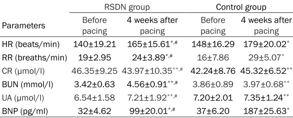
were heard by auscultation, and HR and RR increased. The HR was 140±19.21 beats/
min before surgery and 165±15.61 beats/min after surgery in RSDN group and was 148± 16.29 beats/min before surgery and 179± 20.02 beats/min after surgery in control group. The RR was 19±2.95 breaths/min before sur
-gery and 24±3.89 breaths/min after sur-gery in RSDN group and was 16±7.86 breaths/min before surgery and 29±5.07 breaths/min after surgery in control group. BNP increased after
surgery in both groups (RSDN group: 32±4.62 pg/ml vs. 99±20.01 pg/ml; control group: 37±
6.20 pg/ml vs 187±25.63 pg/ml). Paired t test showed significant differences in above param
-eters before and after surgery. Moreover, mark-ed differences were also notmark-ed between RSDN
group and control group (P<0.05). However, the
kidney function remained unchanged during
the experiment (P>0.05) (Table 1).
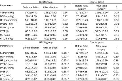
Cardiac function and hemodynamics
Results showed the blood pressure, HR, LVEDS, LVEDD, LVEF, CO and CI at 4 weeks after radio
-frequency ablation (before pacemaker implan
-tation) were comparable to those before radio
-frequency ablation (P>0.05) (Table 2). This im-
plies that RSDN has no influence on the blood pressure, HR, cardiac volume and cardiac func
-tion of healthy dogs.
[image:3.612.90.386.97.216.2]The SBP reduced after heart failure in both groups. SBP reduced from 132±16.42 mmHg
Table 1. Clinical parameters of dogs before and after pacing in both
groups
RSDN group Control group
Parameters pacingBefore 4 weeks after pacing Beforepacing 4 weeks after pacing
HR (beats/min) 140±19.21 165±15.61*,# 148±16.29 179±20.02*
RR (breaths/min) 19±2.95 24±3.89*,# 16±7.86 29±5.07*
CR (μmol/l) 46.35±9.25 43.97±10.35**,# 42.24±8.76 45.32±6.52**
BUN (mmol/l) 3.42±0.63 4.56±0.91**,# 3.86±0.89 3.97±0.68**
UA (μmol/l) 6.54±1.58 7.21±1.92**,# 7.20±2.01 7.35±1.24**
BNP (pg/ml) 32±4.62 99±20.01*,# 37±6.20 187±25.63*
Note: *P<0.05: before vs. after pacing; **P>0.05: before vs. after pacing;#P<0.05:
RSDN vs control group after pacing.
Results
Characteristics of dogs
Results showed there were
no marked differences in
the age, body weight, blood pressure, heart rate,
respi-ration rate and kidney func -tion between RSDN group and control group at
base-line (P>0.05). After RSDN,
the activities, appetite and vital signs remained un-
changed. At 2 weeks
4327 Int J Clin Exp Med 2016;9(2):4324-4331
to 122±10.43 mmHg in RSDN group and from
129±19.56 mmHg to 110±14.42 mmHg in
control group. In addition, the left ventricle was enlarged in both groups. LVEDS increased from
10.9±2.27 to 13.2±2.27 mm in RSDN group
and from 10.2±2.3 to 17.5±4.32 mm in control group. The EF reduced from 63.6±9.23% to 43.89±6.88% in RSDN group and from 66.7±
10.20% to 35.21±5.72% in control group. CO
[image:4.612.92.522.97.383.2]reduced from 3.92±0.69% to 1.82±0.71% in
Table 2. Blood pressure, HR, cardiac volume and cardiac function before and after radiofrequency
ablation in RSDN and control groups
Parameters
RSDN group Control group
Before ablation After ablation P Before false surgery After false surgery P
SBP (mmHg) 132±16.42 128±20.42 0.18 127±20.67 129±19.56 0.24
DBP (mmHg) 78±14.60 78±13.80 0.12 75±15.68 75±15.72 0.19
HR (beats/min) 140±18.34 140±19.21 0.27 142±19.79 148±16.29 0.18
LVEDS (mm) 10.8±3.24 10.9±2.27 0.32 10.8±2.23 10.2±2.31 0.33
LVEDD (mm) 29.7±4.02 29.6±3.04 0.59 27.9±5.68 28.9±3.25 0.5
LVEF (%) 63.6±9.23 67.6±9.23 0.68 67.4±9.28 66.7±10.20 0.21
CO (l/min) 3.94±0.65 3.92±0.69 0.62 3.94±0.72 3.91±0.70 0.42
CI (l/min·Kg) 0.25±0.07 0.23±0.06 0.55 0.27±0.08 0.26±0.08 0.53
Parameters
RSDN group Control group
Before ablation After ablation P Before false surgery After false surgery P
SBP (mmHg) 132±16.42 128±20.42 0.18*,# 127±20.67 129±19.56 0.24*
DBP (mmHg) 78±14.60 78±13.80 0.12*,# 75±15.68 75±15.72 0.19*
HR (beats/min) 140±18.34 140±19.21 0.27*,# 142±19.79 148±16.29 0.18*
LVEDS (mm) 10.8±3.24 10.9±2.27 0.32*,# 10.8±2.23 10.2±2.31 0.33*
LVEDD (mm) 29.7±4.02 29.6±3.04 0.59*,# 27.9±5.68 28.9±3.25 0.50*
LVEF (%) 63.6±9.23 67.6±9.23 0.68*,# 67.4±9.28 66.7±10.20 0.21*
CO (l/min) 3.94±0.65 3.92±0.69 0.62*,# 3.94±0.72 3.91±0.70 0.42*
CI (l/min·Kg) 0.25±0.07 0.23±0.06 0.55*,# 0.27±0.08 0.26±0.08 0.53*
Note: *P>0.05, before vs. after ablation in RSDN group, as well as before vs. after false surgery in control group; #P>0.05: RSDN group vs. control group after ablation or false surgery.
Table 3. Heart function and hemodynamics in RSDN group and control group
RSDN group Control group
Before pacing 4 weeks after pacing Before pacing 4 weeks after pacing
SBP (mmHg) 132±16.42 122±10.43# 129±19.56 110±14.42
DBP (mmHg) 78±13.80 76±15.67# 75±15.72 80±16.04
LVEDS (mm) 10.9±2.27 13.2±2.27# 10.2±2.31 17.5±4.32
LVEDD (mm) 29.6±3.04 30.6±2.85# 28.9±3.25 36.4±8.34
LVEF (%) 63.6±9.23 43.89±6.88# 66.7±10.20 35.21±5.72
CO (l/min) 3.92±0.69 1.82±0.71# 3.91±0.70 1.42±0.46
CI (l/min.Kg) 0.29±0.06 0.12±0.06# 0.26±0.08 0.10±0.05
+dp/dtmax (mmHg/s) 4230.24±687.28 3874.01±523.86# 4248.36±665.21 2437±459.87 -dp/dtmax (mmHg/s) 3211.48±659.68 2645.24±670.35# 3225.42±652.58 1841.56±609.52
LVSP (mmHg) 133±17.26 125±14.32# 131±19.65 114±12.86
LVDP (mmHg) 3.23±1.80 2.99±2.76*,# 3.12±1.96 20.15±4.53
[image:4.612.95.521.439.623.2]RSDN group and from 3.91±0.70 to 1.42±0.46 in control group. The CI reduced from 0.29±0.06 to 0.12±0.06 in RSDN group and from 0.26± 0.08 to 0.10±0.05 in control group. Paired t test showed the heart function after heart failure was significantly different from that before heart failure. Moreover, the reductions in the SBP, LVEF, CO and CO of control group were significantly higher than those of RSDN
group (P<0.05) and the pulse pressure in
con-trol group was markedly lower than in RSDN
group (P<0.05) (Table 3).
+dp/dtmax reduced after heart failure in both groups. +dp/dtmax reduced from 4230.24± 687.28 mmHg/s to 3874.01±523.86 mmHg/s in RSDN group and from 4248.36±665.21 mmHg/s to 2437±459.87 mmHg/s in control group. +dp/dtmax in RSDN group was signifi -cantly higher than in control group (P<0.05).
-dp/dtmax also reduced markedly after heart failure in both groups (P<0.05). -dp/dtmax reduced from 3211.48±659.68 mmHg/s to
2645.24±670.35 mmHg/s in RSDN group and
from 3225.42±652.58 mmHg/s to 1841.56
±609.52 mmHg/s in control group, and the
reduction in RSDN group was significantly lower than in control group (P<0.05). LVSP reduced from 133±17.26 mmHg to 125±14.32 mmHg in RSDN group and from 131±19.65 mmHg to 114±12.86 mmHg, and LVSP in RSDN group was significantly higher than in control group. The LVDP remained unchanged in RSDN group (P>0.05) (3.23±1.80 mmHg vs. 2.99± 2.76 mmHg), but LVDP in control group incre-ased significantly (3.12±1.96 mmHg vs. 20.15± 4.53 mmHg). After heart failure, LVDP in RSDN group was significantly lower than in control
group (P<0.05) (Table 3).
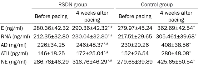
to 290.36±42.32 ng/ml in RSDN group and
from 279.97±45.24 ng/ml to 362.69±42.54 ng/ml in control group. The E increased from 212.35±32.80 ng/ml to 230.04±32.80 ng/ml in RSDN group and from 217.51±29.65 ng/ml to 305.461±39.68 ng/ml in control group. AD increased from 226±34.25 ng/ml to 246± 48.37 in RSDN group and from 230±29.26 ng/ml to 408±38.56 ng/ml in control group. ATII increased from 146±18.25 ng/ml to 172± 25.04 ng/ml in RSDN group and from 152± 26.54 pg/ml to 280±48.08 pg/ml in control group. The NE increased from 286.76±46.29
ng/ml to 316.76±46.29 ng/ml in RSDN group
and from 279.65±39.89 ng/ml to 425.65±
50.54 ng/ml in control group (Table 4). Discussion
Renal sympathetic nerves include afferent and efferent ones. The fiber endings of afferent
nerves are mainly distributed in the proximal ureter, pelvis and regions around the major
ves-sels of the kidney. In addition, there are also
sympathetic nerves in the glomeruli, proximal and distal convoluted tubules and renal
medul-la. The cell body of afferent nerves localizes in
the dorsal root ganglion at T6-L4 and projects to several regions in the central nervous
sys-tem to regulate the efferent signals. The central regulatory region of efferent nerves localizes at the head of ventrolateral medulla, and the axons of efferent nerves cross the T10-L3 sympathetic trunk to celiac ganglion, superior
mesenteric ganglion and aorticorenal ganglion where the neurons are replaced with other
neurons. Postganglionic fibers wrap the renal
artery and are distributed in small vessel, renal cortex, juxtamedullary glomerulus and renal tubules along the renal artery. The renal sympa-Table 4. Sympathetic activities in RSDN group and control group before
and after pacing
RSDN group Control group
Before pacing 4 weeks after pacing Before pacing 4 weeks after pacing
E (ng/ml) 280.36±42.32 290.36±42.32*,# 279.97±45.24 362.69±42.54*
RNA (ng/ml) 212.35±32.80 230.04±32.80*,# 217.51±29.65 305.461±39.68*
AD (ng/ml) 226±34.25 246±48.37*,# 230±29.26 408±38.56*
ATII (pg/ml) 146±18.25 172±25.04*,# 152±26.54 280±48.08*
NE (ng/ml) 286.76±46.29 316.76±46.29*,# 279.65±39.89 425.65±50.54*
Note: *P<0.05 before pacing vs. 4 weeks after pacing; #P<0.05: RSDN vs control group after
pacing.
Sympathetic activities in RSDN group and control group
At 4 weeks after right
ventricular pacing, the peripheral R, E, NE, AD and ATII increased
sig-nificantly, and these
parameters in RSDN
group were
marked-ly lower than in con- trol group (P<0.05).
[image:5.612.91.408.97.201.2]4329 Int J Clin Exp Med 2016;9(2):4324-4331 thetic nerve excitation may stimulate the
juxta-glomerular cells to secret renin, leading to the increased RASS activity. Renin acts on the renal vascular smooth muscle to induce the
contraction of renal vessels, which reduces renal blood flow and glomerular filtration rate
and increases sodium and water retention. The renal sympathetic nerve may also activate the sympathetic center to increase the systemic sympathetic activity [13]. In addition, renin acts
on β receptor to increase the myocardial con -tractility and heart rate and promote myocardi-al hypertrophy, which may compensate the
heart failure at early stage, but finally cause the deterioration of myocardial remodeling and reduce the β receptor density, leading to decompensation and deterioration of heart failure.
Studies have shown that long lasting excessive sympathetic excitation may promote the myo-cardial remodeling and deteriorate cardiac
function, and to inhibit the RASS is able to
suppress the myocardial remodeling during
heart failure [14]. Investigators reveal that renal sympathetic activity increases significantly in mice with heart failure [15]. During the heart failure, renal sympathetic nerves are preferen -tially activated, and renal norepinephrine is
an important prognostic factor of all-cause mortality in heart failure [16]. During heart fail
-ure, the sensitivity of aortic and carotid barore
-flex reduces, but the central sympathetic effer -ent and renal sympathetic activities increase,
and the activity of pulmonary stretch receptors reduces, which compromises the regulation of
renal sympathetic activity. The integration and
processing of afferent signals in the central
nervous system may elevate the sympathetic
efferent activity, especially the renal sympa
-thetic activity. After renal sympa-thetic excita
-tion, postganglionic nerve fibers may release
norepinephrine which acts on juxtaglomerular
cells to promote renin secretion and further increase ATII. Moreover, ATII may further
in-crease the sympathetic activity, leading to a
pathological positive feedback during heart failure. Thus, to block the renal sympathetic nerves may suppress their regulatory effect on the kidney and inhibit this feedback.
Ablation of the renal sympathetic nerves is a new technique used for renal denervation. On the basis of anatomical distribution of renal sympathetic nerves along the renal artery, fem
-oral artery puncture is conducted, and ablation
catheter is inserted to the distal end of renal artery. The radiofrequency energy acts on the
vascular endothelial cells, then the catheter is
withdrawn and rotated for further ablation, leading to ablation at different sites to disrupt the renal sympathetic network. This technique has favorable safety and few complications. It was first used in the therapy of refractory hyper
-tension and its safety and effectiveness have been preliminarily confirmed in studies [17]. Recent studies reveal that heart failure patients may benefit from the ablation of renal sympa
-thetic nerves [6]. In the study of Nozawa et al., ablation of bilateral renal arteries was conduct -ed in rats, and then coronary artery was ligat-ed 2 days later to establish acute myocardial
infarction model. They found that RSDN could significantly improve the sodium and water retention and the left ventricular filling pressure as well as the heart failure of rats with myocar
-dial infarction [18]. Schirmer et al. investigated the therapeutic effect of RSDN on refractory hypertension, and found that RSDN could reduce the left ventricular mass and improve the diastolic function, which were independent of blood pressure change [19]. In 2012, investi
-gators also revealed that RSDN could signifi -cantly reduce the sympathetic activity and
RAAS activity of rats with myocardial infarction and markedly improve the heart failure, and
reverse the myocardial remodeling and sodium
and water retention after myocardial infarction [20]. In a study, 7 patients with NYHA class III-IV (heart failure) were investigated, and results
indicated that the activity tolerance, cardiac
function and symptoms of heart failure were significantly improved in patients at 6 months after RSDN [21].
In the present study, right apex pacing was
introduced to establish heart failure model in dogs and the influence of renal sympathetic radiofrequency ablation on the heart failure
was investigated. Our results showed the renal
blood flow and kidney function remained un-changed after radiofrequency ablation of the
renal sympathetic nerves. The blood pressure,
heart rate, LVESD, LVEDD, CO, CI, left ventricu
dogs developed symptoms (such as loss of
appetite, reduced activities) and signs (pulmo-nary rales, increased heart rate, increased
res-piration rate) of heart failure. However, in dogs with radiofrequency ablation, the symptoms
were improved to a certain extent, the heart rate and respiration rate reduced as compared to control group, and pulmonary rales were also less heard. Heart ultrasonography showed the
LVESD and LVEDD increased in both groups and the left ventricular EF, CO and CI reduced in both groups after introduction of heart failure. However, the increases in LVESD and LVEDD in
control group were higher than in RSDN group,
and the reductions in left ventricular EF, CO
and CI in control group were more obvious as
compared to RSDN group. After introduction of heart failure, the hemodynamics changed sig
-nificantly: LVSP, dp/dtmax and -dp/dtmax re-duced significantly, and LVDP increased mark -edly, but the changes in these parameters were more obvious in control group. In addition, we
further detected the biochemical parameters
related to sympathetic excitation, and results showed the R, E, NE, AD and ATII increased in
both group after heart failure, but the increases
in control group were higher than in RSDN group. This indicates that RSDN inhibits the renal sympathetic activity, reduces NE, R, AT and AD, inhibits the RAAS activation and reduc-es systemic sympathetic activity. In addition, RSDN also suppresses the heart enlargement, compromises the reduced myocardial contrac-tility and inhibit the myocardial remodeling due
right ventricular pacing induced heart failure, leading to the improvement of symptoms of heart failure and cardiac function.
RSDN exerts therapeutic effect on the heart failure, but the specific mechanism is still poor -ly understood. In addition, results about its
therapeutic effect are mainly from experiments in animals, and few studies have been conduct
-ed to investigate the efficacy of RSDN in the therapy of heart failure patients. Thus, more
multicenter studies with large sample size are
required to confirm our findings, standardize the inclusion criteria for RSDN therapy and explore the role of RSDN in the therapy of heart failure [22].
Acknowledgements
This study was supported by the Program of Wu
Jieping Medical Foundation (320.6750.13150).
Disclosure of conflict of interest None.
Address correspondence to: Yingmin Lu, Depart-
ment of Cardiology, Xin Hua (Chongming) Hospital Affiliated to Shanghai Jiao Tong University School of
Medicine, Shanghai 200092, China. E-mail:
References
[1] Chen HZ. Cardiology 2013.
[2] Parati G and Esler M. The human sympathetic
nervous system: Its relevance in hypertension
and heart failure. Eur Heart J 2012; 33:
1058-1066.
[3] Krum H, Sobotka P, Mahfoud F, Bohm M, Esler
M and Schlaich M. Device-based
antihyperten-sive therapy: Therapeutic modulation of the
autonomic nervous system. Circulation 2011; 123: 209-215.
[4] Krum H, Schlaich M, Whitbourn R, Sobotka PA, Sadowski J, Bartus K, Kapelak B, Walton A,
Sievert H, Thambar S, Abraham WT and Esler M. Catheter-based renal sympathetic
denerva-tion for resistant hypertension: A multicentre safety and proof-of-principle cohort study. Lancet 2009; 373: 1275-1281.
[5] Esler MD, Krum H, Sobotka PA, Schlaich MP, Schmieder RE and Bohm M. Renal sympathet -ic denervation in patients with treatment-resis-tant hypertension (The Symplicity HTN-2 Trial): a randomised controlled trial. Lancet 2010; 376: 1903-1909.
[6] Brandt MC, Mahfoud F, Reda S, Schirmer SH, Erdmann E, Bohm M and Hoppe UC. Renal sympathetic denervation reduces left ventricu
-lar hypertrophy and improves cardiac function
in patients with resistant hypertension. J Am Coll Cardiol 2012; 59: 901-909.
[7] Mahfoud F, Urban D, Teller D, Linz D, Stawowy P, Hassel JH, Fries P, Dreysse S, Wellnhofer E, Schneider G, Buecker A, Schneeweis C, Doltra A, Schlaich MP, Esler MD, Fleck E, Bohm M and Kelle S. Effect of renal denervation on left ven
-tricular mass and function in patients with re
-sistant hypertension: data from a multi-centre
cardiovascular magnetic resonance imaging trial. Eur Heart J 2014; 35: 2224-2231b.
[8] Tan LH, Li XG, Guo YZ, Tang XH, Yang K and JIang WH. Effect of renal sympathetic denerva
-tion on left ventricular hypertrophy and inflam
-matory factors in spontaneously hypertensive
rats. J Zhejiang Univ (Med Sci) 2013; 42: 550-555.
[9] Hoffmann BA, Steven D, Willems S and Sydow
K. Renal sympathetic denervation as an
4331 Int J Clin Exp Med 2016;9(2):4324-4331
of ventricular electrical storm in the setting of acute myocardial infarction. J Cardiovasc Electrophysiol 2013; 24: 1175-1178.
[10] Pokushalov E, Romanov A, Corbucci G, Artyo-menko S, Baranova V, Turov A, Shirokova N, Karaskov A, Mittal S and Steinberg JS. A ran
-domized comparison of pulmonary vein isola -tion with versus without concomitant renal
ar-tery denervation in patients with refractory symptomatic atrial fibrillation and resistant hy -pertension. J Am Coll Cardiol 2012; 60: 1163-1170.
[11] Wu J, Zhao T, Shang ZJ, Cong T, Sun YH and
Wang K. The integral and focal cardiac func
-tion of dogs with rapid right ventricular pacing.
Shandong Med J 2013; 53: 44-47.
[12] Shan Y, Tao CM and Zhou LM. A study on the rapid right ventricular pacing induced heart
failure in dogs. Med Inf 2013; 52-53.
[13] Sobotka PA, Krum H, Bohm M, Francis DP and Schlaich MP. The role of renal denervation in the treatment of heart failure. Curr Cardiol Rep 2012; 14: 285-292.
[14] Xu XY, Duan ZZ, Zhao QY and Hu W.
Two-Dimensional Speckle Tracking Imaging Applied to Evaluating Left Ventricular Changes in
Nor-mal Dogs Following Transcatheter Renal Sym-
pathetic Denervation. Guangxi Med J 2014;
1411-1414.
[15] Zhou MH, Zhao QY, Yu SB, Dai ZX, Wang XL, Xiao JP, Yang B and Huang CX. Effects of renal sympathetic denervation on inducibility of atri
-al fibrillation during rapid atri-al pacing. Nat Med J China 2012; 92: 2868-2871.
[16] Petersson M, Friberg P, Eisenhofer G, Lambert G and Rundqvist B. Long-term outcome in rela -tion to renal sympathetic activity in patients
with chronic heart failure. Eur Heart J 2005;
26: 906-913.
[17] Schlaich MP, Hering D, Sobotka P, Krum H, Lambert GW, Lambert E and Esler MD. Effects of renal denervation on sympathetic activa -tion, blood pressure, and glucose metabolism in patients with resistant hypertension. Front Physiol 2012; 3: 10.
[18] Nozawa T, Igawa A, Fujii N, Kato B, Yoshida N, Asanoi H and Inoue H. Effects of long-term re
-nal sympathetic denervation on heart failure after myocardial infarction in rats. Heart Vessels 2002; 16: 51-56.
[19] Schirmer SH, Sayed MM, Reil JC, Ukena C, Linz D, Kindermann M, Laufs U, Mahfoud F and Bohm M. Improvements in left ventricular hy
-pertrophy and diastolic function following renal denervation: effects beyond blood pressure
and heart rate reduction. J Am Coll Cardiol 2014; 63: 1916-1923.
[20] Hu J, Ji M, Niu C, Aini A, Zhou Q, Zhang L, Jiang
T, Yan Y and Hou Y. Effects of renal sympathet
-ic denervation on post-myocardial infarction
cardiac remodeling in rats. PLoS One 2012; 7:
e45986.
[21] Davies JE, Manisty CH, Petraco R, Barron AJ, Unsworth B, Mayet J, Hamady M, Hughes AD, Sever PS, Sobotka PA and Francis DP. First-in-man safety evaluation of renal denervation for chronic systolic heart failure: primary outcome from REACH-Pilot study. Int J Cardiol 2013; 162: 189-192.
[22] Verloop WL, Beeftink MM, Nap A, Bots ML, Velthuis BK, Appelman YE, Cramer MJ, Agema
WR, Scholtens AM, Doevendans PA, Allaart CP
and Voskuil M. Renal denervation in heart fail
-ure with normal left ventricular ejection frac