ISSN Online: 2164-5337 ISSN Print: 2164-5329
DOI: 10.4236/wjcd.2019.911076 Nov. 27, 2019 857 World Journal of Cardiovascular Diseases
Correlation between Serum Uric Acid Level and
Left Ventricular Ejection Fraction in Patients
with Congestive Heart Failure
Mohamed A. W. Ezzat*, Ahmed M. Boghdady, Kassem F. A. Ibrahim, Lotfy H. Abo Dahab
Internal Medicine and Cardiology Department, Faculty of Medicine, Sohag University, Sohag, Egypt
Abstract
Background: One of the risk factors of congestive heart failure that still un-der investigations is hyperuricemia. It’s still debatable whether it’s an inde-pendent risk factor or it’s just a consequence of other disorders associated with cardiovascular diseases like hypertension, diabetes and dyslipidemia. Objective: The aim of our study is to elucidate whether in patients with heart failure serum uric acid level correlates with left ventricular ejection fraction supporting the possibility that the detection of progressive hyperuricemia in these patients may be an indicator of deteriorating cardiac function. Me-thods: We conducted a prospective study that included 124 studied patients and 26 apparently healthy persons at Coronary care unit and Internal Medi-cine Department at Sohag University Hospitals. Studied populations were classified into; * Group “I”: newly diagnosed heart failure, * Group “II”: de-compensated heart failure on regular treatment, * Group “III”: decompen-sated heart failure but stopped their treatment from three months, Group “IV”: control group, healthy and age-matched subjects. We studied the asso-ciation between left ventricular ejection fraction, the severity of congestive heart failure and the serum uric acid levels and the well-known conventional risk factors. Results: The main finding was the significantly higher mean se-rum uric acid levels in patients with congestive heart failure versus apparently healthy persons with “P value = 0.02”. When we adjusted the serum uric acid with other significant risk factors in the univariate analysis which were age, gender and smoking, serum uric acid was an independent risk factor “P value = 0.04”. There was a significant correlation between serum uric acid level and the severity of congestive heart failure “P value < 0.001, correlation coefficient = 0.35”. High rates of serum uric acid levels were recorded in patients with reduced ejection fraction. A uric acid level of 8.45 mg/dl was found to be the most appropriate cut-off point with the sensitivity 62% and the specificity How to cite this paper: Ezzat, M.A.W.,
Boghdady, A.M., Ibrahim, K.F.A. and Da-hab, L.A.B. (2019) Correlation between Serum Uric Acid Level and Left Ventricular Ejection Fraction in Patients with Conges-tive Heart Failure. World Journal of Car-diovascular Diseases, 9, 857-866.
https://doi.org/10.4236/wjcd.2019.911076
Received: October 28, 2019 Accepted: November 24, 2019 Published: November 27, 2019
Copyright © 2019 by author(s) and Scientific Research Publishing Inc. This work is licensed under the Creative Commons Attribution International License (CC BY 4.0).
http://creativecommons.org/licenses/by/4.0/
DOI: 10.4236/wjcd.2019.911076 858 World Journal of Cardiovascular Diseases 78.5%. Conclusion: Higher serum uric acid levels are significantly correlated with the severity of congestive heart failure and left ventricular ejection frac-tion. Serum uric acid is an independent risk factor for congestive heart fail-ure.
Keywords
Uric Acid Level, Congestive Heart Failure
1. Introduction
Uric acid (UA) is the end product of purine breakdown and is excreted by the kidneys. Xanthine oxidase and xanthine dehydrogenase are two enzymes re-sponsible for uric acid breakdown and production. Both enzymes catalyze the oxidation of hypoxanthine to xanthine which is the main enzyme in purine me-tabolism and contribute to the generation of oxygen free radicals which in-creased oxidative stress [1]. In addition, oxidative stress along with nitric oxide disproportion could intensify inflammatory pathways resulting in further in-crease in cytokine production [2].
Moreover, isolated hyperuricemia (irrespective of renal function and admin-istration of drugs) appears to be a marker of altered oxidative metabolism, asso-ciated with tissue hypoxia, which could further damage cardiomyocytes and vascular endothelium. In addition, this can induce disturbances of myocardial contractility, vasoconstriction and may negatively affect the cardiovascular sys-tem and potentially worsen the prognosis in patients with heart failure [3].
Uric acid can also stimulate granulocyte adherence to the endothelium, and peroxide and superoxide free radical production, therefore, uric acid traverses dysfunctional endothelial cells which accumulate as crystal within atherosclerot-ic plaques. These crystals may contribute to local inflammation and plaque pro-gression, and crystal accumulation may be greater in patients with elevated se-rum uric acid concentration. These effects of uric acid can lead to ischemic car-diomyopathy and then post-ischemic heart failure [4].
Chronic heart failure (CHF) is a leading etiology for both morbidity and mortality on a global level, resulting in an increase in both prevalence and health care costs. Recently, our understanding has changed from a mere hemodynamic condition to a much more complicated approach, including neuroendocrine and immune activation. Not only is the cardiovascular system damaged in the long run course of heart failure, but together with peripheral tissues and organs also result in the production of symptoms along with the progression of the disease [5].
DOI:10.4236/wjcd.2019.911076 859 World Journal of Cardiovascular Diseases
2. Methods
2.1. Study DesignOur study was a prospective study that included 124 patients and 26 apparently healthy persons who underwent echocardiography at Internal Medicine and Cardiology Department at Sohag University Hospitals, Sohag governorate, Egypt.
Inclusion criteria:
All studied populations were classified into four groups; • Group “I” patients being newly diagnosed heart failure,
• Group “II” patients with decompensated heart failure on regular treatment, • Group “III” patients with decompensated heart failure but stopped their
treatment from three months,
• Group “IV” control group selected as healthy and age-matched persons from 30 to 70 years old.
Exclusion criteria:
The following groups of patients were excluded from the study on basis of clinical, electrocardiographic, echocardiographic and laboratory parameters: • Liver and kidney diseases, hematological or oncological disorders.
• Patients taking diuretics, multivitamins, alcohol and on any drugs interfering with serum uric acid levels.
• Persons less than 30 years old and above 70 years old.
2.2. Methods Clinical history:
• History of cardiovascular symptoms e.g. chest pain, shortness of breath, pal-pitation, syncope.
• Risk factors of coronary atherosclerosis particularly diabetes mellitus, hyper-tension, Hyperlipidemia, smoking, family history of coronary heart disease, gender, obesity, etc.
• Diabetes Mellitus was defined as fasting plasma glucose ≥ 126 mg/dl and/or random plasma glucose ≥ 200 mg/dl and/or HbAlC ≥ 6.5 or use of glucose lowering medications.
• Hypertension was defined as blood pressure > 140/90 mmHg or the use of Blood pressure lowering medications.
• Dyslipidemia was defined as total cholesterol more than 200 mg/dl, LDL more than 130 mg/dl or HDL less than 65 mg/dl.
• History of previous myocardial infarction.
• History of any previous coronary revascularization procedure (PCI & CABG) and its details.
• Current medication and drug history.
Full clinical examination with special stress on:
DOI: 10.4236/wjcd.2019.911076 860 World Journal of Cardiovascular Diseases formula
BMI = weight (kg)\height 2 (m) • Pulse, blood pressure and respiratory rate.
• Examination of all peripheral pulses, particularly femoral arteries on both groin.
• Careful and complete cardiac and chest examination. Investigations:
1) 12 leads electrocardiography (ECG): Was analyzed for:
• Rate and Rhythm.
• ST segment changes as horizontal or down sloping ST depression 1 mm at the J-point in 2 contiguous leads or upward ST elevation represents myocar-dial injury.
• Pathological Q wave T-wave inversion at least 1 mm deep in 2 continuous leads that have dominant R waves (R/S ratio > 1).
• Pathological Q waves.
2) Echocardiography (ECHO):
Study groups were examined at rest in supine left lateral decubitus position. Analysis according to the American Society of Echocardiography for:
a)The presence of regional wall motion abnormality.
b) Left ventricular end systolic diameter (LVESD), left ventricular end diastolic diameter (LVEDD), interventricular septum diameter and posterior wall diameter.
c) Ejection fraction calculated by M-Mode in parastemal long and short axes or by biplane method according to modified Simpson’s.
• Echocardiography is the most useful, widely available test in the diagnosis of heart failure with preserved ejection fraction (HFpEF) as an LVEF ≥ 50%, consider patients with an LVEF above 40% and less than 50% as heart failure with mid-range ejection fraction (HFmrEF) and consider patients with an LVEF ≤ 40% as heart failure with reduced ejection fraction (HFrEF) [2]. 3) Laboratory investigations:
• Serum creatinine, liver function, Serum uric acid, lipid profile and cardiac enzymes.
• The normal serum uric acid level ranges from 2.4 - 7.4 mg/dL in males whe-reas in females it ranges from 1.4 - 5.8 mg/dL. Hyperuricemia considered when serum uric acid level more than 7.4 mg/dl in males and more than 5.8 mg/dl in females. Serum uric acid level less than 8 mg/dl considered mild elevated, serum uric acid level between 8 mg/dl and 12 mg/dl considered moderate elevated and serum uric acid level more 12 mg/dl considered marked elevated [7].
Ethical aspect
Univer-DOI:10.4236/wjcd.2019.911076 861 World Journal of Cardiovascular Diseases sity, Faculty of Medicine, Egypt.
Statistical analysis
Data demonstrated as mean ± SD for normally distributed continuous va-riables, median (range) for non-normally-distributed continuous variable sand frequencies for categorical variables. Patients were classified into three groups who were newly diagnosed as heart failure or stop treatment of heart failure and control group. Three groups were compared as regard age, gender, body mass index (BMI), diabetes, hypertension, smoking, family history, total cholesterol, LDL, HDL, triglycerides and uric acid. Differences between the groups were as-sessed using Student T-test for continuous parametric variables, Mann-Whitney U-test non-parametric continuous variables and Chi square test for categorical variables. Multivariate logistic regression analysis was performed to assess the effects risk factors on CAD. All statistical analyses were performed using SPSS software version 16 (Chicago, IL, USA) P value < 0.05 indicate significance.
3. Results
The study included 150 persons, 124 patients with congestive heart failure (ei-ther newly diagnosed, uncontrolled on treatment or stop treatment since more than three months) and 26 control persons. The mean age of the studied popula-tion was 48.5 years. Most of the included persons were males, representing 73.38% in studied patients and 61.54% in apparently healthy persons.
Most of the patients group had hyperuricemia (79.83%) while 80.76% of the control group was non hyperuricemic.
The mean uric acid level recorded a mean of 8.87 mg/dl ranging from 4 mg/dl to 14 mg/dl in studied patients and 5 mg/dl ranging from 3.5 mg/dl to 9 mg/dl in apparently healthy persons. The mean of uric acid level of group who stop treatment, group regular on treatment, and group who newly diagnosed was 8.87, 8.35 and 7.1 respectively.
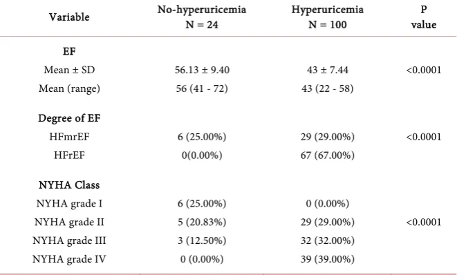
According to NYHA classification, 30.77%, 40% and 27.27% of group who stop treatment, group regular on treatment, and group who newly diagnosed respectively were grade IV. The majority of the patients with serum uric acid level > 12 mg/dl (88.37%) were heart failure grade IV and 11.63% were heart failure grade III. About 50.88% of Patients with serum uric acid level between 8 to 12 mg/dl were heart failure grade III and 43.86% were heart failure grade II and patients with serum uric acid level between 6 to 8 mg/dl, 38.10% of them were heart failure grade II, 19.05% were heart failure grade I, and 14.29% were heart failure grade III. Hyperuricemic studied populations with serum uric acid level < 6 mg/dl, the majority of them (58.62%) had no dyspnea whereas 3.45% had heart failure grade IV. The level of serum uric acid was increasing with more severity of congestive heart failure especially in patients with reduced ejection fraction. The more advanced NYHA class the more increase in the number of hyperuricemic patients.
DOI: 10.4236/wjcd.2019.911076 862 World Journal of Cardiovascular Diseases EF of group who stop treatment was 42%, and that of group who regular on treatment was 43%, the mean EF of group who newly diagnosed was 44% and the mean EF of apparently healthy persons was 65% (Tables 1-4).
4. Discussion
One of the risk factors of CHF that still under investigations is hyperuricemia. It’s still debatable whether it’s an independent risk factor or it’s just a conse-quence of other disorders associated with cardiovascular diseases like hyperten-sion, diabetes and dyslipidemia [8].
[image:6.595.208.539.253.424.2]The possible effect of hyperuricemia on cardiovascular health may be related
Table 1. Demographic data of the studied population and uric acid level.
Variable Patients (124) Control (26)
Gender
Males 91(73.38%) 16 (61.54%)
Females 33 (26.61%) 10 (38.46%)
Hyperuricemia
No Hyperuricemia 25 (20.16%) 21 (80.76%)
Hyperuricemia 99 (79.83%) 5 (19.23%)
Uric acid level
Mean ± SD 8.43 ± 2.95 5.55 ± 1.48
Mean (range) 8.87 (4 - 14) 5 (3.5 - 9)
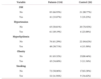
Table 2. Risk factors among studied population.
Variable Patients (124) Control (26)
DM
No 83 (66.93%) 21 (80.77%)
Yes 41 (33.07%) 5 (19.23%)
Hypertension
No 63 (50.81%) 20 (76.92%)
Yes 61 (49.19%) 6 (23.08%)
Hyperlipidemia
No 76 (61.29%) 22 (84.62%)
Yes 48 (38.71%) 4 (15.38%)
Obesity
No 81 (65.32%) 23(88.46%)
Yes 43 (34.68%) 3 (11.54%)
Smoking
No 72 (58.06%) 17(65.38%)
[image:6.595.201.540.456.721.2]DOI:10.4236/wjcd.2019.911076 863 World Journal of Cardiovascular Diseases Table 3. Comparison among the groups of studied populations as regard EF, NHYA clas-sification.
Variable
Newly diagnosed
N = 55
On treatment N = 30
Stop treatment
N = 39
Healthy
N = 26 P value
EF
Mean ± SD 44.65 ± 7.52 41.7 ± 7.97 42.67 ± 6.82 64.5 ± 4.81 <0.0001
Mean (range) 44 (28 - 58) 43 (25 - 55) 42 (22 - 55) 65 (57 - 72)
P1 = 0.40, P2 = 1.00, P3 < 0.0001, P4 = 1.00, P5 < 0.0001, P6 < 0.0001
Degree of EF
Preserved EF 4 (7.27%) 0 (0.00%) 0 (0.00%) 19 (73.08%) <0.0001
Mid-range EF 18 (32.73%) 11 (36.67%) 12 (30.77%) 7 (26.92%)
Reduced EF 33 (60.00%) 19 (63.33%) 27 (69.23%) 0
P1 = 0.32, P2 = 0.21, P3 < 0.0001, P4 = 0.61, P5 < 0.0001, P6 < 0.0001
NYHA Class
No dyspnea 0 (0.00%) 0 (0.00%) 0 (0.00%) 26 (100%)
Dyspnea with extra effort 0 (0.00%) 0 (0.00%) 0 (0.00%) 0 <0.0001
Dyspnea with normal effort 20 (36.36%) 13 (43.33%) 11 (28.2%) 0
Dyspnea with little effort 20 (36.36%) 5 (16.67%) 16 (41.03%) 0
Dyspnea at rest 15 (27.27% 12 (40.00%) 12 (30.77%) 0
P1 = 0.09, P2 = 0.37, P3 < 0.0001, P4 = 0.11, P5 < 0.0001, P6 < 0.0001
P1 compared Newly diagnosed & on treatment, P2 compared Newly diagnosed & Stop treatment, P3 com-pared Newly diagnosed & apparently healthy persons, P4 comcom-pared on treatment & Stop treatment, P5 compared on treatment & apparently healthy persons, P6 compared Stop treatment & apparently healthy persons, EF = ejection fraction, NYHA = New York Heart Association.
Table 4. Relation of uric acid level to EF and NYHA classification of the patients groups.
Variable No-hyperuricemia N = 24 Hyperuricemia N = 100 value P
EF Mean ± SD Mean (range)
56.13 ± 9.40 56 (41 - 72)
43 ± 7.44 43 (22 - 58)
<0.0001
Degree of EF HFmrEF HFrEF 6 (25.00%) 0(0.00%) 29 (29.00%) 67 (67.00%) <0.0001 NYHA Class NYHA grade I NYHA grade II NYHA grade III NYHA grade IV
6 (25.00%) 5 (20.83%) 3 (12.50%) 0 (0.00%) 0 (0.00%) 29 (29.00%) 32 (32.00%) 39 (39.00%) <0.0001
[image:7.595.208.539.510.709.2]DOI: 10.4236/wjcd.2019.911076 864 World Journal of Cardiovascular Diseases to its association with endothelial dysfunction, anti-proliferative effects, high oxidative stress, generation of free radicals and thrombus formation, all pro-moting atherosclerosis and its consequences [9].
The controversy in the literature about the effect of hyperuricemia on cardi-ovascular health triggered us trying to study this association.
Our study included 124 studied patients and 26 apparently healthy persons. All studied populations were classified into four groups being newly diagnosed with CHF, on regular treatment for CHF or who stop treatment of CHF, and they selected to our study in addition to number of healthy age-matched subjects served as controls. We studied the association between left ventricular ejection fraction, the severity of congestive heart failure and the serum uric acid levels and the well-known conventional risk factor that include age, sex, BMI, positive family history of IHD, smoking, hypertension, diabetes and dyslipidemia.
The majority of the studied patients in this research were hyperuricemic with percentage of 79.83% while 80.76% of the control group was non hyperuricemic. The mean of uric acid level of group who stop treatment, group regular on treatment, and group who newly diagnosed was 8.87 mg/dl, 8.35 mg/dl and 7.1 mg/dl respectively while the mean of uric acid level of apparently healthy per-sons was 5 mg/dl. These results coincided with a study was conducted in the Faculty of Medicine, Cairo University, 2015 included 778 studied patients and 148 of healthy age-matched subjects served as controls. The authors found that the majority of studied patients were hyperuricemic with percentage of 63.5% (mean uric acid level 7.9 md/dl) and the opposite of this occurred to apparently healthy persons where 92% were non hyperuricemic (the mean of uric acid level was 4.4 mg/dl [10].
There was higher association between hyperuricemia and hypertension, di-abetes, dyslipidemia with higher BMI versus normouricemic patients and these results were in agreement with the study of Seo Young Kim, 2006” [11].
The pathogenic role of uric acid in hypertension, and also insulin resistance that present in association with metabolic syndrome has a causative role in de-veloping hyperuricemia; all these factors may explain our results.
Our results revealed that there was a strong association between high serum uric acid levels and the severity of congestive heart failure. This was in agree-ment with a study by Deveci et al. conducted on 1012 patients and they found significantly higher mean serum uric acid in patients with coronary artery dis-eases (6.3 ± 2.0 vs. 5.4 ± 1.4, P value < 0.001) [12].
An Egyptian study conducted at Mansoura University, 2017 about uric acid level and coronary artery disease found significant higher mean serum uric acid levels in patients with CAD versus free patients (7.43 ± 2.95 vs. 5.55 ± 1.48, P value = 0.02) with significant correlation between serum uric acid level and the severity of CAD (P value < 0.001, r = 0.35) [13].
DOI:10.4236/wjcd.2019.911076 865 World Journal of Cardiovascular Diseases severity of congestive heart failure and this was close to results of Deveci et al.; they found a uric acid level of 6.8 mg/dl to be the most accurate cut-off with 40% sensitivity and 87.5% specificity [12].
A study carried at University of Palermo, Italy, included 1118 patients classi-fied to three groups patients with systolic and diastolic dysfunction (EF < 50% and E wave < A wave; n = 40), those with isolated diastolic dysfunction (EF > 50% and E wave < A wave; n = 24) and those that had anormal left ventricular function (EF > 50% and E wave > A wave, n = 54). The results showed higher values of serum uric acid in patients with diastolic dysfunction with or without systolic dysfunction compared to patients with normal ventricular function (6.54 ± 0.72 vs. 5.42 ± 0.78, p = 0.0116) and with increasing serum uric acid there was a proportional reduction of EF [14].
5. Conclusion
Our study concluded that more increase in serum uric acid levels is significantly associated with more severe congestive heart failure and more reduced left ven-tricular ejection fraction. Serum uric acid may be considered as an independent risk factor for CHF “P value = 0.04” and serum uric acid level is significantly as-sociated with the severity of congestive heart failure “P value < 0.001”. A uric acid level of 8.45 mg/dl was found to be the most appropriate cut-off point with the sensitivity 62% and the specificity 78.5%.
Conflicts of Interest
The authors declare no conflicts of interest regarding the publication of this paper.
References
[1] McMurray, J.J.V., Cherubini, A., Ble, A., Bos, A.J.G., Maggio, M., Dixit, V.D., et al. (2014) Uric Acid and Inflammatory Markers. European Heart Journal, 27, 1174-1181. https://doi.org/10.1093/eurheartj/ehi879
[2] Anker, S.D., Granner, D.K., Mayes, P.A. and Rodwell, V.W. (2012) Harper’s Illu-strated Biochemistry. Molecular Physiology.
[3] Adamopoulos, S., Anker, S.D., Auricchio, A., Böhm, M., Dickstein, K., et al. (2012) ESC Guidelines for the Diagnosis and Treatment of Acute and Chronic Heart Fail-ure. European Heart Journal, 33, 1787-1847.
[4] Fang, J. and Alderman, M.H. (2015) Serum Uric Acid and Cardiovascular Mortality the NHANES I Epidemiologic Follow-Up Study 1971-1992. National Health and Nutrition Examination Survey. JAMA, 283, 2404-2410.
[5] Dickstein, K., Krishnan, E., Chen, L. and Schumacher, H.R. (2012) Serum Uric Acid and Cardiovascular Disease: Recent Developments, and Where Do They Leave Us.
The American Journal of Medicine, 118, 816-826. https://doi.org/10.1016/j.amjmed.2005.03.043
DOI: 10.4236/wjcd.2019.911076 866 World Journal of Cardiovascular Diseases [7] Lotan, C., Benjamin, E.J., Go, A.S., Arnett, D.K., Blaha, M.J., Cushman, M., et al. (2015) Heart Disease and Stroke Statistics 2015 Update: A Report from the Ameri-can Heart Association. Circulation, 131, e29-322.
[8] Harzand, A., Palacio, A., Verma, S., Jones, J., Hare, J., et al. (2016) Uric Acid as a Predictor of All-Cause Mortality in Heart Failure: A Meta-Analysis. Congestive Heart Failure, 17, 25-30.https://doi.org/10.1111/j.1751-7133.2011.00200.x
[9] Deveci, T., Takeishi, Y., Arimoto, T., Okuyama, H., Nozaki, N., Hirono, O., et al. (2013) Hyperuricemia Associated with High Cardiac Event Rates in the Elderly with Chronic Heart Failure. Journal of Cardiology, 47, 219-228.
[10] Sun, T., Furumoto, T., Tsuchihashi-Makaya, M., Goto, K., Goto, D., Yokota, T., et al. (2014) Hyperuricemia Predicts Adverse Outcomes in Patients with Heart Failure.
International Journal of Cardiology, 151, 143-147.
[11] Gotsman, I., Keren, A., Lotan, C. and Zwas, D.R. (2012) Changes in Uric Acid Le-vels and Allopurinol Use in Chronic Heart Failure: Association with Improved Sur-vival. Journal of Cardiac Failure, 18, 694-701.
https://doi.org/10.1016/j.cardfail.2012.06.528
[12] Deveci, O.S., Kabakci, G., Okutucu, S., Tulumen, E., et al. (2010) The Association between Serum Uric Acid Level and Coronary Artery Disease. Clin Pract, 64, 900–907.
[13] Coutinho Tde, A., Turner, S.T., Peyser, P.A., Bielak, L.F., Sheedy, P.F. and Kullo, I.J. (2007) Associations of Serum Uric Acid with Markers of Inflammation, Metabolic Syndrome, and Subclinical Coronary Atherosclerosis. American Journal of Hyper-tension, 20, 83-89.https://doi.org/10.1016/j.amjhyper.2006.06.015