Author’s Accepted Manuscript
Neural signatures of recognition memory in 10- to
12-month-old infants
Szilvia Linnert, Brigitta Tóth, Márton Nagy,
Eugenio Parise, Ildikó Király
PII:
S0028-3932(17)30317-2
DOI:
http://dx.doi.org/10.1016/j.neuropsychologia.2017.08.023
Reference:
NSY6473
To appear in:
Neuropsychologia
Received date: 28 February 2017
Revised date:
6 August 2017
Accepted date: 18 August 2017
Cite this article as: Szilvia Linnert, Brigitta Tóth, Márton Nagy, Eugenio Parise
and Ildikó Király, Neural signatures of recognition memory in 10- to
12-month-old
infants,
Neuropsychologia,
http://dx.doi.org/10.1016/j.neuropsychologia.2017.08.023
This is a PDF file of an unedited manuscript that has been accepted for
publication. As a service to our customers we are providing this early version of
the manuscript. The manuscript will undergo copyediting, typesetting, and
review of the resulting galley proof before it is published in its final citable form.
Please note that during the production process errors may be discovered which
could affect the content, and all legal disclaimers that apply to the journal pertain.
1 Neural signatures of recognition memory in 10- to 12-month-old infants
Szilvia Linnert1, Brigitta Tóth2, Márton Nagy 3,4, Eugenio Parise1, Ildikó Király3
1, Department of Psychology, Lancaster University, Lancaster, United Kingdom
2, Institute of Cognitive Neuroscience and Psychology, Research Centre for Natural Sciences, Hungarian Academy of Sciences, Budapest, Hungary
3, Department of Cognitive Psychology, Eötvös Loránd University, Budapest, Hungary
4, Doctoral School of Psychology, Eötvös Loránd University, Budapest, Hungary
Corresponding author: Szilvia Linnert Department of Psychology
Fylde Collage Lancaster University Lancaster
United Kingdom LA1 4YF
2 Abstract
Understanding memory mechanisms is crucial in the study of infant social and cognitive development. Here, we show that the Nc ERP component, known to reflect frequency-related attentional and/or memory processes, is a good candidate to investigate infant recognition memory. Previous paradigms have only investigated the effect of frequency during on-line stimulus presentation, but not during stimulus encoding. In this paper, we present a novel method for measuring the neural correlates of recognition memory and the ‘degree’ of familiarity in 10- to 12-month-old infants. During a familiarization phase, two images were presented frequently, while another two images were presented infrequently to the infants. In the test phase, the infrequent familiar, the frequent familiar, and the novel stimuli, were all presented with equal probability. We found larger Nc amplitudes following the familiar stimuli compared to the novel ones. The ‘degree’ of familiarity, on the other hand, did not modulate the
Nc amplitude. These results can only be explained with memory-related processes, since in our paradigm the on-line presentation frequency did not vary. Furthermore, the lack of familiarization frequency effect suggests that the Nc might be a neural correlate of declarative memory.
Keywords
3 Neural signatures of recognition memory in 10- to 12-month-old infants
The declarative memory, the capacity to recall and recognize facts and events, is fundamental to human cognition. It emerges by the end of the first year of life (Bauer, Wenner, Dropik, & Wewerka, 2000), and is crucial in social and cognitive development. Learning culturally relevant information and establishing social relationship relies on declarative memory. For instance, it is possible to hypothesize that the capacity to strengthen social bonds or the ability to develop a certain attachment, is directly affected by the capacity to store information about that specific social partner (Bushnell, 2001). Learning how to use new artifacts relies largely on teaching episodes that need to be stored in memory (Casler & Kelemen, 2005; Tomasello, 1999) and often lasts a lifetime (cf. Gergely & Csibra, 2006). Therefore, memory plays a direct and crucial role in social and cognitive development. Research on memory development has provided evidence on long-term recall in children well before speech onset, demonstrating that this capacity shows marked variability in the first year despite its relevance for infants’ social and cognitive development (Carver
& Bauer, 1999).
There is some evidence that very young infants are capable of forming and recollecting complex memories that include what-where-when components of the original events (Richmond & Nelson, 2009; but see also Koski, Olson, & Newcombe, 2013). However, investigating declarative memory represents a challenge in developmental research. An effective way would be to combine behavioral and neuroscience methods, such as the electroencephalogram (EEG) (Bauer, Wiebe, Carver, Waters, & Nelson, 2003; Carver, Bauer, & Nelson, 2000). Nevertheless, this requires the identification of a memory-related EEG signature, such as an event-related potential (ERP) component.
Carver, Bauer, and Nelson (2000) and Bauer, Wiebe, Carver, Waters, and Nelson (2003) found differences in the ERP responses to pictures of previously experienced “old” sequences and never-before-experienced “new” sequences in 9
month-old infants. Bauer et al. (2003) analyzed the Negative central (Nc) ERP component – a negative component over frontal and central scalp areas with a
4 Norcia, 1981). Novel stimuli elicited greater Nc amplitude compared to familiar images, indicating that the infants encoded the events. This result suggests that the Nc ERP component is a reliable indicator of some sort of recognition or familiarity in infants. Furthermore, the Nc component has been shown to be sensitive to socially relevant stimuli, such as the mother’s face (de Haan & Nelson, 1997, 1999) and the
infants' favorite toy (de Haan & Nelson, 1999).
Though the Nc amplitude has been previously suggested to be sensitive to memory processes, the on-line stimulus presentation frequency also seems to modulate it. Courchesne et al. (1981) presented images of female faces with different frequency rates (88% standard vs. 12% oddball trials). The Nc component was significantly larger for the oddball compared to the standard face. The authors have suggested two interpretations. First, the Nc might reflect differences in stimulus presentation frequency: the larger Nc amplitude reflects the detection of rare stimuli. Such on-line frequency change monitoring indicates a bottom-up, attentional process. Second, the Nc amplitude might reflect the strength of the memory trace. The frequently presented stimulus became more and more familiar during the experiment. Therefore, a smaller Nc amplitude reflects stronger memory trace for the frequent event, and a higher Nc amplitude reflects weaker memory trace for the rare event. This second explanation suggests a top-down, memory-related process. Nevertheless, according to both explanations the Nc seems to be a neural correlate of some sort of frequency based encoding process (Ackles, 2008).
5 either the frequent familiar or the novel images. Such differences might signal either memory-related processes (familiar vs. novel difference) or the effect of presentation frequency (frequent vs. infrequent difference). On the other hand, the results also indicated that the on-line presentation frequency alone does not modulate the Nc amplitude. A control group of infants was familiarized with images that were not used later in the oddball procedure. Therefore, all stimuli were new for this group as they were presented with frequent novel, infrequent novel, and infrequent trial-unique stimuli. The Nc amplitude did not differ between stimulus types, indicating that the Nc amplitude cannot simply reflect bottom-up, presentation-frequency dependent, attention or orientation effects (cf. also Ackles, 2008). Therefore, top-down factors, such as detection of something being familiar, might modulate the Nc amplitude.
In all previous experiments, the effect of frequency on the Nc has been investigated only by manipulating the on-line stimulus presentation probability. But according to the memory-based explanation of the Nc (Courchesne et al., 1981), the Nc amplitude might rather be sensitive to a frequency-based stimulus encoding process (Ackles, 2008). Memories with a what-where-when component are encoded based on a unique, single event, whereas familiarity-based recognition doesn’t necessarily rely on a single exposure. Previous studies on the Nc have not investigated the differential contribution of stimulus frequency to memory processes. We propose to systematically vary both the frequency and the familiarity components of the stimuli during the encoding (familiarization phase), while keeping constant the on-line stimuli presentation frequency (test phase). Such a manipulation allows us to study the effect of previously encountered frequency differences - in other words, the ‘degree’ of familiarity - on the amplitude of the Nc component, with no confound of
on-line presentation frequency differences.
6 infrequent familiar stimuli) effects can only be ascribed to the familiarization phase, revealing the role of memory encoding. As a result, any modulation of the Nc component can only depend on the ‘degree’ of stimuli familiarity (memory-related
top-down processes) and cannot reflect a basic change due to frequency monitoring (bottom-up attentional processes). We expect Nc amplitude differences between the frequent familiar and novel stimuli. However, the question is whether the Nc amplitudes for the infrequent familiar stimuli would be more similar to the frequent familiar or novel images.
Methods
Participants
The experiment took place in the EEG Lab at the Department of Cognitive Psychology, Eötvös Loránd University, Budapest, Hungary. All parents were informed about the experiment before they volunteered and signed a consent form. The Ethical Committee of the Institute of Psychology at the ELTE University approved the research project and the experimental procedure. Fifteen infants (6 females), full-term and normally developing, were included in the final sample. The mean age was 11 months 15 days (SD = 24.56 days). Eleven additional infants participated, but were excluded from the final sample, because they did not provide enough artifact free segments to compute the ERPs (N=6); because of equipment failure (N = 3) or fussiness (N = 2). Infants were together with their parent during the experiment and they received a toy for volunteering.
Stimuli and materials
eye-7 movement data. We calculated the average fixation time (the quotient of total looking time and number of fixations on the toy) for each object and there were no significant differences from the overall average fixation for the toys used in the EEG experiment (one-sample t-tests, test value = 69.9 ms, p < .05). This indicates that none of the toys presented in the EEG experiment was more interesting for the infants, ruling out saliency related effects.
The stimuli in the EEG experiment were colored pictures of 8 toys with a hand holding them against a black background (height: 400 pixels, width: 300 pixels). We included the hand to suggest a social context and increase infants’ attention. During the experiment the stimuli appeared on a black background in the center of a 17” LCD
computer screen with a 60 Hz refresh rate and 1280 x 1024 pixels resolution at a viewing distance of approximately 70 cm. We used E-prime 2.0 software (Psychology Software Tools, Pittsburgh, PA) for stimuli presentation. We monitored the infant’s
8
Figure 1. Stimuli. The two frequent, two infrequent and two novel images were randomly chosen for each participant.
Procedure
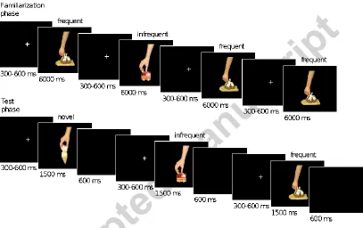
[image:9.595.96.503.271.527.2]Figure 2 shows the schematic diagram of the experimental design. The experiment consisted of two phases: a familiarization and a test (EEG) phase. Each infant saw six images, randomly chosen out of the set of eight. In the familiarization phase, we familiarized the participants with four images, and in the test phase, they saw two additional, randomly selected pictures as novel stimuli.
Figure 2. Trial structure in the familiarization and test phases. In the familiarization phase the frequent stimuli were presented in 75%, while the infrequent stimuli were presented in 25% of the trials. In the test phase all three stimulus types were shown with equal probability.
9 ms. The familiarization phase lasted approximately 3 minutes. The presentation order was pseudorandom with the following constraints: we delivered the trials in three blocks, in each block we presented the infrequent images once, and the frequent images three times. Within a block, the order of the presented images was randomized. During a 5-10 minutes long break between the experimental phases, the infants could play.
In the test phase, we presented all four previously shown images together with two novel stimuli. We displayed all frequent familiar, infrequent familiar and novel stimuli with equal probability (33.3%). We presented the stimuli in blocks of six trials where all images appeared once. The presentation order was random within a block. A single trial started with a white fixation cross displayed in the middle of the black screen for a random period between 300 - 600 ms followed by a stimulus presentation for 1500 ms. The screen was blank for 600 ms following the stimulus presentation. The test phase ended when the child became fussy or their attention could no longer be directed to the screen. During this phase, we recorded both EEG and video (synchronized with the EEG).
EEG recording and analysis
To record the EEG, we used an EGI (Electrical Geodesics Incorporated, Eugene, OR) GES300 EEG system with NetAmps300 amplifier and 128 channel HydroCel Geodesic Sensor Nets (HCGSN). The EEG was recorded with NetStation software (Electrical Geodesics Incorporated, Eugene, OR). We kept the impedances below 75 kOhm. During the recording, the EEG was referenced to vertex (Cz). The sampling rate was 1000 Hz, with an antialiasing low-pass filter of 400 Hz. We recorded vertical eye movements from the electrodes above the eyes (channels 8 and 25 on HCGSN net).
10 automatically rejected eye-blinks and bad channels if the average amplitude in an 80 ms moving window was exceeding ±55 μV at EOG channels or ±200 μV at any other
channel. In addition, each individual epoch was visually inspected and we rejected further epochs or channels. If an epoch contained less than 13 bad channels, we interpolated the signal of the bad channels using the remaining channels for spherical spline interpolation. We rejected those epochs where more than 13 channels contained artifacts. We excluded all infants with less than eight artifact free segments per condition. For each infant and for each condition (frequent familiar, infrequent familiar and novel) we computed average ERPs on artifact free segments. The number of average artifact free segments per infant didn’t differ between the three conditions (frequent familiar: M = 17.27, SD = 6.90, range: 8 to 29; infrequent familiar: M = 17.20; SD = 6.61, range: 10 to 30; novel: M = 17.25, SD = 7.03, range: 8 to 32; ANOVA F(2, 28) = 0.006; p = .994; η2 < .001). We used the average voltage in the 100 ms pre-stimulus period to baseline correct the ERPs and re-referenced them to average reference. We used EEGLAB (v. 9.0.5.6b) toolbox with 30 Hz low-pass display filter to display grand average ERPs.
We defined the Nc component (based on the grand average waveforms) as the most prominent negative component peaking between the 300 - 700ms post-stimulus interval. Based on Reynolds and Richards (2005) we defined a frontal midline ROI, including the Fz and the surrounding electrodes (4, 10, 16, 18, 19) and a central midline ROI, including Cz and the surrounding electrodes (7, 31, 55, 80, 106). We computed the mean amplitudes between 360 - 560 ms post-stimulus intervals for the two ROIs and the three stimulus conditions separately. We selected the time window based on visual inspection of the grand average waveforms (see Figure 3). We used SPSS software (version 22) for statistical analysis of the Nc amplitude, performing a 3 x 2 ANOVA with Grand averages (frequent familiar, infrequent familiar and novel) and Area (frontal and central ROIs) as within subjects’ factors on the mean Nc
amplitudes. When necessary, we adjusted the p-values using the Greenhouse-Geisser correction for violations of the assumption of sphericity.
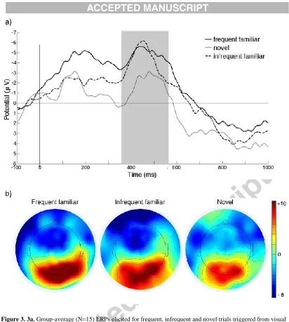
11 Figure 3 shows the grand average ERP waveforms and its scalp distribution for the three types of stimuli. Figure 4 shows the mean amplitudes and standard errors. We found a significant main effect of Stimulus type (F(2, 28) = 3.84; p = .034,
η2 = .215).The Nc amplitudes were larger for frequent familiar (M = -4.43 μV, SD = 3.63 μV) and infrequent familiar stimuli (M = -3.91 μV, SD = 5.28 μV), compared to
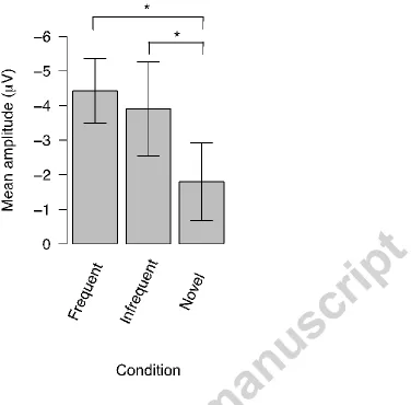
novel stimuli (M = -1.80 μV, SD = 4.36 μV). The post-hoc t-tests revealed that the Nc response evoked by novel stimuli was smaller compared to both frequent familiar (t(14) = 2.58, p = .022) and infrequent familiar (t(14) = 2.254, p = .041) stimuli. The difference between frequent and infrequent familiar stimuli was not significant (t(14) = 0.52, p = .629). There was no significant main effect of Area (F(1, 14) = .115; p
= .739, η2 = .008) or interaction of Area by Stimulus type (F(2, 28) = .637; p = .536,
η2 = .044). Additionally, we ran non-parametric tests for the main results we found.
12
13
Figure 4. Mean Nc amplitudes for frontal and central ROIs collapsed together. The columns represent the mean Nc amplitudes, the error bars represent SE. The Nc amplitude for the novel images differed both from the Nc amplitudes for the frequent and infrequent stimuli.
The Nc amplitude difference changes with age (Bauer et al., 2006). In order to test whether in our sample the Nc amplitude difference was modulated by age, we split the sample into two groups, according to the infants’ age (younger group: N = 8;
older group: N = 7). We ran an additional, 3 x 2 x 2 ANOVA with Stimulus type (frequent familiar, infrequent familiar and novel) and Area (frontal and central ROIs) as within subjects factors and Age (young and old) as a between subjects factor. We found only a significant main effect of stimulus type (F(2, 26) = 4.282; p = .025, η2 = .248). There was no main effect of Age (F(1, 13) = 2.968; p = .109, η2 = .186), neither Age by Stimulus type interaction (F(2, 28) = 1.77; p = .189, η2 = .12).
14 The present study aimed to investigate the differential contribution of the presentation frequency on memory processes. We manipulated the degree of familiarity in 10 to 12-month-old infants and measured the Nc ERP component in a passive viewing recognition task. Unlike other studies, where the effect of on-line presentation frequency was tested, we manipulated the presentation frequency during the familiarization (memory encoding) phase. During the test phase, we compared event-related brain activity elicited by images with different degrees of familiarity (infrequent familiar, frequent familiar and novel), but with equal on-line presentation frequency. This allowed us to measure memory-related top-down differences, rather than low-level effects of on-line presentation frequency. The results revealed that the amplitude of the Nc component over the midline frontal and central electrodes was clearly sensitive to a familiarity - novelty distinction. The Nc amplitude was higher following familiar images compared to novel pictures. Yet, the presentation frequency difference that originated in familiarization did not modulate the amplitude of the Nc component.
Usually, a larger Nc amplitude is associated with novel or less frequent stimuli (Bauer et al., 2003; Courchesne et al., 1981; Reynolds & Richards, 2005), whereas we found larger Nc amplitudes for familiar relative to novel images. Nevertheless, some studies have also found more negative Nc amplitudes following familiar stimuli (DeHaan & Nelson, 1997, 1999). Below, we will discuss three factors that can possibly modulate the differences in Nc amplitudes: (1) attention allocation processes; (2) effect of repeated presentation; (3) contextual effects of stimulus presentation.
15 encoded familiar) stimulus” (Roder et al., 2000, pp. 502). Encoding processes could
also change with age. Bauer et al. (2006) interpreted the change of the Nc effect between 9 and 10 months as differential allocation of processing resources between familiar and novel images. While 9-month-old infants devoted more attention to the novel stimuli, 10-month-olds devoted more attention to the familiar stimuli (Bauer et al., 2006). This explanation also indicates the role of attention during encoding. In our study, age didn’t modulate the Nc differences between stimulus types. On the other
hand, a process that allocates attention to partially encoded familiar stimuli could explain our results, as all the toys were novel to the infants at the beginning of the experiment. Nevertheless, it is important to emphasize that such attention-allocation requires top-down processes, because it is based on recognition.
The way frequency differences were implemented in previous studies could also be a factor modulating the direction of the Nc effect. Some studies presented the novel stimuli repeatedly (e.g. Bauer et al., 2006, 2003; de Haan & Nelson, 1997, 1999), while in other studies the novel stimuli differed on each trial (Nelson & Collins, 1992; Nelson & Collins, 1991; Reynolds & Richards, 2005; Richards, Reynolds, & Courage, 2010). A trial-unique stimulus appears only once in an experiment, therefore, the frequency of such item is very low. Wiebe et al. (2006) showed familiar-repeated, novel-repeated and trial-unique novel stimuli with equal probability to 9 month-olds and compared the Nc amplitudes from the early and late trials. Repeated presentation decreased the Nc amplitude from early to late trials, while the Nc did not change for the trial-unique novel stimuli. In this case, a larger Nc for trial-unique novel stimuli might simply be an effect of decreased Nc amplitude following the repeatedly presented stimuli. In our study this cannot be the case, because all stimulus types were presented repeatedly.
Finally, the context in which the stimuli are presented can also modulate the Nc amplitude. de Haan and Nelson (1997) presented the mother’s face and a stranger’s face to 6-month-old infants. The Nc amplitude was larger for the mother’s face only if the stranger’s face was dissimilar compared to the mother’s (Experiment
1). If the two faces were similar to each other, the Nc amplitude was more negative for the stranger’s (novel) face (Experiment 3). The authors suggested that the context
16 & Nelson, 1997). The role of context is also supported by other studies in infants (Ackles & Cook, 1998; Reynolds & Richards, 2005) and in adults (Richmond, Colombo, & Hayne, 2007). Infants cannot be given any task instructions, therefore the question arises, whether they interpreted the procedure as a recognition task or not, and the context in which the stimuli appear might modulate this aspect. Repeated presentation of novel stimuli might facilitate the recognition of familiar objects, whereas trial-unique novel stimuli might capture more attention than the repeatedly presented familiar ones.
Overall, there is emerging consensus that a larger Nc amplitude reflects stronger allocation of attention. However, as we have shown above, several factors modulate the process of capturing attention. The familiarity effect in our study is necessarily based on the prior image presentation (familiarization phase) and cannot be the result of different presentation frequency during the test phase. Therefore, in this study the amplitude of Nc reflects recognition memory processes. A stronger attention orientation response to the familiar images seems to be rational.
We did not find a difference in terms of the Nc magnitude between frequent familiar and infrequent familiar stimuli. However, in our study even the infrequent stimuli were presented 3 times for 6 seconds (18 seconds overall) during the familiarization phase. Posing the question, is a single presentation sufficient for recognition? In infant memory research, it is a challenge to separate familiarity-based recognition (with no what-where-when components of the original event) from declarative-memory based recognition (when the original event is actually recalled). Memories with a what-where-when component are encoded based on a unique, single event, whereas familiarity-based recognition does not necessarily rely on a single exposure. Our results indicate that the Nc component is not sensitive to presentation frequency during encoding – at least for the presentation frequencies we used. This suggests that the Nc does not merely signal the degree of familiarity and that the Nc component might also be a valid candidate to signal declarative memory processes. However, further studies should investigate this question using only a single exposure of an ‘event’.
17 sensitive to familiarity or recognition and it relates to top-down processes. The lack of difference between frequent and infrequent familiar stimuli could mean that the Nc component is related to declarative memory. The present study and the novel paradigm we introduced, is an initial step toward the use of the Nc component as an electrophysiological signature that can be used to tease apart whether recognition memory in infants, an essential and distinctive component of human social development, is based upon specific or general memories.
Acknowledgement
I.K. received funding from KMOP-4.2.1/B-10-2011-0002.
E.P. is a researcher in the International Centre for Language and Communicative Development (LuCiD) at Lancaster University. The support of the Economic and Social Research Council (ES/L008955/1) is gratefully acknowledged.
References
Ackles, P. K. (2008). Stimulus novelty and cognitive-related ERP components of the infant brain. Perceptual and Motor Skills, 106(1), 3–20.
http://doi.org/10.2466/pms.106.1.3-20
Ackles, P. K., & Cook, K. G. (1998). Stimulus probability and event-related potentials of the brain in 6-month-old human infants: a parametric study.
International Journal of Psychophysiology, 29(2), 115–143. http://doi.org/10.1016/S0167-8760(98)00013-0
Bauer, P. J., Wenner, J. A, Dropik, P. L., & Wewerka, S. S. (2000). Parameters of remembering and forgetting in the transition from infancy to early childhood.
Monographs of the Society for Research in Child Development, 65(4), i–vi, 1-204. http://doi.org/10.1016/j.imlet.2014.04.001
18 behavioral and electrophysiological indices. Psychological Science, 14(6), 629– 35. http://doi.org/10.1046/j.0956-7976.2003.psci_1476.x
Bauer, P. J., Wiebe, S. A, Carver, L. J., Lukowski, A. F., Haight, J. C., Waters, J. M., & Nelson, C. a. (2006). Electrophysiological indexes of encoding and behavioral indexes of recall: examining relations and developmental change late in the first year of life. Developmental Neuropsychology, 29(2), 293–320.
http://doi.org/10.1207/s15326942dn2902_2
Bushnell, I. W. R. (2001). Mother’s face recognition in newborn infants: learning and
memory. Infant and Child Development, 10(1–2), 67–74. http://doi.org/10.1002/icd.248
Carver, L. J., & Bauer, P. J. (1999). When the event is more than the sum of its parts: 9-month-olds’ long-term ordered recall. Memory, 7(2), 147–74.
http://doi.org/10.1080/741944070
Carver, L. J., Bauer, P. J., & Nelson, C. A. (2000). Associations between infant brain activity and recall memory. Developmental Science, 3(2), 234–246.
http://doi.org/10.1111/1467-7687.00116
Casler, K., & Kelemen, D. (2005). Young children’s rapid learning about artifacts.
Developmental Science, 8(6), 472–480. http://doi.org/10.1111/j.1467-7687.2005.00438.x
Courchesne, E., Ganz, L., & Norcia, A. M. (1981). Event-related brain potentials to human faces in infants. Child Development, 52(3), 804–811. Retrieved from http://www.ncbi.nlm.nih.gov/pubmed/7285651
de Haan, M., & Nelson, C. (1997). Recognition of the mother’s face by six-month-old
infants: a neurobehavioral study. Child Development, 68(2), 187–210. http://doi.org/10.1111/j.1467-8624.1997.tb01935.x
de Haan, M., & Nelson, C. (1999). Brain activity differentiates face and object processing in 6-month-old infants. Developmental Psychology, 35(4), 1113–21. http://doi.org/10.1037/0012-1649.35.4.1113
Fantz, R. L. (1964). Visual Experience in Infants: Decreased Attention to Familiar Patterns Relative to Novel Ones. Science, 146(3644), 668–670.
19 Gergely, G., & Csibra, G. (2006). Sylvia’s recipe: The role of imitation and pedagogy
in the transmission of cultural knowledge. Oxford: Berg Publisher.
Hunter, M. A., Ross, H. S., & Ames, E. W. (1982). Preferences for familiar or novel toys: Effect of familiarization time in 1-year-olds. Developmental Psychology,
18(4), 519–529. http://doi.org/10.1037/0012-1649.18.4.519
Koski, J., Olson, I. R., & Newcombe, N. S. (2013). Tracking the eyes to see what children remember. Memory, 21(3), 396–407.
http://doi.org/10.1080/09658211.2012.735241
Nelson, C. A., & Collins, P. F. (1991). Event-related potential and looking-time analysis of infants’ responses to familiar and novel events: Implications for
visual recognition memory. Developmental Psychology, 27(1), 50–58. http://doi.org/10.1037/0012-1649.27.1.50
Nelson, C. A, & Collins, P. F. (1992). Neural and behavioral correlates of visual recognition memory in 4- and 8-month-old infants. Brain and Cognition, 19(1), 105–21. http://doi.org/10.1016/0278-2626(92)90039-O
Reynolds, G. D., Courage, M. L., & Richards, J. E. (2010). Infant attention and visual preferences: converging evidence from behavior, event-related potentials, and cortical source localization. Developmental Psychology, 46(4), 886–904. http://doi.org/10.1037/a0019670
Reynolds, G. D., & Richards, J. E. (2005). Familiarization, Attention, and Recognition Memory in Infancy: An Event-Related Potential and Cortical Source Localization Study. Developmental Psychology, 41(4), 598–615. http://doi.org/10.1037/0012-1649.41.4.598
Richards, J. E. (2003). Attention affects the recognition of briefly presented visual stimuli in infants: an ERP study. Developmental Science, 6(3), 312–328. http://doi.org/10.1111/1467-7687.00287
Richmond, J., Colombo, M., & Hayne, H. (2007). Interpreting visual preferences in the visual paired-comparison task. Journal of Experimental Psychology:
20 Richmond, J., & Nelson, C. A. (2009). Relational memory during infancy: Evidence
from eye tracking. Developmental Science, 12(4), 549–556. http://doi.org/10.1111/j.1467-7687.2009.00795.x
Roder, B. J., Bushneil, E. W., & Sasseville, A. M. (2000). Infants’ Preferences for
Familiarity and Novelty During the Course of Visual Processing. Infancy, 1(4), 491–507. http://doi.org/10.1207/S15327078IN0104_9
Tomasello, M. (1999). The cultural ecology of young children’s interactions with objects and artifacts. In E. Winograd, R. Fivush, & W. Hirst (Eds.), Ecological approaches to cognition: Essays in honor of Ulric Neisser (pp. 153–170). Mahwah, NJ: Erlbaum.
Wiebe, S. A., Cheatham, C. L., Lukowski, A. F., Haight, J. C., Muehleck, A. J., & Bauer, P. J. (2006). Infants’ ERP Responses to Novel and Familiar Stimuli
Change Over Time: Implications for Novelty Detection and Memory. Infancy,
9(1), 21–44. http://doi.org/10.1207/s15327078in0901_2 Highlights
The Nc amplitudes were larger following familiar images compared to novel ones.
This effect is based on memory-related processes and not due to online presentation frequency.
The degree of familiarity did not modulate the Nc amplitude.