A Thesis Submitted for the Degree of PhD at the University of Warwick Permanent WRAP URL:
http://wrap.warwick.ac.uk/99039
Copyright and reuse:
This thesis is made available online and is protected by original copyright. Please scroll down to view the document itself.
Please refer to the repository record for this item for information to help you to cite it. Our policy information is available from the repository home page.
Complexes
A Thesis Submitted for the Degree of Doctor of Philosophy
James Paul Charles Coverdale MChem (Hons)
Supervisors: Prof. Peter J. Sadler, FRS
Prof. Martin Wills
University of Warwick, Department of Chemistry
Acknowledgements i
Declaration ii
Abstract iii
Conferences and courses attended iv
Abbreviations v
Chapter 1 – Introduction
1.1 Asymmetric transfer hydrogenation 4
1.1.1 The Noyori ruthenium bifunctional catalyst 4
1.1.2 Osmium transfer hydrogenation catalysts 7
1.2 Metals in medicine 9
1.2.1 Historical use of metallodrugs 9
1.2.2 The discovery of cisplatin 11
1.2.3 Platinum group metals in medicine 14
1.3 Cancer 18
1.3.1 The origin of cancer 18
1.3.2 Current treatment of cancer 21
1.4 Catalytic therapies 22
1.4.1 Oxidative catalytic therapies 23
1.4.2 Reductive catalytic therapies 24
1.5 Aims 26
2.1 Materials 41
2.1.1 Metal salt aquation analysis 42
2.1.2 Synthesis of dimer precursors 42
2.2 Instrumentation 46
2.2.1 Nuclear magnetic resonance spectroscopy (NMR) 46
2.2.2 Electronic absorption spectroscopy (UV-Vis) 46
2.2.3 Elemental analysis 46
2.2.4 Mass spectrometry 47
2.2.5 ATR Fourier-transform infrared (FT-IR) spectroscopy 47
2.2.6 Gas chromatography (GC) 47
2.2.7 X-ray crystallography 48
2.2.8 Inductively-coupled plasma spectroscopy (ICP) 48
2.2.9 Flow Cytometry 49
2.3 Methods 50
2.3.1 Transfer hydrogenation kinetics 50
2.3.2 Biological studies (in vitro cell culture) 51
2.4 References 56
Chapter 3 - Asymmetric catalytic osmium sulfonamide complexes
3.1 Introduction 58
3.2 Experimental 61
3.2.1 Materials 61
3.2.2 Synthesis of diamine sulfonamide ligands 62
3.2.5 Catalytic reactions 76
3.2.6 Density Functional Theory (DFT) calculations 78
3.3 Results 79
3.3.1 Synthesis and characterisation of novel osmium(II) catalysts 79
3.3.2 X-ray crystallography 81
3.3.3 Storage stability of osmium sulfonamide complexes 83
3.3.4 Solution stability of osmium sulfonamide complexes 84
3.3.5 Asymmetric transfer hydrogenation 86
3.3.6 Density Functional Theory (DFT) 88
3.4 Discussion 91
3.4.1 Structure elucidation of a novel osmium(II) pre-catalyst 91
3.4.2 X-ray crystallography 92
3.4.3 Transfer hydrogenation catalysis 93
3.4.4 Density Functional Theory (DFT) 97
3.5 Conclusions 98
3.6 References 99
Chapter 4 - Contrasting biological activities of Os(II) / Ir(III) complexes
4.1 Introduction 104
4.2 Experimental 106
4.2.1 Materials 106
4.2.2 Synthesis of iridium sulfonamide complexes 12-17 107
4.2.3 Conversion of 1,4-NADH to NAD+ 113
4.3 Results 118
4.3.1 Synthesis of Ir(III) sulfonamide complexes 118
4.3.2 Conversion of 1,4-NADH to NAD+ 120
4.3.3 Partition coefficient determination (Log P) 122
4.3.4 Antiproliferative activity and metal accumulation 123
4.3.5 Metallodrug accumulation in cancer cells 129
4.3.6 Cellular metal distribution 136
4.3.7 Generation of reactive oxygen species and superoxide 137
4.3.8 Combination with L-buthionine sulfoximine 139
4.4 Discussion 140
4.4.1 Synthesis and stability of Ir(III) sulfonamide complexes 140
4.4.2 Hydrophobicity, metal accumulation and anticancer activity 141
4.4.3 Multi-cell line antiproliferative activity screening 143
4.4.4 Contrasting cellular accumulation of Os / Ir complexes 150
4.4.5 Contrasting cellular metal distribution of Os / Ir complexes 153
4.4.6 Redox-targeting mechanism of sulfonamide complexes 154
4.5 Conclusions 156
4.6 References 158
Chapter 5 - Osmium(II)-catalysed in cell asymmetric transfer hydrogenation
5.1 Introduction 165
5.2 Experimental 167
5.2.1 Materials 167
5.2.4 In-cell reduction of pyruvate to lactate 173
5.3 Results 174
5.3.1 Reduction of pyruvic acid using formic acid 174
5.3.2 Aqueous-phase reduction of pyruvate 175
5.3.3 Transfer hydrogenation reactions in cancer cells 178
5.3.4 In-cell reduction of pyruvate to lactate 183
5.3.5 In-cell hydrogenation using N-formylmethionine 186
5.3.6 Mechanism of action for formate-activated catalysis 188
5.4 Discussion 194
5.4.1 Transfer hydrogenation as a new mechanism of action 194
5.4.2 Asymmetric reduction of pyruvate in cells 195
5.4.3 Enzymatic activation of N-formylmethionine 199
5.5 Conclusions 201
5.6 References 202
Chapter 6 - In vivo studies of organo-osmium and organo-iridium complexes
6.1 Introduction 206
6.2 Experimental 210
6.2.1 Materials 210
6.2.2 Synthesis of a rhodamine ligand and Os complex 18 211
6.2.3 In vivo studies and husbandry of zebrafish 213
6.2.4 Acute toxicity assessment in zebrafish 213
6.2.5 Fluorescence imaging in zebrafish 214
6.3.2 ROS induction in whole-mount zebrafish 217
6.3.3 Synthesis and properties of a fluorescent Os complex 219
6.4 Discussion 223
6.4.1 Acute aquatic toxicity of osmium complexes 223
6.4.2 In vivo generation of reactive oxygen species 224
6.5 Conclusions 226
6.6 References 227
Chapter 7 - Conclusions and future work
7.1 Conclusions 234
7.2 Future work 240
7.2.1 Increasing the water-solubility of sulfonamide catalysts 240
7.2.2 Enhancement of anti-cancer efficacy and in cell catalysis 241
7.2.3 Sulfonamide ligand modification 242
7.2.4 Further utilisation of the zebrafish (Danio rerio) model 243
7.2.5 Involvement of the mitochondrial electron transport chain 244
7.3 References 245
Acknowledgements
Since first exploring the chemistry of osmium(II) complexes during my master’s
degree, I was fortunate enough to continue my research as part of a PhD at Warwick
University. To Professor Peter J. Sadler and Professor Martin Wills; I am immensely
grateful for the opportunity, and the invaluable guidance and support you have offered
me since I first started working in your labs in 2013.
Nothing in science comes easily, and I would like to thank Dr. Isolda Romero-Canelón
and Dr. Carlos Sanchez-Cano for helping me become a better researcher; combining
long hours, psycho-working style, and maintaining carefully-placed optimism. I have
been lucky enough to work with you both around the world – Grenoble (France) and
Guangzhou (China) – and I’m sure that we will remain close in the future; if not
through science, then through food, gin and tonic. To Hannah, and our fantastic
technicians Ji and Bindy; thank you for your time, energy, laughter and patience, both
inside and outside of tissue culture. Since I began working in biology (sorry, Abraha),
I have undoubtedly made some friends for life. I have been lucky enough to work
alongside so many people from around the world; Khatija, Joan, Robbin, Mariana,
Carol, Chloe and Yasmin – you have all helped to shape my research into the final
form that has come together in this thesis. Thank you all for your encouragement, food
breaks and expertise. I also would not have completed my research without the support
of the Warwick Chemistry staff. Particularly, thank you to Dr. Lijiang Song and Phil
Aston, who patiently endured my over-enthusiasm for ICP samples.
From beyond the lab, I would most like to thank my family and friends. Your
unwavering love and support over the last three years have allowed me to follow my
Declaration
I hereby declare that except where specific reference is made to other sources, the
work contained in this thesis is the original work of the author. It has been composed
by myself and has not been submitted, in whole or in part, for any other degree,
diploma, or other qualification.
Some of the work presented in this thesis has been published:
1. J. P. C. Coverdale, C. Sanchez-Cano, G. J. Clarkson, R. Soni, M. Wills, P. J.
Sadler, Chem. Eur. J., 2015, 21, 8043.
2. J. P. C. Coverdale, I. Romero-Canelón, C. Sanchez-Cano, G. J. Clarkson, M.
Wills, P. J. Sadler, Nature Chemistry, Manuscript accepted.
James P. C. Coverdale
Abstract
Half-sandwich complexes of ruthenium, iridium, and more recently osmium, have
shown promise as anticancer agents. Many of these ‘piano-stool’ complexes appear to
target the redox balance in cells. Separately, similar complexes have been investigated
for the catalysis of hydrogenation reactions, with many examples achieving high
turnover frequencies and enantioselectivities. This thesis is concerned with achieving
in cell catalysis to increase drug potency and generate selectivity for cancer cells.
A series of eighteen Os(II) and Ir(III) complexes, of the type [M(ηx-arene)(diamine)]
(Os-arene: p-cymene, biphenyl, or m-terphenyl; Ir-arene: Cp*, CpxPh, or CpxBip), were
synthesised and fully characterised. The structures were derived from a Ru(II) transfer
hydrogenation catalyst [Ru(η6-p-cymene)(TsDPEN)], TsDPEN = N
-tosyl-diphenyl-ethylenediamine. The complexes were isolated as 16-electron amido catalysts, which
were highly stable in solution and upon storage, unlike their 16-electron Ru(II)
counterparts, and were highly active for asymmetric transfer hydrogenation of
ketones. Os complexes afforded enantiomerically-pure alcohols with high conversion
and enantioselectivity (> 99%) at rates exceeding those of the existing Ru catalyst.
Two Os and Ir complexes were explored for the conversion of NADH to its oxidised
form (NAD+) under physiologically-relevant conditions. Antiproliferative activities
determined in 14 human cell lines correlated with experimentally-determined
hydrophobicities. Typically, Os catalysts were found to be more active than their Ir
counterparts, though were internalised by cancer cells to a lesser degree, suggestive of
a more potent in-cell mechanism of action. Structural modifications identified an
apparent inert site of substitution on the sulfonamide substituent. Furthermore, their
Acute in vivo toxicities were determined in zebrafish, and all compounds investigated
exhibited lower toxicities than the Pt anticancer drug, cisplatin. The complexes were
shown to generate reactive oxygen species (ROS) in cancer cells, and similarly
generated ROS in zebrafish.
Transfer hydrogenation catalysis was explored under physiologically-relevant
conditions using sodium formate as a biologically-compatible hydride source.
Osmium complexes catalysed the reduction of pyruvate, a key metabolite in cells, to
either L-lactate or D-lactate, selectively (ca. 83% ee). Upon co-administration of the
catalyst and sodium formate, cancer cell proliferation was decreased by up to 13×
(relative to cells treated with the catalyst alone), while no sodium formate effect was
determined in non-cancerous cells. Importantly, the treatment of cells with a particular
enantiomer of the Os catalyst and sodium formate facilitated the in cell reduction of
pyruvate to D-lactate, providing, to the best of my knowledge, the first example of a
synthetic catalyst carrying out asymmetric transfer hydrogenation chemistry in cells.
Conferences and courses attended
1. Bruker AVANCE/TopSpin NMR Course (Coventry, UK). January 2015.
2. Transferable skills in science (University of Warwick, UK). June 2015.
3. Postgraduate Chemistry Symposium (University of Warwick, UK). June 2015.
4. 10th International School of Organometallic Chemistry (Camerino, Italy).
September 2015. Poster presentation.
5. RSC Dalton Division Joint Interests Group Meeting (University of Warwick,
UK). March 2016. Poster presentation.
6. Postgraduate Chemistry Symposium (University of Warwick, UK). June 2016.
Abbreviations
ATH Asymmetric Transfer Hydrogenation
Bip Biphenyl
BMEN 1,2-bis(4-methoxyphenyl)ethylenediamine
Bs Benzenesulfonyl
BsDPEN Benzenesulfonyl diphenylethylenediamine
(L)-BSO L-Buthionine sulfoximine
CBP Carboplatin (cis
-diamine(cyclobutane-1,1-dicarboxylate-O,O')platinum(II))
CDDP Cisplatin (cis-diamminedichloroplatinum(II))
COSY Correlation Spectroscopy
Cp* 1,2,3,4,5-pentamethylcyclopentadienyl
CpxPh Tetramethyl(phenyl)-cyclopentadienyl
CpxBiPh Tetramethyl(biphenyl)-cyclopentadienyl
CTR1 Copper transporter protein 1
d Doublet (NMR)
dd Doublet of doublets (NMR)
ddw Doubly deionized water
DFT Density Functional Theory
DMEM Dulbecco’s Modified Eagle Medium
DNA Deoxyribonucleic Acid
DPEN 1,2-diphenylethylenediamine
equiv. Equivalents
Fb 4-fluorobenzenesulfonyl
FbDPEN 4-fluorobenzenesulfonyl diphenylethylenediamine
FDA Food and Drug Administration (U.S.A.)
GSH Glutathione
HBSS Hanks' Balanced Salt Solution
HR-MS High resolution mass spectrometry
IC50 Half-maximal inhibitory concentration
ICP-OES Inductively-coupled plasma optical emission spectroscopy
ICP-MS Inductively-coupled plasma mass spectrometry
LC50 Half-maximal lethal concentration
λ(ex/em) Wavelength (excitation / emission)
m Multiplet
m-terp 1,3-diphenylbenzene (meta-terphenyl)
MS Mass spectrometry
m/z Mass-to-charge ratio
NAD+ Nicotine adenine dinucleotide (oxidised)
1,4-NADH Nicotine adenine dinucleotide (reduced)
Ns 4-nitrobenzenesulfonyl
NsDPEN 4-nitrobenzenesulfonyl diphenylethylenediamine
NMR Nuclear magnetic resonance
P-gp P-glycoprotein
P21 (p21) Tumour suppressor gene 21 (tumour suppressor protein 21)
P53 (p53) Tumour suppressor gene 53 (tumour suppressor protein 53)
PBS Phosphate-buffered saline
ROS Reactive oxygen species
RPMI-1640 Roswell Park Memorial Institute 1640
s Singlet
SG Singapore (zebrafish embryos)
SRB Sulforhodamine B
SrbDPEN
2-(3-diethylamino-6-diethylazaniumylidene-xanthen-9-yl)-5-(N-(2-amino-1,2-diphenyl-ethyl)sulfonoyl)-benzenesulfonate
TCA Trichloroacetic acid
Ts Tosyl (4-methylbenzenesulfonyl)
TsBMEN 4-toluenesulfonyl-1,2-bis(4-methoxyphenyl)ethylenediamine
TsDPEN 4- toluenesulfonyl-1,2-diphenylethylenediamine
TU Tübingen (zebrafish embryos)
UV-Vis Ultraviolet-Visible spectroscopy
v/v Volume / volume (percentage solution)
Cell lines studied in this thesis:
A2780 Human ovarian carcinoma
A2780cis Human ovarian carcinoma (cisplatin-resistant)
A549 Human lung carcinoma
HCT116 Human colon carcinoma
HCT116-p21-/- Human colon carcinoma (P21-deficient)
HCT116-p53-/- Human colon carcinoma (P53-deficient)
HEPG2 Human hepatocellular carcinoma
MCF7 Human breast adenocarcinoma
OE19 Human oesophageal carcinoma
PC3 Human prostate adenocarcinoma
SK-OV-3 Human ovarian adenocarcinoma
SW626 Human ovarian adenocarcinoma
HOF Human ovarian fibroblasts (non-cancerous primary cell line)
Chapter 1
1.
Introduction
This thesis is concerned with the synthesis of novel Os(II) organometallic complexes
of the general structure [Os(η6-arene)(diamine)] which are active asymmetric transfer
hydrogenation (ATH) catalysts. For comparison, structurally-similar Ir(III) η5-Cp*
complexes (Cp* = 1,2,3,4,5-cyclopentamethyl) were also synthesised. The complexes
are derived from the reputable ruthenium(II) chemistry pioneered by Professor Ryoji
Noyori. The catalytic cycle begins with the transference of hydride from a sacrificial
donor molecule (such as formic acid) to the catalyst, forming a metal-hydride complex
which can then go on to donate the hydride to a substrate acceptor molecule, affording
the product in a highly enantioselective manner and with high conversion (Figure
1.3).1, 2 Ruthenium(II) arene chlorido pre-catalysts of the general structure [Ru(η6
-arene)Cl(diamine)] are well-established,1 however the apparent inertness of Os(II)
allows the active 16-electron catalytic species [Os(η6-arene)(diamine)] to be directly
obtained, rather than isolation of a pre-catalyst which must first dissociate chloride. It
is envisaged that the greater stability of osmium(II) transfer hydrogenation catalysts
compared to ruthenium(II) will reduce catalyst degradation (trace Ru(0) is known to
contaminate reduction products), while direct use of the active catalyst may be
advantageous, achieving higher catalytic rates.
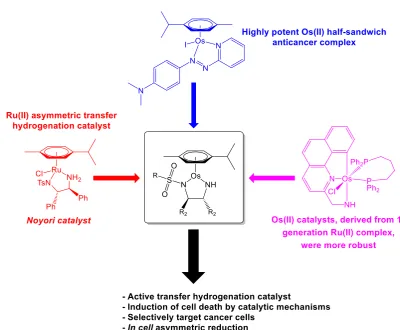
Highly potent Os(II) half-sandwich anticancer complexes have previously been
identified, achieving nanomolar antiproliferative activities towards cancer cells in a
non-catalytic manner.3 Conversely, a catalytic metallodrug may be obtained by
combining structural components from a known asymmetric hydrogenation catalyst
and a potent anticancer complex in a single structure (Figure 1.1). In vitro catalysis
may specifically target unique biochemical traits of a particular disease (e.g. the
The osmium(II) catalysts designed in this thesis are explored initially for the catalysis
of simple aromatic ATH substrates (based on acetophenone) under forcing conditions
in organic solvents (formic acid : triethylamine azeotrope), before exploring both the
catalytic oxidation (NADH to NAD+) and reduction (pyruvate to lactate) of important
biomolecules under physiologically-relevant conditions (aqueous-phase reduction,
bio-compatible hydride source). Ultimately, the catalysts are employed to carry out in
cell reductions with high enantioselectivity, making use of new bio-orthogonal
[image:20.595.112.513.326.656.2]chemistry as a new strategy for inducing cell death.
Figure 1.1. Rational design of Os(II) transfer hydrogenation catalysts for the treatment of cancer, based
1.1
Asymmetric transfer hydrogenation
Transfer hydrogenation reactions are a relatively recent advance in hydrogenation
chemistry. This involves the relocation of hydrogen from a sacrificial donor molecule
to an acceptor (target) transition metal complex, thereby providing a controlled route
to reduce organic targets, and safer alternative to reactions involving the use of
hydrogen gas under high pressure conditions.1, 5 In the mid-20th century, iridium
hydride complexes were shown to catalyse the reduction of unsaturated ketones to
alcohols, using isopropanol as a sacrificial hydride donor.6
1.1.1 The Noyori ruthenium bifunctional catalyst
Prof. Noyori is often considered to be the pioneer of asymmetric transfer
hydrogenation. His work, concerning catalysts capable of affording
enantiomerically-pure alcohols (and later, imines) with high conversion, won the 2001 Nobel Prize in
Chemistry. First-generation catalysts [Ru(BINAP)Cl2(diamine)] (BINAP =
2,2'-bis(diphenylphosphino)-1,1'-binaphthyl) demonstrated high turnover frequency and
enantioselectivity (> 99%) for the reduction of ketones (Figure 1.2a).7 Studies
demonstrated that the mechanism of hydrogenation was non-classical and occurred in
an outer-sphere process, rather than the traditional [2+2] mechanism (Figure 1.2).8
(a)
(b)
Figure 1.2. (a) First and second-generation Ru transfer hydrogenation catalysts, developed by Noyori
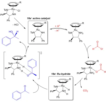
non-The second-generation bifunctional half-sandwich complexes improved catalytic
efficiency,7 even though they share structural similarity with the first-generation
catalysts in the chiral diamine (Figure 1.2a).1, 2, 9-20 The catalytic mechanism for
Noyori-type bifunctional catalysts has been studied in great depth, both
experimentally and computationally.9, 20-22 The complexes must first dissociate the
chloride ligand, and are deprotonated by a base, forming the 16-electron complex
[M(arene)(TsDPEN)], the active catalytic species. Note that the M2+ complex is still
neutral overall due to the formation of two anionic nitrogen atoms on the bidentate
ligand, which is reflected by the shorter metal-nitrogen bond length in the x-ray
crystallographic structures. Upon addition of a sacrificial hydride donor (commonly
formic acid or 2-propanol)2, 15 the basic nitrogen atom is re-protonated, and a
metal-hydride bond is formed, along with the elimination of the oxidised sacrificial molecule
(carbon dioxide, if the donor was formic acid).2, 15-26 The reverse process can then
occur with the target hydrogen acceptor, transferring hydrogen from the hydride
catalyst to the target via a 6-membered transition state (Figure 1.3). The entire catalytic
process is considered outer sphere,21-26 since neither the acceptor or donor molecules
have any direct interaction with the metal centre. The system is reversible, and will
carry out the thermodynamically favourable reduction. This can be influenced by the
selection of formic acid as a hydride source, since after donation of hydrogen, carbon
dioxide gas is released, effectively making the reaction irreversible.
The success of the Noyori transfer hydrogenation catalysts is down to the reaction
achieving high enantiomeric excess when prochiral acceptor molecules are used. The
geometry of the transition state, and subsequent enantioselectivity, is directed by the
chirality of the bidentate diamine17 and stabilised by favourable -interactions with
Figure 1.3. General mechanism for the asymmetric transfer hydrogenation of ketones using the Noyori 2nd generation bifunctional catalyst (hydrogen donor: formic acid; hydrogen acceptor: acetophenone).
Wills et al. modified the diamine component of the Noyori catalyst by alkylation of
either nitrogen atom, forming a covalent “tether” between the diamine and the η6-arene
of the catalyst.27, 28 This modification stabilised the catalyst further by constraining the
pseudo-octahedral geometry of the chlorido pre-catalyst to be retained in the
16-electron active catalyst, resulting in higher reaction turnover frequencies and high
conversions, whilst retaining the high enantioselectivity of the original Noyori
catalyst.28-30 Electron-rich amino-substituted ketones were readily reduced under
aqueous conditions using the tethered ruthenium catalysts, however
The analogous iridium(III) and rhodium(III) TsDPEN bifunctional catalysts were soon
reported, from which other structurally-derived transfer hydrogenation catalysts were
developed.5, 32-38 In particular, the Rh(III) catalyst was found to be the most efficient
catalyst for aqueous reduction of ketones using sodium formate as a hydride source.32
Transfer hydrogenation catalysts using the bidentate ligand TsCYDN
(N-tosyl-1,2-cyclohexadiamine) have been developed, and some out-perform the catalytic rate of
TsDPEN analogues.35 TsCYDN catalysts have also been used to carry out asymmetric
reductions in water with sodium formate without the need for an inert atmosphere.39
Noyori-type catalysts have also been immobilised on supports and polymers for both
continuous-flow systems and ease of recyclability. The supported catalyst retained
high catalytic activity with no loss of enantioselectivity.39-42
1.1.2 Osmium transfer hydrogenation catalysts
More recently, osmium transfer hydrogenation catalysts have been explored, despite
osmium often being considered catalytically-inferior to other platinum group metals
(Figure 1.4).43 Osmium catalysts were largely pioneered by Baratta et al. who have
worked extensively in the field of pincer complexes,6 achieving extraordinarily high
rates of reduction (up to 106 h-1 TOF) at very low catalyst loadings (0.005 mol%).44-48
Other osmium catalysts containing a ferrocene moiety were found to out-perform
ruthenium analogues, achieving up to 3 × 10⁵ h-1 TOF (Figure 1.4).49
Various bidentate ligands have been explored to produce both chiral and achiral
osmium catalysts.50-52 Complexes with N,O-chelating ligands achieved high
enantioselectivities, including cis-aminoindanol53, 54 and L-α-amino carboxylate55
complexes. Other N,N-ligands, including iminopyridine,56 and pybox (pybox =
Os-pincer Os-ferrocene Os-pybox
Figure 1.4. Various osmium transfer hydrogenation catalysts. Pincer complexes,44, 47 complexes
conjugated to ferrocene,49 and pybox ligands57 all demonstrated high activity and enantioselectivity for
the transfer hydrogenation of ketones.
Osmium derivatives of the Noyori first-generation ATH catalyst were explored, and
were demonstrated to be more robust. [OsCl2(diphosphine)(2-aminomethylpyridine)]
catalysed the rapid transfer hydrogenation of acetophenone (TOF 5.7 × 10⁵ h-1), an
order of magnitude greater than the ruthenium analogues.47 Hydride complexes of
osmium formed in situ have also been investigated for the transfer hydrogenation of
acetophenone, showing high catalytic activity but low enantioselectivity.43 Hydride
complex mer-[OsHCl(CO)(PR3)3] achieved 70% conversion with 3.5% ee in
isopropanol (356 K).58 As well as the reduction of ketones to alcohols, osmium
complexes have been used to catalyse the reduction of imines to amines,43, 59
hydrogenation of olefins,60 and the hydrogenation of esters to their corresponding
alcohols, though were typically out-performed by their ruthenium analogues.61
Osmium catalysts have also been investigated for hydrogen-borrowing reactions.62
Such reactions are highly similar to transfer hydrogenation chemistry, however the
reagent itself acts as the hydrogen donor. A molecule (typically an alcohol) donates
hydrogen to the catalyst molecule and is oxidised to the corresponding ketone, which
can undergo a subsequent reaction (e.g. with an amine to form an imine). Hydrogen is
1.2
Metals in medicine
Inorganic chemistry provides a wealth of novel solutions to pharmaceutical
challenges. Where organic compounds are typically restricted to linear, trigonal or
tetrahedral coordination, the introduction of a metal centre allows access to new
geometries, such as square planar, trigonal bipyramidal and octahedral. The central
cobaltion in cobalamin (Vitamin B12) provides structure and functionality unmatched
by an organic molecule. Inorganic metal ions can also access a vast range of oxidation
states. For example, osmium may exist from -2 to +8. Since redox processes are key
to many cellular pathways,63 the tuneable redox potential of a metal-based drug may
be advantageous to clinical success.
1.2.1 Historical use of metallodrugs
Metals have played an important role in treatments of diseases and illnesses over the
ages. Gold has been used in medicine since ancient times, and may be traced back as
early as 2500 BC. Preparations were particularly common in ancient China and India,
and were used to treat a range of illnesses, ranging from diabetes mellitus to memory
loss and asthma.64 Since the 1920s, gold thiolates such as sodium
2-(auriosulfanyl)-3-carboxypropanoate (sodium aurothiomalate) and tetraacetyl-β-d-thioglucose gold(I)
triethylphosphine (Auranofin) have been used for the treatment of rheumatoid
arthritis, and are currently being explored for their anticancer activities.65 Gold(I)
N-heterocyclic carbene (NHC) metallodrugs have also earned popularity as potential
anticancer agents. Many Au complexes have been shown to be potent TrxR
(Thioredoxin reductase) inhibitors. An example is the di-carbene gold complex
Sodium aurothiomalate Auranofin MC-3
Figure 1.5. Gold thiolates used for the treatment of rheumatoid arthritis (sodium aurothiomalate and
Auranofin) alongside MC3, a potent apoptosis-inducing gold(I) N-heterocyclic carbene complex.66
Silver has also been used medicinally for thousands of years.67 The ancient Romans
and Greeks were known to have stored water in silver vessels, and made use of the
antimicrobial properties of silver nitrate, which continues to be used during the modern
day treatment of severe skin burns.68 The antibacterial activity of silver ions is now
well established,67 and commonly feature in personal hygiene products. Recently,
alkyl phosphine complexes of group 11 metals (copper, silver and gold) have been
investigated for their in vitro antitumor activity, with mechanistic studies identifying
interactions of the gold and silver complexes with thioredoxin reductase, while the
copper complexes lead to proteasome inhibition.69
In 1907, a compound of arsenic was first synthesised and subsequently tested for
activity against spirochaetes, a group of organisms found to cause syphilis, infections
from which were highly prevalent in the early 20th century.70 The compound, named
“606” after its preparation number (also known as Arsphenamine or Salvarsan)
continued to be widely used until penicillin became readily available after World War
II.70 In fact, a study conducted in 2005 reported the structure of “606” as a mixture of
two structural isoforms, both of which led to the formation of the active compound
1.2.2 The discovery of cisplatin
In 1965, physicist Barnett Rosenberg (Michigan State University) observed inhibition
of cell division as well as filamentous growth, up to 300× the usual length, in E. coli.72
Electrolysis products formed from his platinum mesh electrodes and salt solution were
found to be Peyrone’s compound, cis-[PtCl2(NH3)2], first synthesised in 1845.73 The
compounds molecular structure was elucidated in 1893 by Werner,73, 74 and almost a
century later, was found by Rosenberg to exhibit potential as an anticancer agent in
rats, as reported in his 1969 publication: “Platinum compounds: a new class of potent
anti-tumor agents”.72 Peyrone’s compound (cis-diaminedichloroplatinum(II) /
cisplatin) was first administered to a human patient in 1971, and became clinically
available in 1978 under the brand name Platinol®.73 The discovery of a
pharmaceutically active metal-containing compound was so significant in that an
entirely new area of bio-inorganic research emerged, involving the development of
other novel metallodrugs. Cisplatin is also commonly found in combination therapies,
such as administration with the anticancer drug paclitaxel (for the treatment of ovarian,
breast, lung, head and neck cancers), fluorouracil (5FU, for the treatment of oral
cancers)75 and metformin (for the treatment of lung adenocarcinoma).74, 76
Cisplatin has since been used in the successful treatment of various cancers, including
testicular, lung, breast, cervical and prostate cancers.74 It is the mainline treatment for
ovarian cancer, a highly prominent female gynaecological cancer with high mortality
rates, often associated with late-stage diagnosis and poor detection.76 However,
platinum-resistant ovarian cancers are common, through either intrinsic or acquired
resistance.77-79 For this reason, this thesis will largely focus on a human ovarian
carcinoma cell line (A2780) allowing for direct comparison between novel catalytic
The DNA-targeting mechanism of action of cisplatin is commonly accepted. Cellular
Pt uptake occurs via copper transporter proteins as well as a contribution from passive
diffusion. The chloride ligands are labile, and once inside the cell, the lower
intracellular chloride concentration favours chloride dissociation, allowing stepwise
hydrolysis to afford the di-aqua complex [Pt(NH3)2(OH2)2]2+ which can form
1,2-intra-strand cross-links with purine bases; typically, N7 of guanine (Figure 1.6). If the
DNA cannot be successfully repaired, high-mobility group (HMG) proteins recognise
and bind platinated DNA,80 causing either apoptotic or necrotic cell death, depending
on the cisplatin exposure time.76, 81 The cis geometry of cisplatin is important, since
its trans isomer is inactive as intra-strand adducts cannot form. While DNA adduct
formation lacks specificity for cancer cells, metal complexes are commonly
multi-targeting, which may increase cancer cell selectivity.82, 83 Though only ca. 1% of
cisplatin reaches the nucleus, the complex is also known to interact with many other
biomolecules. The tripeptide GSH (Glu-Cys-Gly) is present at millimolar (1-10 mM)
concentrations in mammalian cells, and has been shown to highly influence the
efficacy of cisplatin.84 GSH also chelates Cu2+ causing up-regulation of the copper
transporter Ctr1, increasing Pt uptake. Conversely, GSH-Pt adducts may also facilitate
Pt efflux via the multidrug resistance protein 2 (MRP2) efflux pump.84, 85
Figure 1.6. Cisplatin undergoes step-wise hydrolysis inside cells due to the lower cytosolic chloride
Despite the widespread use of cisplatin, dosage is limited due to the side effects in
normal tissue; causing nausea, vomiting, hair loss, nephrotoxicity, neurotoxicity, and
hearing loss.74 Cisplatin resistance is also of clinical concern, which may be either
intrinsic or acquired. Resistance mechanisms may include decreased cellular uptake,
or increased efflux by membrane-bound transport ATPases such as MDR-1/2.74, 86 To
reduce off-target effects, Carboplatin was introduced in the 1980s, and possesses a
bidentate carboxylate in the place of the two chlorido ligands.73 Carboplatin is used
for the treatment of lung, head and neck cancers, and does not exhibit the nephrotoxic
effects of cisplatin, which may be associated with the lower reactivity and subsequent
slower rate of DNA binding. The longer retention half-life of 30 hours (compared to
1.5-3.6 h for cisplatin) means that effects are longer-lasting. Other cisplatin-derived
compounds (Figure 1.7) that have reached the clinic or clinical trials include
Oxaliplatin (1999),87 Heptaplatin (2005), Nedaplatin (1996) and Lobaplatin (2004).88
Cisplatin Carboplatin Oxaliplatin
Heptaplatin Nedaplatin Lobaplatin
Figure 1.7. Six platinum(II) anticancer drugs. Cisplatin, carboplatin and oxaliplatin are established
[image:30.595.117.513.460.673.2]1.2.3 Platinum group metals in medicine
The six elements of the platinum group (Ru, Os, Rh, Ir, Pt, Pd) are chemically very
similar. It is perhaps unsurprising that after the successes of Pt therapies, interests into
the design of Ru complexes have grown exponentially over the last few decades. A
Ru(III) complex, NKP-1339 (sodium trans-[tetrachloridobis(1H-indazole)ruthenate]),
the sodium salt of KP1019 (Figure 1.8), is currently in phase II clinical trials.89 The
planar arrangement of chloride ligands around the octahedral Ru centre shows
structural similarity to the square planar geometry of cisplatin. NKP-1339, like
cisplatin, induces G2/M arrest and apoptosis in cancer cells.90 However unlike
cisplatin, NKP-1339 disrupts the redox balance of the cell by generation of reactive
oxygen species (ROS),91 and also appears to target the endoplasmic reticulum.90 In
fact, the generation of ROS by metal complexes in cells is well documented, providing
an alternative mechanism of action to DNA binding.92-95 The contrasting mechanism
provides evidence that both the chemical and biological properties of the complex may
be modified by careful selection of suitable ligands, paired with a particular metal.
The analogous cis-osmium(III) complexes, derived from KP1019, have been prepared
by the 1-electron reduction of precursor Os(IV) complexes.96 The osmium complexes
were found to not interact with amino acids or model nucleotides (such as
5’-guanosine monophosphate). While the Os(III) complexes exhibited lower
antiproliferative activities than those of the parent Ru(III) compound, they showed
selectivity for human melanoma cells (FemX), while the antiproliferative activities
determined using KP1019 were comparable between all cell lines investigated.97
Interestingly, though trans-[tetrachlorobis(1H-indazole)ruthenate(III)] (KP1019) and
its trans-Os(III) isomer are known, at the time of writing, the cis-Ru(III) isomer is not
KP-1019 NAMI-A
Figure 1.8. Anticancer Ru(III) complexes in clinic trials: (a) KP-1019,91 (b) NAMI-A.98
A structurally similar ruthenium complex, NAMI-A (Figure 1.8) has been extensively
studied.89, 99, 100 The complex entered a phase I/II clinical study, in combination with
gemcitabine, for the treatment of non-small cell lung cancers, however was only
moderately tolerated in patients.98 Similarly to KP1019, the osmium analogues of
NAMI-A have also been investigated, prepared stepwise from osmium tetroxide.101
As a third-row transition metal with slower exchange kinetics, osmium was previously
discounted as a useful metal centre for a metallodrug. The osmium complexes did not
undergo hydrolysis of the chloride ligands, unlike the ruthenium centre in NAMI-A,
yet showed approximately an order of magnitude greater activity than the ruthenium
counterparts.101 In contrast, replacement of Ru for Os in KP1019 decreased
antiproliferative activity, indicating that trends anticancer activity cannot be predicted
upon metal exchange.
While NAMI-A and KP1019 were synthesised by selection of small-molecule ligands
with properties that would benefit the overall complex, ruthenium complexes of
on staurosporine have been synthesised (Figure 1.9).102 These complexes exhibit high
selectivity for particular tyrosine-protein kinases, specifically for the Abelson tyrosine
kinase (Abl) which is known to be highly involved with cell growth processes. In fact,
the complex was found to be inactive against a range of other kinases (e.g. PKCα).102
Interestingly, the IC50 (half-maximal inhibitory concentration) of the Ru complex was
found to be ca. 10× lower than that of the free ligand (2 nM compared to 25 nM)
towards the Abl, reinforcing the importance of the metal centre.
In recent years, “sandwich” structures have gained substantial interest. Such
complexes are identified by the coordination of one (or many) η5 or η6- arene(s) to a
central metal, which can act to stabilise the overall complex as well as providing a
lipophilic region. Since the synthesis of the organo-iron compound, Ferrocene [Fe(η5
-C5H5)2] in 1951,103 structurally-derived sandwich complexes of ruthenium, osmium
and iron have been conjugated to the anticancer compound Tamoxifen, increasing the
potency towards breast cancer cells (Figure 1.9). Iron-conjugates of ferrocene were
found to be more active than Os and Ru counterparts, demonstrating that the role of
the ferrocene moiety was not purely structural.104
Protein kinase inhibitors (M = Ru)
Tamoxifen derivatives (M = Fe / Ru / Os)
Half-sandwich “piano-stool” complexes [M(ɳ6-arene)(bidentate)(monodentate)] only
possess one metal-coordinated arene, and typically adopt pseudo-octahedral geometry.
Early examples of half-sandwich complexes with anticancer properties are the Ru
compound RM-175 and its Os analogue, AFAP-51, with the general structure[M(ɳ6
-biphenyl)(ethylenediamine)Cl]+ (Figure 1.10).105 Though the chemistry of Ru(II) and
Os(II) ions is often considered similar, the Os complex was found to be 6 more potent
than the Ru analogue. RM-175 (Ru) however showed high in vivo activity, leading to
a reduction in metastases which was not found in the Os case.105 In this example, there
are meaningful biological implications resulting from the fine-tuning of the redox and
kinetics by of the different metal centres. Sadler et al. developed a series of osmium
and ruthenium azo/imino-pyridine complexes of the general formula [M(ɳ6
-arene)(azo/imino-pyridine)(X)]+ where X = Cl or I, that show high activity against
cancer cells,106 which were found to be higher for iodido complexes than their chloride
analogues.3 The complexes were shown to exhibit a new mechanism of action that
differed from that of existing platinum therapies, since these complexes did not appear
to undergo aquation nor bind to DNA.3 The complexes also retained activity in
Pt-resistant cells.107 One Os complex in particular (FY26; Figure 1.10), has been shown
to delay growth of human colon cancer xenografts in mice, with negligible toxicity.108
RM-175 AFAP-51 FY-26
1.3
Cancer
In the UK, someone dies from cancer every two minutes.109 Cancer is one of the most
significant cause of death around the world,110 and is particularly prevalent in
developed countries. In Britain, cancer is now expected to affect around 1 in 2 people,
with the lifetime risk for men slightly exceeding that for women.109, 111Cancers may
result from inherited genetic traits, but it is estimated that up to 80% are caused by
external factors such as radiation, carcinogenic chemicals or viruses.112 Many cancers
that were previously considered highly lethal (such as testicular cancer, Hodgkin's
lymphoma, and some leukemias) are now effectively treatable with anticancer drugs.86
However, while highly sensitive screening methods are detecting many cancers before
they have metastasized, the battle against malignant cancers is expected to remain a
key part of modern medicine.86
1.3.1 The origin of cancer
The six hallmarks of cancer, according to Hanahan and Weinberg, are
growth-suppressor evasion, growth signaling self-sufficiency, becoming active for invasion
and metastasis, enabling replicative immortality, the induction of angiogenesis and
resisting cell death.113
In normal cells, proto-oncogenes code for proteins that play an important role in many
cellular processes such as cell division, cell differentiation, and cell death.
Over-production of these proteins leads to a loss of control over such cellular processes.
Proto-oncogenes may become activated by mutations that either increase the
expression level or activity to become oncogenes,114 which may result in causing
controlled cell proliferation. A key tumor suppressor gene (P53) encodes the
transcriptional regulator protein p53, the absence of which greatly increases the
occurrence of tumors.112 After exposure to DNA-damaging agents, expression of p53
is increased, resulting in cell cycle arrest and apoptosis. Both the activity of the p53
protein and transcription of the P53 gene are negatively regulated by the E3 ubiquitin
ligase, Mdm2 (Figure 1.11). Mutations in P53 are in fact the most common genetic
alteration in human cancers.115 Such mutations include: (a) deletion of the
carboxyl-terminal domain, preventing p53 tetramer formation, (b) amino acid alteration in the
DNA binding domain, preventing downstream gene activation, (c) overexpression of
MDM2, stimulating the degradation of p53.116
Figure 1.11. Simplified p53 pathway (adapted from A. J. Levine and M. Oren, 2009).117 P53 is
[image:36.595.165.477.392.664.2]Differences in the physiology and biochemistry of cancer cells compared to normal
cells may facilitate selective targeting by novel anticancer agents. Physiologically,
tumors typically are commonly hypoxic,118 and have low extracellular pH as a result
of lactate secretion.119 These traits are commonly observed as microenvironments
which are chemically-distinct from healthy tissue, and have led to interest in
developing inactive pro-drugs that become activated by tumor physiology.120 Such
compounds typically contain N-oxides, quinones or transition metals, which are
activated under hypoxic conditions. Tirapazamine, a di-N-oxide compound,
demonstrated up to 200× more cytotoxicity in hypoxic cells.121
Biochemically, the metabolism of glucose in cancer cells differs from that in healthy
cells.122 Rapidly-proliferating cells require more ATP, a cellular energy source. While
healthy cells metabolize glucose to pyruvate (via glycolysis) and subsequently oxidize
pyruvate to carbon dioxide by oxidative phosphorylation, the ‘Warburg Effect’
summarises the observation that cancer cells display a preference for the reduction of
pyruvate to lactate, rather than oxidation to carbon dioxide.123Only in the absence of
oxygen would healthy cells utilise lactate production as a means of acquiring energy,
since ATP production by glycolysis is far less efficient than by oxidative
phosphorylation (2 molecules of ATP, compared to 36 molecules per glucose
molecule).4 For this reason, the consumption of glucose is significantly higher in
cancer cells.124 This glucose-dependence makes glycolysis a target for anticancer drug
design, and in fact, many glycolytic inhibitors (2-deoxyglucose, lonidamine,
3-bromopyruvate, Imatinib) have been explored for use as anticancer agents.124
Furthermore, the availability of cellular reducing agents (e.g. NADH or NADPH) has
been associated with the rate of proliferation,125 making redox-based strategies an
1.3.2 Current treatment of cancer
Three treatment options are well established in the clinic, and are usually used in
combination to achieve the best outcome for the patient. Surgical excision of a tumour
is most commonly employed after size-reduction using radiotherapy or chemotherapy.
Many existing chemotherapy agents target cellular replication processes, by damaging
DNA or affecting key pathways involved with cell growth. Many of these targets are
not unique to cancer cells, and drugs have little selectivity for cancerous cells over
healthy cells that also have rapid rates of proliferation, such as the hair follicles, bone
marrow and gastrointestinal tract cells;128 resulting in a range of side-effects.
New targeted approaches to chemotherapy are required that exploit the unique
biochemical properties of cancer cells. While cancer-specific drug delivery strategies
can reduce off-target effects, research into future novel anticancer compounds may
identify other chemotherapeutic targets, such as the previously discussed hypoxic
environment,120, 121 high rate of glycolysis,124 and increased levels of oxidative
stress129 in cancer cells. By combining targeting monoclonal antibodies with an
anticancer drug, antibody-drug conjugates increase the selectivity of existing
therapies. Trastuzumab emtansine is the first FDA-approved conjugate, for use against
human epidermal growth factor receptor 2 positive (HER2+) breast cancers.130 Recent
studies have also explored the potential of kinase inhibitors. Kinases are known to be
crucial in many cell signalling pathways, including those associated with cancer (such
as proliferation and cell survival mechanisms).131 While highly potent (nanomolar
IC50), many of the inhibitors investigated lacked specificity for a particular kinase.132
In addition, drug carriers; for example nanoparticles, liposomes and polymeric
conjugates, have also been explored to improve delivery of cytotoxic agents
1.4
Catalytic therapies
Though absorption, metabolism and excretion mechanisms of current pharmaceuticals
have been thoroughly studied, the damage that may occur at high concentrations is a
common clinical concern. By designing catalytic therapies, dosage could be
dramatically reduced while achieving the same efficacy. For many diseases, including
cancer, the redox balance in cells is known to differ from normal levels.63, 134 Cellular
redox homeostasis is mainly regulated by two co-factors; nicotine adenine
dinucleotide (NAD), and flavin adenine dinucleotide (FAD), which catalytically
transport hydride. Chemical mimics that can catalytically generate and transfer
redox-active molecules may play a crucial role in future pharmaceuticals.
Catalysts are well-established in chemical synthesis to achieve reaction rates that
would not be possible in their absence. Inorganic catalysts allow access to a range of
redox potentials and coordination numbers. Ligands may be stabilising,
substrate-directing, or participate in the catalytic cycle, and allow for the fine-tuning of the
properties. However, many inorganic catalysts require inert atmospheres (to prevent
metal oxidation) and particular solvents to circumvent catalyst deactivation. Advances
are being made in the development of water-compatible catalysts,5 which are
advantageous when considering the development of catalytic medicines, which would
be required to maintain activity in an aqueous cellular environment, and indeed many
successful examples have been reported. Artificial protease complexes of cobalt(III)
and copper(II) have been shown to catalytically cleave bovine serum albumin and
myoglobin.135 Suzuki-Miyaura cross-couplings and allylcarbamate cleavages have
been carried out inside cells using palladium(0) nanoparticles.136 Ruthenium catalysts
have been used to uncage an active anticancer drug in HeLa cells,137 and to uncouple
1.4.1 Oxidative catalytic therapies
The catalytic generation of reactive oxygen species (ROS), such as hydroxyl radicals
OH• and superoxide O2•- has been frequently identified for cells exposed to metal
complexes.56, 139 This is particularly important for the treatment of many conditions,
including cancer, Alzheimer’s disease and Parkinson’s disease; where abnormal
cellular redox balance is observed, offering a promising therapeutic target.140
Methodical design and modification of ligands around the metal centre allows for fine
tuning of the metal centre chemistry, to access physiologically-relevant redox couples.
Inhibition of thioredoxin reductase (TrxR), an enzyme known to be important in the
regulation of cellular redox levels, has been demonstrated using ruthenium(II) arene
complexes.141 Ruthenium azopyridine complexes have been shown to catalytically
oxidise glutathione (GSH), an important anti-oxidant molecule in cells, to glutathione
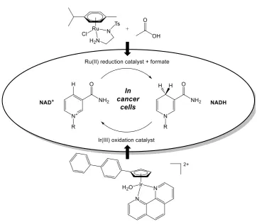
disulphide (GSSG).93 Iridium(III) catalysts have been successfully used catalytically
oxide NADH,142 and shown to increase the NAD+/NADH ratio inside cancer cells
(Figure 1.12).143 While osmium(II) iminopyridine and azopyridine complexes have
been also shown to generate reactive oxygen species in cells, the oxidation was not
catalytic, and only iminopyridine complexes were capable of NADH oxidation
(forming NAD+)by abstraction of hydride, while azopyridine complexes could not.106
Superoxide dismutase (SOD) mimics have also been studied to catalyse the conversion
of the superoxide radical to dioxygen and hydride peroxide, since SOD is considered
one of the cell’s primary defence mechanisms against oxidative stress.144-146 Many
transition metal-catalysed examples have been described, including OsO4,144 while
more recent reports have explored the high activity of manganese(II) complexes. In
1.4.2 Reductive catalytic therapies
Transfer hydrogenation of NAD+ to its reduced form (NADH) have been extensively
studied using inorganic catalysts of ruthenium and iridium, in combination with
sodium formate (or hydrogen gas).149, 150 NADH mimics have also been used for
reduction reactions in aqueous solution, and hydrogenation (regeneration) of the
coenzyme mimics, using sodium formate, has been successfully described.151
Non-asymmetric ruthenium N,N-bidentate piano-stool complexes
[Ru(arene)Cl(tosyl-diamine)] have been used to reduce NAD+ using sodium formate in an aqueous
model,152 and later inside cancer cells using a non-toxic concentration of sodium
[image:41.595.137.500.382.698.2]formate (Figure 1.12).129
Figure 1.12. Modulation of the NAD+/NADH ratio inside cancer cells by: (a) transfer hydrogenation
Stereoselective ruthenium(II) complexes have been assessed for both catalytic
efficiency and antiproliferative activity in cells. Interestingly, highly potent complexes
were found to be poor transfer hydrogenation catalysts, while promising catalysts were
less effective against cancer cells.153 Half-sandwich rhodium(III) Cp* complexes have
been shown to reduce NAD+ to NADH by 1H-NMR in a model system, and can also
rapidly convert pyruvate to lactate, using sodium formate as a hydride source, though
the reduction is not enantioselective. In competition experiments, the rhodium
complexes preferentially reduced NAD+ over pyruvate.154 Pyruvate reduction using
ruthenium(II) bipyrimidine and iridium(III) phenanthroline complexes has also been
studied by 1H-NMR,143 however no previous examples have demonstrated reduction
of pyruvate by an osmium catalyst. Furthermore, at the time of writing, no examples
have reported the selective reduction of pyruvate to L- or D-lactate with high
enantioselectivity, nor the reduction of pyruvate inside cells.
The work in this thesis builds on the aforementioned examples to explore the
antiproliferative activities of Os(II) and Ir(III) transfer hydrogenation catalysts against
cancer cells, which are compared to current non-catalytic Pt therapies. Utilising the
enantioselective properties of the catalyst, modulation of intracellular L- or D-lactate
concentrations by asymmetric reduction of pyruvate inside cells are carried out using
novel Os(II) complexes, generating selectivity for cancer cells over healthy cells.Such
complexes are based on racemic Ru(II) catalysts previously described to carry out the
reduction of NAD+ to 1,4-NADH inside cells (using sodium formate as a hydride
source). Furthermore, the reverse osmium(II)-catalysed reaction is explored by
catalysing the oxidation of the co-enzyme and cellular hydride source 1,4-NADH to
NAD+ in a model system under biologically-relevant conditions (pH 7.4,
1.5
Aims
This thesis aims to explore the novel catalytic and anticancer properties of 16-electron
osmium(II) sulfonamide complexes of the general formula [Os(ɳ6-arene)(diamine)],
where the ɳ6-arene is para-cymene, biphenyl or meta-terphenyl, and the diamine
substituent contains a sulfonamide with various R group substituents.
• Asymmetric transfer hydrogenation of ketones by an Os(II) catalyst
Structurally similar ruthenium 16-electron complexes of the general form [Ru(ɳ6
-arene)(diamine)] have previously been identified as the active species in the catalytic
cycle for the transfer hydrogenation of ketones. In Chapter 3, the osmium complexes
are assessed for their activity, including degree of enantioselectivity, for the transfer
hydrogenation of asymmetric ketones (derived from acetophenone) to assess primary
catalytic activity in organic solvent systems.
• Conversion of the co-enzyme NADH to NAD+ by an Os(II) catalyst
In Chapter 4, the osmium(II) (and structurally-similar iridium(III)) sulfonamide
catalysts are explored as catalysts for the oxidation of NADH, an important biological
source of hydride. Furthermore, the primary anticancer activities are investigated in
range of human cell lines to examine activity trends. As the compounds contain a
xenobiotic element, cellular metal accumulation mechanisms and pathways, and
cellular distribution patterns are reported, using representative compounds from the
osmium(II) and iridium(III) series. The oxidation of NADH is known to generate
• Asymmetric reduction of pyruvate in cancer cells by an Os(II) catalyst
Chapter 5 further explores the catalytic potential of the complexes in vitro by
co-administering the catalyst with a source of hydride (sodium formate) to enhance
antiproliferative potency. This effect is examined in both cancer cells and healthy cells
(primary fibroblasts). Both enantiomers of Os(II) sulfonamide complex [Os(p
-cymene)(TsDPEN)] are explored for their ability to catalyse the asymmetric reduction
of pyruvate to L- or D-lactate in cells and in model systems, which are studied using
time-dependent 1H-NMR and enzymatic assays. Enzymes that are known to be
over-expressed in certain cancer cells are also explored to selectively generate hydride in
situ, further increasing the selectivity of the compounds for cancer cells.
• Exploring the toxicity and mechanism of Os(II) complexes in zebrafish
Lead osmium(II) anticancer complexes are explored in Chapter 6 for the first time in
vivo using the zebrafish model (Danio rerio) to evaluate both acute aquatic toxicity as
described in OECD guideline 236. Data are compared to results obtained with
currently available anticancer drugs and other osmium(II) complexes which have
shown clinical promise. Additionally, since production of reactive oxygen species was
observed in cells, generation of ROS was investigated in whole-mount zebrafish to
determine whether the observation is maintained in a whole live animal. ROS were
visualised using confocal microscopy and a green-fluorescent ROS probe. To confirm
the specific role of the metal complex, a red-fluorescent Os(II) complex derived from
the sulfonamide series was designed, synthesised, fully characterised, and its
biological activities assessed before co-imaging Os(II) complex (red) and ROS
1.6
References
1. R. Noyori and S. Hashiguchi, Acc. Chem. Res., 1997, 30, 97-102.
2. S. Hashiguchi, A. Fujii, J. Takehara, T. Ikariya and R. Noyori, J. Am. Chem.
Soc., 1995, 117, 7562-7563.
3. Y. Fu, A. Habtemariam, A. M. B. H. Basri, D. Braddick, G. J. Clarkson and
P. J. Sadler, Dalton Trans., 2011, 40, 10553-10562.
4. M. G. Vander Heiden, L. C. Cantley and C. B. Thompson, Science, 2009,
324, 1029-1033.
5. X. Wu and J. Xiao, Chem. Commun., 2007, 2449-2466.
6. D. Wang and D. Astruc, Chem. Rev., 2015, 115, 6621-6686.
7. T. Ohkuma, Proc. Jpn. Acad. Ser. B Phys. Biol. Sci., 2010, 86, 202-219.
8. C. A. Sandoval, T. Ohkuma, K. Muñiz and R. Noyori, J. Am. Chem. Soc.,
2003, 125, 13490-13503.
9. S. E. Clapham, A. Hadzovic and R. H. Morris, Coord. Chem. Rev. , 2004,
248, 2201-2237.
10. S. Gladiali and E. Alberico, Chem. Soc. Rev., 2006, 35, 226-236.
11. J. Václavík, P. Šot, B. Vilhanová, J. Pecháček, M. Kuzma and P. Kačer,
Molecules, 2013, 18, 6804-6828.
12. T. Ikariya and A. J. Blacker, Acc. Chem. Res., 2007, 40, 1300-1308.
13. C. Wang, X. Wu and J. Xiao, Chem. Asian J., 2008, 3, 1750-1770.
14. A. Robertson, T. Matsumoto and S. Ogo, Dalton Trans., 2011, 40,
10304-10310.
15. A. Fujii, S. Hashiguchi, N. Uematsu, T. Ikariya and R. Noyori, J. Am. Chem.
16. T. Ikariya, S. Hashiguchi, K. Murata and R. Noyori, Org. Synth., 2005, 82,
10-17.
17. T. Ikariya, K. Murata and R. Noyori, Org. Biomol. Chem., 2006, 4, 393-406.
18. N. Uematsu, A. Fujii, S. Hashiguchi, T. Ikariya and R. Noyori, J. Am. Chem.
Soc., 1996, 118, 4916-4917.
19. M. Yamakawa, H. Ito and R. Noyori, J. Am. Chem. Soc., 2000, 122,
1466-1478.
20. R. Noyori, M. Yamakawa and S. Hashiguchi, J. Org. Chem., 2001, 66,
7931-7944.
21. D. A. Alonso, P. Brandt, S. J. M. Nordin and P. G. Andersson, J. Am. Chem.
Soc., 1999, 121, 9580-9588.
22. C. P. Casey and J. B. Johnson, J. Org. Chem., 2003, 68, 1998-2001.
23. K.-J. Haack, S. Hashiguchi, A. Fujii, T. Ikariya and R. Noyori, Angew.
Chem., Int. Ed.. 1997, 36, 285-288.
24. P. Brandt, P. Roth and P. G. Andersson, J. Org. Chem., 2004, 69, 4885-4890.
25. J. W. Handgraaf and E. J. Meijer, J. Am. Chem. Soc., 2007, 129, 3099-3103.
26. P. A. Dub and T. Ikariya, J. Am. Chem. Soc., 2013, 135, 2604-2619.
27. R. Hodgkinson, V. Jurčík, A. Zanotti-Gerosa, H. G. Nedden, A. Blackaby, G.
J. Clarkson and M. Wills, Organometallics, 2014, 33, 5517-5524.
28. R. Soni, K. E. Jolley, G. J. Clarkson and M. Wills, Org. Lett., 2013, 15,
5110-5113.
29. D. Morris, A. Hayes and M. Wills, J. Org. Chem., 2006, 71, 7035-7044.
30. A. Hayes, D. Morris, G. Clarkson and M. Wills, J. Am. Chem. Soc., 2005,
31. R. Soni, T. H. Hall, B. P. Mitchell, M. R. Owen and M. Wills, J. Org. Chem.,
2015, 80, 6784-6793.
32. X. Wu, X. Li, A. Zanotti-Gerosa, A. Pettman, J. Liu, A. J. Mills and J. Xiao,
Chem. Eur. J., 2008, 14, 2209-2222.
33. T. Ohkuma, N. Utsumi, M. Watanabe, K. Tsutsumi, N. Arai and K. Murata,
Org. Lett., 2007, 9, 2565-2567.
34. T. Thorpe, J. Blacker, S. M. Brown, C. Bubert, J. Crosby, S. Fitzjohn, J. P.
Muxworthy and J. M. J. Williams, Tetrahedron Lett., 2001, 42, 4041-4043.
35. K. Murata, T. Ikariya and R. Noyori, J. Org. Chem., 1999, 64, 2186-2187.
36. X. Sun, G. Manos, J. Blacker, J. Martin and A. Gavriilidis, Org. Process Res.
Dev., 2004, 8, 909-914.
37. Z. M. Heiden and T. B. Rauchfuss, J. Am. Chem. Soc., 2009, 131,
3593-3600.
38. C. Li, B. Villa-Marcos and J. Xiao, J. Am. Chem. Soc., 2009, 131,
6967-6969.
39. X. Wu, D. Vinci, T. Ikariya and J. Xiao, Chem. Commun., 2005, 4447-4449.
40. W. Wang and Q. Wang, Chem. Commun., 2010, 46, 4616-4618.
41. X. Huang and J. Y. Ying, Chem. Commun., 2007, 1825-1827.
42. C. M. Zammit and M. Wills, Tetrahedron: Asymmetry, 2013, 24, 844-852.
43. G. Chelucci, S. Baldino and W. Baratta, Acc. Chem. Res., 2015, 48, 363-379.
44. W. Baratta, S. Baldino, M. J. Calhorda, P. J. Costa, G. Esposito, E.
Herdtweck, S. Magnolia, C. Mealli, A. Messaoudi, S. A. Mason and L. F.
Veiros, Chem. Eur. J., 2014, 20, 13603-13617.
45. W. Baratta, G. Bossi, E. Putignano and P. Rigo, Chem. Eur. J., 2011, 17,
46. W. Baratta, F. Benedetti, A. Del Zotto, L. Fanfoni, F. Felluga, S. Magnolia,
E. Putignano and P. Rigo, Organometallics, 2010, 29, 3563-3570.
47. W. Baratta, M. Ballico, A. Del Zotto, K. Siega, S. Magnolia and P. Rigo,
Chem. Eur. J., 2008, 14, 2557-2563.
48. A. Acosta-Ramirez, M. Bertoli, D. G. Gusev and M. Schlaf, Green Chem.,
2012, 14, 1178-1188.
49. E. Putignano, G. Bossi, P. Rigo and W. Baratta, Organometallics, 2012, 31,
1133-1142.
50. W. N. O Wylie, A. J. Lough and R. H. Morris, Organometallics, 2011, 30,
1236-1252.
51. S. E. Clapham and R. H. Morris, Organometallics, 2005, 24, 479-481.
52. R. Castarlenas, M. A. Esteruelas and E. Oñate, Organometallics, 2008, 27,
3240-3247.
53. J. W. Faller and A. R. Lavoie, Organometallics, 2002, 21, 3493-3495.
54. J. W. Faller and A. R. Lavoie, Org. Lett., 2001, 3, 3703-3706.
55. D. Carmona, F. J. Lahoz, P. García-Orduña, L. A. Oro, M. P. Lamata and F.
Viguri, Organometallics, 2012, 31, 3333-3345.
56. Y. Fu, R. Soni, M. J. Romero, A. M. Pizarro, L. Salassa, G. J. Clarkson, J. M.
Hearn, A. Habtemariam, M. Wills and P. J. Sadler, Chem. Eur. J., 2013, 19,
15199-15209.
57. E. Vega, E. Lastra and M. P. Gamasa, Inorg. Chem., 2013, 52, 6193-6198.
58. C. Schlünken, Miguel A. Esteruelas, Fernando J. Lahoz, Luis A. Oro and H.
Werner, Eur. J. Inorg. Chem., 2004, 2004, 2477-2487.
59. M. Rosales, J. Castillo, A. González, L. González, K. Molina, J. Navarro, I.