Original Article
Long non-coding RNA SNHG1 promotes cell
proliferation and invasion by competitively
binding to miR-145 in breast cancer
Xiao-Bo Wu1*, Rui Wang2*, Wen-Song Wei1
1Breast Disease Center, The Third Hospital of Nanchang, Nanchang 330009, China; 2Department of Thyroid,
Breast Surgery, The First People’s Hospital of Lianyungang, Lianyungang 222000, China. *Co-first auther. Received September 4, 2017; Accepted March 14, 2018;Epub June 15, 2018; Published June 30, 2018
Abstract: Long non-coding RNAs (lncRNA) exert critical functions in the development and progression of breast cancer (BCa). In this study, we investigated the role of lncRNA SNHG1 in BCa. We found that SNHG1 was overex-pressed in BCa tissues and cell lines, and increased SNHG1 expression was closely associated with poor prognosis of BCa patients. Further experiments revealed that knockdown of SNHG1 inhibited BCa cell proliferation, migration, and invasion in vitro, and suppressed BCa xenograft growth in vivo. Through bioinformatic analysis and luciferase reporter assay, we further identified that SNHG1 could bind to miR-145 at predicted binding sites. miR-145 was poorly expressed and negatively correlated with SNHG1 levels in BCa tissues. In summary, these data demonstrate that SNHG1 could act as an “oncogene” for BCa through negative regulation of miR-145.
Keywords: Long non-coding RNA, SNHG1, breast cancer, miR-145, prognosis
Introduction
Breast cancer (BCa) is the leading cause of cancer-related death among the female popu-lation worldwide [1]. In 2015, approximately 40,290 BCa-related deaths occurred in the USA [2]. The incidence of BCa is rising at an alarming rate, imposing an enormous econom-ic burden on health care system [3]. Therefore, exploration of novel useful targets for BCa diag-nosis and treatment is very important.
Long noncoding RNAs (lncRNAs) are a class of transcribed RNA molecules over 200 nucleo-tides with no protein-coding capacity [4]. Lnc- RNAs have recently gained widespread atten-tion because aberrant expression of lncRNAs may potentially alter basic cellular biological processes and contribute to tumorigenesis [5]. An increasing number of dysregulated lncRNAs in BCa have been identified in recent years. For example, overexpression of CCAT2, CRNDE, and HOXA-AS2 is capable of enhancing the abil-ities of proliferation and invasion of BCa cells [6-8].
Several studies have reported that small nucle-olar RNA host gene 1 (SNHG1), one kind of
lncRNA located on human chromosome 11q- 12.3, displays increased expression in wide spectrum of human cancers, including hepato-cellular carcinoma [9, 10] and glioma [11]. However, up to now, little has been known about the alteration and functional significance of SNHG1 in BCa.
In the present study, we investigated the ex- pression pattern, biological function, and un- derlying mechanism of SNHG1 in BCa, and sug-gest that SNHG1 might be a potential novel di- agnostic biomarker and therapeutic target for BCa.
Materials and methods
Tissue specimens and patient characterization
collected samples were verified by two experi -enced pathologists. The samples were rapidly frozen in liquid nitrogen and then stored at -80°C. The investigation project was approved by the Ethics Committee of the First People’s Hospital of Lianyungang.
Cell culture and transfection
BCa cell lines MDA-MB-231, MDA-MB-468, and MCF-7 were cultured in Dulbecco’s modified Eagle’s medium (DMEM; Hyclone, USA) supple-mented with 10% fetal bovine serum (FBS; Invitrogen). The normal human breast cell line MCF-10A was incubated in DMEM/F12 (1:1) (Hyclone). All cells were cultured at 37°C in a humidified atmosphere with 5% CO2.
Three small interfering RNAs (siRNAs) targeting SNHG1 (si-SNHG1-1, 5’-CCTTAAAGTGTTAGC-
Taq (TaKaRa, Dalian, China) was then used to conduct qRT-PCR on an ABI 7900 system (Applied Biosystems, Foster City, CA, USA). The sequences of the primers are summa- rized in Table 2. The qRT-PCR results were analyzed by the 2-ΔΔCt method [12] and nor-
malized to U6 snRNA or GAPDH mRNA expres- sion.
CCK-8 assay
[image:2.612.91.382.94.454.2]Cell proliferation was documented every 24 h for 4 days using the Cell Counting Kit-8 (CCK-8; Dojindo, Rockville, MD, USA). Cells were seed-ed in 96-well plates at 2 × 10³ cells per well. 10 μl of CCK-8 solution was added into each well and incubated for another 2 hours at 37°C. Absorbance values were detected at a wave-length of 450 nm using a microplate reader (Bio-Rad, Hercules, CA, USA).
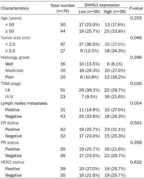
Table 1. Correlation between SNHG1 expression and clinicopatho-logical features of BCa patients
Characteristics Total number (n=74) SNHG1 expression P-value
Low (n=36) High (n=38)
Age (years) 0.255
< 50 30 17 (23.0%) 13 (17.6%)
≥ 50 44 19 (25.7%) 25 (33.8%)
Tumor size (cm) 0.046
< 2.5 47 27 (36.5%) 20 (27.0%)
≥ 2.5 27 9 (12.2%) 18 (24.3%)
Histology grade 0.396
Well 16 10 (13.5%) 6 (8.1%)
Moderate 38 18 (24.3%) 20 (27.0%)
Poor 20 8 (10.8%) 12 (16.2%)
TNM stage 0.035
I-II 51 29 (39.2%) 22 (29.7%)
III-IV 23 7 (9.5%) 16 (21.6%)
Lymph nodes metastasis 0.054
Positive 31 11 (14.9%) 20 (27.0%)
Negative 43 25 (33.8%) 18 (24.3%)
ER status 0.501
Positive 42 19 (25.7%) 23 (31.1%)
Negative 32 17 (23.0%) 15 (20.3%)
PR status 0.358
Positive 35 19 (25.7%) 16 (21.6%)
Negative 39 17 (23.0%) 22 (29.7%)
HER2 status 0.632
Positive 39 20 (27.0%) 19 (25.7%)
Negative 35 16 (21.6%) 19 (25.7%)
ER, estrogen receptor; PR, progesterone receptor; HER2, human epidermal receptor 2.
AGACACAGAT-3’, si-SNHG1- 2, 5’-GATTAAGACACTGGGA- GCCAATGAA-3’ and si-SNH- G1-3, 5’-GGGAGCCAATGAA- ACAGCAGTTGAG-3’) and sc- rambled negative control si- RNA were synthesized by Shanghai Genepharma Co., Ltd. (Shanghai, China). BCa cells were seeded into six-well plates, and transfecti- on was performed using Li- pofectamine 2000 (Invitro- gen). 48 hours post-trans-fection, transfection effica-cy was analyzed by real-time quantitative RT-PCR (qRT- PCR).
RNA extraction and qRT-PCR
Transwell assay
Cell migration and invasion assays were per-formed in a 24-well transwell plate with 8-μm polyethylene terephthalate membrane filters (Costar, Corning, MA, USA). Cells in serum-free medium were added to the upper chambers with either uncoated or Matrigel-coated mem-branes. Lower chamber was filled with medium containing 10% FBS. After 24 h of incubation, the non-migrated cells on the upper sides of the filters were scraped off, and the cells locat -ed in the lower filters were fix-ed with 4% para
-experimental procedures were approved by the Ethics Committee of the First People’s Hospital of Lianyungang.
Bioinformatics prediction and dual-luciferase reporter assay
The potential microRNA binding sites of SNH- G1 predicted by computer-aided algorithms were obtained from starBase v2.0 (http://star-base.sysu.edu.cn/mirLncRNA.php). The puta-tive miR-145 target binding sequence in SNHG1 and its mutant of the binding sites were
ampli-Table 2. The sequences of the primers
Gene name Primer sequences
SNHG1 Forward primer TAACCTGCTTGGCTCAAAGGG
SNHG1 Reverse primer CAGCCTGGAGTGAACACAGA
GAPDH Forward primer CGAGATCCCTCCAAAATCAA
GAPDH Reverse primer TTCACACCCATGACGAACAT
miR-145 RT primer GTCGTATCCAGTGCAGGGTCCGAGGTATTCGCACTGGATACGACAGGGAT
U6 RT primer GTCGTATCCAGTGCAGGGTCCGAGGTATTCGCACTGGATACGACAAAATA
miR-145 Forward primer GTCCAGTTTTCCCAGGA
miR-145 Reverse primer GTGCAGGGTCCGAGGT
U6 Forward primer CTCGCTTCGGCAGCACATATACT
[image:3.612.89.443.78.480.2]U6 Reverse primer ACGCTTCACGAATTTGCGTGTC
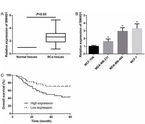
Figure 1. SNHG1 is upregulated in BCa and predicts poor prognosis. A. The expression of SNHG1 in BCa tissues was increased than that in adjacent non-tumor tissues. B. Expression of SNHG1 in BCa cell lines was significantly increased than that in normal MCF-10A cells. Data are presented as the mean ± SD from at least three independent experiments. *P < 0.05. C. BCa patients with high-expressing SNHG1 showed a significantly reduced overall survival (P=0.028).
formaldehyde, and then st- ained with 0.1% crystal viol- et. Migrated or invaded cells were counted in five randomly chosen fields per well under a microscope.
In vivo tumorigenesis assay
[image:3.612.91.375.231.474.2]fied and inserted into downstream of the fire-fly luciferase gene in a pGL3promoter vector (Promega, Madison, WI, USA), named as: SNHG1-WT or SNHG1-MUT, respectively. For dual-luciferase assay, the luciferase reporter gene vector with SNHG1-WT or SNHG1-MUT, together with miR-145 mimics or mimics con-trol, were co-transfected into BCa cells. At 48 h after transfection, luciferase activities were determined using the Dual Luciferase Reporter Assay System (Promega) and normalized to Renilla luciferase.
Statistical analysis
Each experiment was performed in triplicate and repeated at least three times. The data are expressed as the mean ± SD. Kaplan-Mei- er plots and log-rank tests were used for sur-vival analysis. All statistical analyses were performed using SPSS version 18.0 software (SPSS Inc., Chicago, IL, USA) and Graphpad Pri-
sm (version 6.01) software (GraphPad Software, Inc., La Jolla, CA, USA). P value < 0.05 is consid-ered significant.
Results
SNHG1 is upregulated in BCa and predicts poor prognosis
[image:4.612.93.523.73.393.2]In order to fully understand the functions of SNHG1 in BCa, we first examined expression of SNHG1 in 74 pairs of BCa tissues and their adjacent non-cancerous tissues by qRT-PCR. We observed that, as demonstrated in Figure 1A, SNHG1 levels were markedly higher in 74 BCa tissues in their counterparts. We then examined SNHG1 expression in BCa cell lines, and found that SNHG1 was more highly expressed in BCa cell lines (MDA-MB-231, MDA-MB-468 and MCF-7) than that of in the normal human breast cell line MCF-10A (Figure 1B).
To further explore the clinical significance of SNHG1 in BCa, the association between SNHG1 expression and the clinicopathological status of 74 BCa patients was analyzed. BCa patients were divided into a high expression group (≥ mean, n=38) and a low expression
[image:5.612.92.524.74.196.2]group (< mean, n=36) on the basis of the cutoff value of SNHG1 expression. As indicated in
Table 2, increased SNHG1 expression level in BCa tissues was significantly associated with tumor size (P=0.046) and TNM stage (P=0.035). Kaplan-Meier analysis revealed that high SN- HG1 expression was closely related to a poorer overall survival in BCa patients (log-rank test,
P=0.028; Figure 1C).
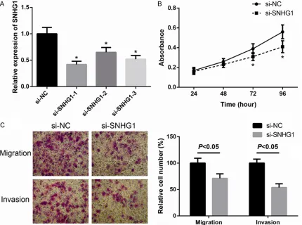
SNHG1 promotes the proliferation, migration and invasion of BCa cells
Since our clinical data showed that SNHG1 was high expressed in BCa, we further investigated its oncogenic features and effects on BCa cell lines. MCF-7 cells were transiently transfected with three siRNAs targeting SNHG1, which effi -ciently silenced endogenous expression of SNHG1 (Figure 2A). si-SNHG1-1 caused the lowest level of SNHG1 and was accordingly selected for further study. CCK-8 assay were performed to assess the role of SNHG1 in BCa cell proliferation and the results showed that the BCa cells transfected with si-SNHG1 grew slower compared to control cells (Figure 2B). Furthermore, as shown in Figure 2C, MCF-7 cells with reduced SNHG1 expression exhibited decreased migration and invasion compared to control cells. These findings indicate that down -regulation of SNHG1 inhibits BCa cell prolifera-tion, migraprolifera-tion, and invasion.
SNHG1 promotes growth of BCa xenografts in vivo
To confirm the above data in vivo, MCF-7 cells stably transfected with sh-SNHG1 or sh-NC were injected subcutaneously into nude mice, respectively. Xenografts tumor volume was measured every three days after a palpable
Figure 3. SNHG1 promotes the growth of BCa xenografts in vivo. A. Tumor volume was calculated every 3 days. B. Four days after implantation, tumors were excised and weighted. Data are presented as the mean ± SD (n=6 per group). *P < 0.05.
[image:5.612.90.288.253.554.2]tumor formed, and mice were killed four weeks after cell implantation. As shown in Figure 3A, tumors derived from the sh-SNHG1 group grew at a slower rate than sh-NC group, and the tumor weight in the sh-SNHG1 group was sig-nificantly less than sh-NC group (Figure 3B). Thus, silencing of SNHG1 could markedly inhib-it tumorigenesis of BCa cells in vivo.
SNHG1 functions as a ceRNA for miR-145 in BCa cells
Recent studies have reported lncRNAs could act as molecular sponges or ceRNAs to regu-late the biological functions of miRNAs. To fur-ther clarify the molecular mechanisms of SNHG1 on the biological phenotypes of BCa cells, we searched for the target miRNAs using starBase v2.0 (http://starbase.sysu.edu.cn/ mirLncRNA.php). miR-145 was thus selected as one of the candidate targets of SNHG1, because of the putative target sequences, as shown in Figure 4A. To further confirm whether SNHG1 is a functional target for miR-145, lucif-erase activity assay was performed. Through the luciferase activity assay, we found that miR-145 mimics markedly inhibited SNHG1-WT reporter activity, while it had nearly no inhibito-ry effect on the SNHG1-MUT reporter activity (Figure 4B). Additionally, we found that inhibi-tion of SNHG1 led to remarkably increased expression of miR-145in MCF-7 cells (Figure 4C).
miR-145 expression is negatively associated with SNHG1 expression in BCa tissues
To further determine the association between SNHG1 and miR-145 in BCa, we examined
still remains dismal. The etiology of BCa involves a complex interplay of various factors. Recently, accumulating studies have indicated that lncRNAs play important roles in regulating various cellular processes [13] and act as driv-ers of tumor suppressive and oncogenic func-tions in the development and progression of BCa [14]. Therefore a better understanding of correlation between lncRNAs and tumor etiolo-gy will provide novel therapeutic targets for the early diagnosis and treatment of BCa. SNHG1 is a recently discovered lncRNA, which has not yet been extensively explored in human BCa. In the present study, we studied the role of SNHG1 in BCa progression. Based on the results of qRT-PCR, we discovered that the ex- pression of SNHG1 was significantly increased in BCa tissues and cell lines, which is consis-tent with earlier studies in colorectal carcino- ma [15] and prostate cancer [16]. Further, we explored the association between SNHG1 expression and clinical characteristics of BCa patients, and observed that patients with high-ly expressed SNHG1 had more aggressive char-acteristics and unfavorable prognosis.
[image:6.612.91.376.73.184.2]In order to highlight the function of SNHG1 in BCa, we further explored the critical roles of SNHG1 in the progression of BCa by loss-of-function analysis. Our data showed that knock-down of SNHG1 contributed to significant inhi -bition of BCa cell proliferation, migration, and invasion in vitro. Additionally, downregulation of SNHG1 could inhibit the BCa tumorigenesis in murine model. Taken together, these findings suggest that SNHG1 might function as a poten-tial oncogene to promote BCa development and progression.
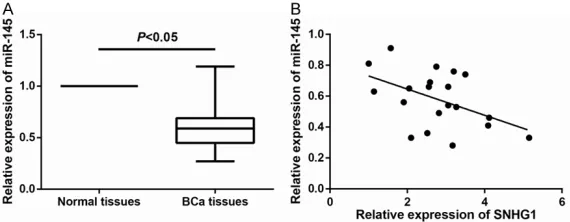
Figure 5. miR-145 expression is negatively associated with SNHG1 expres-sion in BCa tissues. A. Expresexpres-sion of miR-145 in BCa tissues was reduced compared to that in adjacent non-tumor tissues. B. Pearson correlation anal-ysis shows a negative association between SNHG1 and miR-145 expression in BCa tissues.
expression of miR-145 in BCa tissues and normal tissues by qRT-PCR. As shown in Figure 5A, miR-145 expression was significantly reduced in BCa tissues compared with that in normal tissues. Furthermore, as shown in Figure 5B, miR-145 expression was negative-ly correlated with the relative SNHG1 expression in BCa tis-sues (r=-0.478, P=0.033).
Discussion
The ceRNA hypothesis is gaining attention. This hypothesis suggests that lncRNA could func-tion as a competing endogenous RNA (ceRNA) or a molecular sponge in regulating the ex- pression patterns and biological functions of miRNA [17]. It has been reported that miR-101-3p is a direct target of SNHG1 in non-small cell lung cancer [18], indicating that SNHG1 might be a natural sponge for miRNAs. In the present study, miRNA complementary base pairing with SNHG1 was predicted by bioinfor-matics method, and we discovered miR-145 might form complementary base pairing with SNHG1. MiR-145 was previously shown to be a tumor repressor, including BCa [19, 20], and we therefore speculated that SNHG1 might pro-mote BCa progression in a miR-145-dependent manner.
In summary, the present study offers the first direct investigation of a relationship between SNHG1 and BCa progression. We also observed that SNHG1 might function as a ceRNA to attenuate the endogenous function of miR-145 in BCa. However, other possible mechanisms by which SNHG1 participates in BCa remain to be further explored.
Disclosure of conflict of interest
None.
Address correspondence to: Wen-Song Wei, Bre- ast Disease Center, The Third Hospital of Nanch- ang, Nanchang 330009, China. Tel: 18079109828; E-mail: [email protected]
References
[1] Torre LA, Bray F, Siegel RL, Ferlay J, Lortet-Tieu-lent J and Jemal A. Global cancer statistics, 2012. CA Cancer J Clin 2015; 65: 87-108. [2] DeSantis CE, Fedewa SA, Goding Sauer A,
Kramer JL, Smith RA and Jemal A. Breast can-cer statistics, 2015: convergence of incidence rates between black and white women. CA Cancer J Clin 2016; 66: 31-42.
[3] Li Y, Li J, Wang Y, Zhang Y, Chu J, Sun C, Fu Z, Huang Y, Zhang H, Yuan H and Yin Y. Roles of cancer/testis antigens (CTAs) in breast cancer. Cancer Lett 2017; 399: 64-73.
[4] Wang KC and Chang HY. Molecular mecha-nisms of long noncoding RNAs. Mol Cell 2011; 43: 904-914.
[5] Yan X, Hu Z, Feng Y, Hu X, Yuan J, Zhao SD, Zhang Y, Yang L, Shan W, He Q, Fan L, Kan-dalaft LE, Tanyi JL, Li C, Yuan CX, Zhang D,
Yuan H, Hua K, Lu Y, Katsaros D, Huang Q, Montone K, Fan Y, Coukos G, Boyd J, Sood AK, Rebbeck T, Mills GB, Dang CV and Zhang L. Comprehensive genomic characterization of long non-coding RNAs across human cancers. Cancer Cell 2015; 28: 529-540.
[6] Wu ZJ, Li Y, Wu YZ, Wang Y, Nian WQ, Wang LL, Li LC, Luo HL and Wang DL. Long non-coding RNA CCAT2 promotes the breast cancer growth and metastasis by regulating TGF-beta signal-ing pathway. Eur Rev Med Pharmacol Sci 2017; 21: 706-714.
[7] Huan J, Xing L, Lin Q, Xui H and Qin X. Long noncoding RNA CRNDE activates Wnt/beta-catenin signaling pathway through acting as a molecular sponge of microRNA-136 in human breast cancer. Am J Transl Res 2017; 9: 1977-1989.
[8] Fang Y, Wang J, Wu F, Song Y, Zhao S and Zhang Q. Long non-coding RNA HOXA-AS2 pro-motes proliferation and invasion of breast can-cer by acting as a miR-520c-3p sponge. Onco-target 2017; 8: 46090-46103.
[9] Zhang M, Wang W, Li T, Yu X, Zhu Y, Ding F, Li D and Yang T. Long noncoding RNA SNHG1 pre-dicts a poor prognosis and promotes hepato-cellular carcinoma tumorigenesis. Biomed Pharmacother 2016; 80: 73-79.
[10] Zhang H, Zhou D, Ying M, Chen M, Chen P, Chen Z and Zhang F. Expression of long non-coding RNA (lncRNA) small nucleolar RNA host gene 1 (SNHG1) exacerbates hepatocellular carcinoma through suppressing miR-195. Med Sci Monit 2016; 22: 4820-4829.
[11] Wang Q, Li Q, Zhou P, Deng D, Xue L, Shao N, Peng Y and Zhi F. Upregulation of the long non-coding RNA SNHG1 predicts poor prognosis, promotes cell proliferation and invasion, and reduces apoptosis in glioma. Biomed Pharma-cother 2017; 91: 906-911.
[12] Livak KJ and Schmittgen TD. Analysis of rela-tive gene expression data using real-time quantitative PCR and the 2 (-Delta Delta C(T)) Method. Methods 2001; 25: 402-408.
[13] Clark MB and Mattick JS. Long noncoding RNAs in cell biology. Semin Cell Dev Biol 2011; 22: 366-376.
[14] Cerk S, Schwarzenbacher D, Adiprasito JB, Stotz M, Hutterer GC, Gerger A, Ling H, Calin GA and Pichler M. Current status of long non-coding RNAs in human breast cancer. Int J Mol Sci 2016; 17.
[15] Sun X, Wang Z and Yuan W. Down-regulated long non-coding RNA SNHG1 inhibits tumor genesis of colorectal carcinoma. Cancer Bio-mark 2017; 20: 67-73.
cancer. Biochem Biophys Res Commun 2017; 487: 146-152.
[17] Poliseno L, Salmena L, Zhang J, Carver B, Haveman WJ and Pandolfi PP. A coding-inde -pendent function of gene and pseudogene mRNAs regulates tumour biology. Nature 2010; 465: 1033-1038.
[18] Cui Y, Zhang F, Zhu C, Geng L, Tian T and Liu H. Upregulated lncRNA SNHG1 contributes to progression of non-small cell lung cancer through inhibition of miR-101-3p and activa-tion of Wnt/beta-catenin signaling pathway. Oncotarget 2017; 8: 17785-17794.
[19] Zhao H, Kang X, Xia X, Wo L, Gu X, Hu Y, Xie X, Chang H, Lou L and Shen X. miR-145 sup-presses breast cancer cell migration by target-ing FSCN-1 and inhibittarget-ing epithelial-mesenchy-mal transition. Am J Transl Res 2016; 8: 3106-3114.