Expression profi le of the
SOX9
gene in the testes
of sexually immature and mature male goats
(
Capra hircus
), and its potential infl uence
on postnatal testis development
I. Szatkowska
1, M. Jedrzejczak-Silicka
1, A. Dybus
1, B. Wiszniewska
2,
J. Udala
1, D. Zaborski
1, J. Wojcik
1, T. Stankiewicz
1, W.S. Proskura
1*
1West Pomeranian University of Technology, Szczecin, Poland 2Pomeranian Medical University, Szczecin, Poland
*Corresponding author: [email protected]
ABSTRACT: The aim of this study was to compare the expression levels of the SOX9 (SRY-box 9) gene in the testes of 18 White improved male goats (Capra hircus) divided into three age groups (one, 10 and 15 months of age with seven, eight and three individuals per group, respectively). Abnormalities in testis development were observed in three individuals from the group of 10-month-olds. Additionally, differences in SOX9 expression that may affect the process of testis maturation and testicular spermatogenic activity were investigated among individuals. The expression of the SOX9 gene in testicular tissues was analysed using qPCR. Maximal SOX9 expression was observed in 10-month-old males with normal testes, while expression was significantly reduced in the same age group of males with abnormal testes. Abnormalities in testis development were associated with low parameters of semen quality. The lowest expression levels of SOX9 were observed in 15-month-old goats. The results of the present study indicate that SOX9 expression changes significantly during the development of male goats.
Keywords: gene expression; goat; gonads; testis development; sexual maturation; SOX9; SRY
The molecular mechanism of testis development and differentiation in the prenatal period, involv-ing genes such as SRY (Sex Determining Region Y gene), SOX9 (SRY-box 9 gene), WT1 (Wilms Tumor 1 gene), SF1 (Splicing Factor 1 gene), GATA4 (GATA Binding Protein 4 gene) and AMH (Anti-Mullerian Hormonegene), is well documented (Kanaiet al. 2005; Plottonet al. 2012). However, the effects of these gene products on postnatal gonadal matura-tion, how their expression is regulated at different developmental stages, as well as their relation-ships with other factors are poorly understood. It is known that SOX9 plays a key role in the main-tenance of testicular status in mice (Barrionuevo et al. 2009; Barrionuevoet al. 2012). A deficiency in SOX9 expression in adult life may lead to vari-ous abnormalities, from gonadal dysgenesis to
transdifferentiation into an ovary (Barrionuevo et al. 2009; Barrionuevo et al. 2012; Plotton et al. 2012). Experiments on SOX9 conditional null mutant mice (Barrionuevoet al. 2009) confirmed that SOX9 plays a crucial role in embryonic sex determination. Also, SOX9 together with SOX8 (SRY-box 8 gene) is essential for spermatic cord formation during early testis development. The formation of the spermatic cord is fundamental to the maintenance of spermatogenesis in the adult testis, and its absence leads to infertility in adult males (Barrionuevoet al. 2009; Jianget al. 2013).
As described above, it is known that SOX9 is criti-cal for proper development of gonads in mammals such as mice. Th us, we hypothesised that atypical expression of the SOX9 gene might be linked to abnormalities in testis development and to poor
men parameters in goats. Th e fi rst aim of the present study was to compare the expression levels of the
SOX9 gene in the testes of sexually immature and mature male goats. Th e second goal was to corre-late SOX9 expression in individual goats with testis maturation and testicular spermatogenic activity.
MATERIAL AND METHODS
The study was approved by the Local Ethics Committee for Animal Research in Szczecin, and complied with the Polish regulations and guidelines for experiments on animals. A total of 18 White improved male goats (Capra hircus) were divided into three age groups (one, 10 and 15 months of age with seven, eight and three individuals per group, respectively). In order to exclude potential female-to-male sex-reversed goats, the sex was confi rmed by the presence of the SRY gene using PCR. Th e primer sequences used for this purpose are given in Table 1.
Testicular tissues collected post mortem into test tubes containing RNAlater® (Sigma Aldrich) were used for the SOX9 expression analysis. Total RNA was isolated using the Total RNA kit (A&A Biotechnology, Gdynia, Poland), quality was as-sessed by 1% agarose gel electrophoresis and the RNA was quantifi ed fl uorometrically using a Qubit fl uorimeter and the Quant-iTTM RNA BR Assay Kit
(Invitrogen GmbH, Germany). Approximately 1 μg of total RNA was processed for reverse transcription using a First Strand cDNA Synthesis Kit (Th ermo Fisher Scientifi c, Lafayette, USA). Th e relative quan-tity of SOX9 was assessed using the KAPATM SYBR®
qPCR Kit (Kapa Biosystems, USA) in the Rotor-Gene instrument (Qiagen GmbH, Germany). Th e primer pairs used for qPCR are given in Table 1. Th e real-time PCRs were performed with
single-stranded cDNA (40 ng) and all samples were run in triplicate in reactions containing 12.5 μl of SybrGreen PCR Master Mix, 200nM of each primer and H2O, up to a fi nal volume of 15 μl. Prior to the analysis of SOX9 expression levels, the expression stability of the two reference genes (ACTB – Actin Beta gene and UXT –Ubiquitously-Expressed, Prefoldin-Like Chaperone gene) for testicular tis-sue was determined using geNorm v. 3.5 software.
SOX9 expression levels were normalised to the UXT
gene. For relative transcript quantifi cation, standard curves for the reference gene (UXT) and the gene of interest (SOX9)were generated using a four-fold serial dilution of cDNA. Th e amplifi cation effi ciency for UXT and SOX9 was 104% and 102%, respec-tively. Th e relative expression of the gene of interest was analysed using the 2–ΔΔC
T method (Livak and
Schmittgen 2001). Th e relative fold change in SOX9
expression among age groups was tested with the Kruskal-Wallis non-parametric ANOVA.
Testicular tissue sections collected post mortem
were fixed in Bouin’s liquid and embedded in paraf-fin blocks. Serial sections stained with the Periodic acid-Schiff stain (PAS) were used for histological evaluation of the testes.
At the age of seven months, all eight bucks from the group of 10-month-olds were subjected to semen evaluation. Semen was collected by elec-troejaculation during the short photoperiod (mid-September to mid-December). Th e analysis was carried out four times at 3-week intervals. The electroejaculated semen was evaluated immediately after collection and the following parameters were determined according to routine methods: ejaculate volume (within an accuracy of 0.1 cm3),
[image:2.595.62.534.618.726.2]percent-age progressive motility (within an accuracy of 5%), sperm concentration (in Burker’s chamber), the per-centage of spermatozoa with primary and secondary
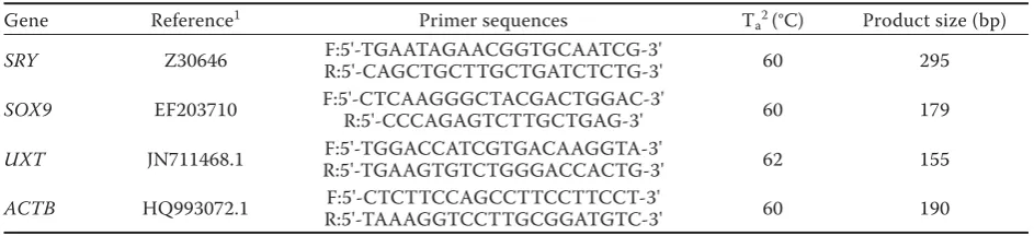
Table 1. Primer sequences used for PCR and qPCR
Gene Reference1 Primer sequences T
a2 (°C) Product size (bp)
SRY Z30646 F:5'-TGAATAGAACGGTGCAATCG-3'
R:5'-CAGCTGCTTGCTGATCTCTG-3' 60 295
SOX9 EF203710 F:5'-CTCAAGGGCTACGACTGGAC-3'R:5'-CCCAGAGTCTTGCTGAG-3' 60 179
UXT JN711468.1 R:5'-TGAAGTGTCTGGGACCACTG-3'F:5'-TGGACCATCGTGACAAGGTA-3' 62 155
ACTB HQ993072.1 R:5'-TAAAGGTCCTTGCGGATGTC-3'F:5'-CTCTTCCAGCCTTCCTTCCT-3' 60 190
10% nigrosin staining. Testis size was expressed in terms of scrotal circumference (measured with a tape measure to the nearest 0.5 cm) and testicu-lar volume calculated from the measurements of length, width and thickness (using a calliper accu-rate to 0.1 cm). Th ese calculations were performed after deducting scrotal skin-fold thickness (Th ibier and Colchen-Bourlaud 1972).
RESULTS
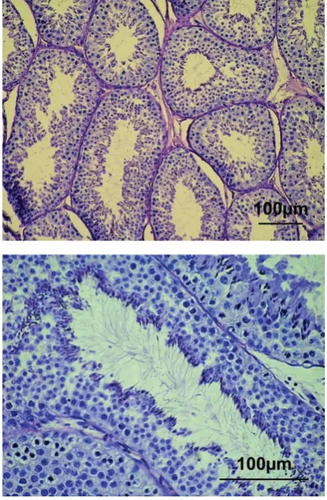
The presence of the SRY gene was confirmed in all individuals (Figure 1). The data on the relative quantification of SOX9 gene expression in the stud-ied individuals are reported in Table 2. Statistical analysis revealed a significant relationship be-tween age and the relative level of SOX9 expression (P = 0.0092). The relative quantity of SOX9 gene expression in the group of 1-month-old bucks was morphological abnormalities (Blom 1981) and the
[image:3.595.303.533.97.448.2]percentage of spermatozoa with intact acrosomes (Saacke and White 1972). Morphological abnor-malities and acrosome integrity were assessed using
[image:3.595.63.294.98.218.2]Figure 1. Confi rmation of Y chromosome presence by electrophoretic separation of the PCR products ampli-fi ed using primers for the SRY gene; lanes 1 and 8 (DNA ladder), lanes 2–6 (males), lane 7 (blank sample)
Table 2. Relative quantity of SOX9 gene expression in the testicles of White improved goats of diff erent ages
Age (months) Individual ΔCt SD RQ
1
A1 5.76 0.32 1.16 B1 5.06 0.14 1.89 C1 3.54 0.14 5.43 D1 4.33 0.36 3.14 E1 6.07 0.00 0.94 F1 6.06 0.20 0.95 G1 7.33 0.27 0.39
x̅ 5.45 0.21 1.44
10
A10 0.91 0.34 33.59 B10 2.40 0.28 11.96 C10 0.49 0.14 44.94 D10 0.19 0.38 55.33 E10 0.76 0.14 37.27
x̅ 0.95 0.27 32.67 F101 5.11 0.17 1.83
G101 4.97 0.41 2.01
H101 4.33 0.37 3.14
x̅ 4.80 0.33 2.26
15
A152 5.98 0.08 1.00
B15 5.61 0.33 1.29 C15 5.89 0.09 1.06
x̅ 5.83 0.20 1.11
Ct = threshold cycle, RQ = relative quantity of gene expres-sion, SD = standard deviation
1Individuals with abnormally developed testicles 2Calibrator sample
[image:3.595.62.291.385.699.2]22.7-fold lower than that in the group of 10-month-olds (P = 0.0137) and almost equal to that observed in the group of 15-month-old bucks (P = 1).
The mean relative quantity of SOX9 gene expres-sion in 10-month-old goats was estimated from
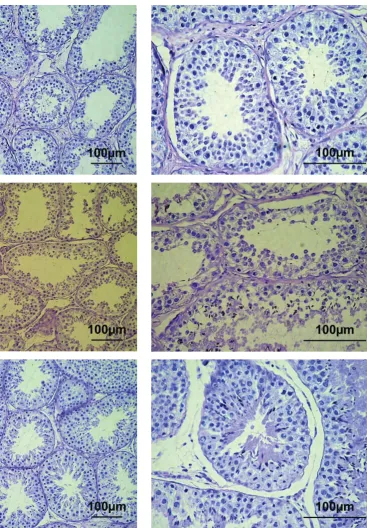
in-dividuals with normal testis morphology, typical of bucks at the post-pubertal stage. In five individu-als, the normal structure of seminiferous tubules (characteristic of the functionally active testes) with complete spermatogenesis was observed in the
[image:4.595.153.521.170.699.2]tological sections (Figure 2). In three goats (animals No. F10, G10 and H10), abnormalities of the sper-matogenic epithelium with undifferentiated cells lacking nuclei and that were desquamating into the lumen were found in the seminiferous tubules (Figure 3). Moreover, morphological alterations in-dicating oligospermia or complete absence of the final differentiation of spermatozoa were visible. In these three bucks, the expression level of the
SOX9 gene was considerably lower as compared to the normal bucks (Table 2).
DISCUSSION
The present study shows a relatively low level of
SOX9 gene expression in 1-month-old bucks. At this age, the gonads are undergoing the onset of the intensive division of gonocytes and differentiation into prespermatogonia. According to Montazer-Torbatiet al. (2010), the highest expression of SOX9
in goats is found during the perinatal period, after which it decreases drastically. It is likely that this is caused by the activation of oestrogen receptor alpha, which negatively regulates SOX9 expres-sion (Kaoet al. 2012). Thus, it can be concluded that age differences in the group of 1-month-old bucks (30 ± 3 days) could be the main reason for the inter-individual variations in SOX9 expression levels. Moreover, no histopathological alterations of the testes were observed, which could have been associated with SOX9 expression. The testicular parenchyma in all bucks from this age group con-tained sex cords, formed by immature sustentacular cells (Sertoli cells) and gonocytes. The results of experiments performed on conditional null mu-tant mice (Barrionuevoet al. 2009) showed that inactivation of the SOX9 gene after the stage of sex determination neither disturbs the process of testis differentiation or sex cord formation during the embryonic period, nor induces abnormalities in testis morphology or their initial spermatogenic activity within the first two months of the postna-tal period. Based on the study of XY(Sry–),Ods/+
mice, Qin and Bishop (2005) concluded that SOX9
is sufficient for normal testis development during the embryonic and early postnatal periods.
The highest levels of SOX9 expression were ob-served in the normally developed 10-month-old males (Table 2). In these bucks, a distinct lumen in the seminiferous tubules and all generations of
germinal cells, including spermatozoa, were found in the seminiferous epithelium. Sustentacular cells were fully differentiated, whereas interstitial cells (Leydig cells) were found in the form of clusters in the interstitial tissue (Figure 2). On the other hand, in the abnormally developed testes of 10-month-old males, dystrophy of spermatogenic epithe-lial cells, oligospermia, developmentally arrested spermatocytes, and damaged spermatogenic epi-thelium with undifferentiated cells without nuclei which were desquamating into the lumen, were ob-served. These changes were associated with low values of measured parameters of the collected semen (Table 3) and a considerably lower level of
[image:5.595.305.531.512.699.2]SOX9 gene expression as compared to the males with normally developed testes (Table 2). A simi-lar post-pubertal arrest of testes development was observed in SOX9 conditional null mice, in which the spermatogenic epithelium showed reduced cel-lularity with a decreased number of spermatids. In addition, approximately two-thirds of the tubules contained spermatogonia and early spermatocytes, while the remaining tubules were completely devoid of these (Barrionuevoet al. 2009). A very similar phenotype was described for the XY(Sry–),Ods/+ mice, in which the testicular level of SOX9 protein was reduced by about 75% (Qin and Bishop 2005). These authors proposed that the aforementioned
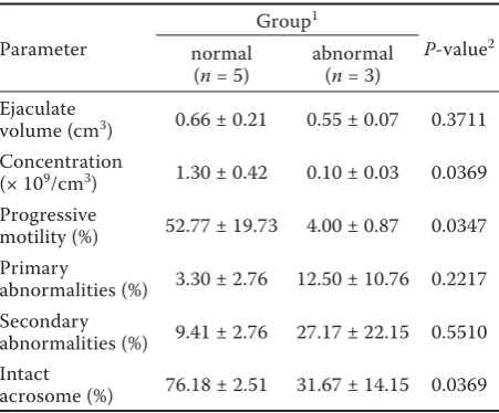
Table 3. Comparison of semen parameters between bucks with normal and abnormal testis development in the group of 10-month-old animals
Parameter
Group1
P-value2
normal (n = 5)
abnormal (n = 3) Ejaculate
volume (cm3) 0.66 ± 0.21 0.55 ± 0.07 0.3711
Concentration
(× 109/cm3) 1.30 ± 0.42 0.10 ± 0.03 0.0369
Progressive
motility (%) 52.77 ± 19.73 4.00 ± 0.87 0.0347 Primary
abnormalities (%) 3.30 ± 2.76 12.50 ± 10.76 0.2217 Secondary
abnormalities (%) 9.41 ± 2.76 27.17 ± 22.15 0.5510 Intact
acrosome (%) 76.18 ± 2.51 31.67 ± 14.15 0.0369
1Individuals were classifi ed into two groups based on the
histopathological picture of testis tissue sections collected
post mortem after semen evaluation
phenotype could result from the lack of SOX9 in adult sustentacular cells. This can lead to the aber-rant expression of genes and proteins important for the structural integrity of testicular tubules such as extracellular matrix components and molecules involved in the formation of sustentacular-susten-tacular or sustensustentacular-susten-tacular-germ cell contacts, as well as junctional molecules. Thus, the lack of SOX9 in adult sustentacular cells impairs their functional interaction with germ cells and causes dysregula-tion of the spermatogenic cycle leading to sterility (Barrionuevoet al. 2009).
The group of mature 15-month-old bucks was characterised by the lowest level of SOX9 gene ex-pression among the three age groups investigated. The inter-individual differences were negligible in this group, indicating that the expression of the
SOX9 gene was quite stable in sexually mature male goats, which is in accordance with the results of Montazer-Torbatiet al. (2010).
The SOX9 gene belongs to the SOX (SRY-related HMG box) family of transcription factors. The SOX9 protein contains several conserved struc-tural elements including a C-terminal transacti-vation domain, a dimerization domain, and two nuclear export signals. The HMG domain binds to consensus sites in the regulatory regions of its target genes, and thus is the most important with respect to target gene expression. SOX9 is a factor with multiple functions in organogenesis (Bhandari et al. 2012), among which testis induc-tion and formainduc-tion are crucial for interpreting the results of the present study. A discussion regarding the transcriptional activity of this determinant of the male developmental pathway should focus on two important aspects. The first concerns target gene activation, involved in testis initiation, dif-ferentiation and development. The second one is linked to how testis-specific expression of SOX9 is itself regulated. Both problems seem to be equally complex. The initiation and regulation of SOX9 expression, restricted in the testis to precursor and, subsequently, differentiating and differentiated sus-tentacular cells, occur via a testis-specific and very long testis-specific enhancer of Sox9 (TES) enhanc-er, well described in mice and humans (Sekido and Lovell-Badge 2008; Benko et al. 2011). In its central part, a highly-conserved region of 1.4 kb (a core region – TESCO) was identified, which contains consensus sites for multiple transcription factors. They can be classified as positive and negative
reg-ulators (SRY, SF1, FGF9, FGFR2, GATA4, FOXL2 and ER alpha), and the list is regularly updated (Barrionuevo et al. 2012). An analogous situation is observed in the case of potential target genes of SOX9. Although it is accepted that its major target is the anti-Mullerian hormone (AMH) promoter, the latest research conducted on rats has shown that there are over 100 genes with consensus bind-ing sites for SOX9 (Bhandari et al. 2012).
Th e main regulator initiating SOX9 transcription-al activity via TESCO in the primitive mtranscription-ale gonads is SRY. Its major role is SOX9 activation (together with SF1), which once initiated, is maintained through-out pre- and postnatal life. It is accepted that SRY is only transiently expressed during the determination of the bipotential gonad. Th erefore, other factors must be involved in maintaining SOX9 expression. A recent study by Montazer-Torbati et al. (2010) directly challenges this model and shows that, in goats, SRY transcript levels, which peak at 36 days post-coitum (dpc), are maintained until about the fi rst month after birth before drastically decreas-ing in sexually mature individuals. Consequently, it can also activate SOX9 transcription in the fi rst weeks of postnatal life. At 3–4 months of age, the developmental stage of the gonads of male goats is associated, fi rst and foremost, with the onset of intensive gonocyte divisions and their further dif-ferentiation into prespermatogonia. Th e high AMH levels (induced by SOX9) maintained at this time inhibit their entry into meiotic divisions, simulta-neously inhibiting the proliferation of interstitial cells (Montazer-Torbati et al. 2010). Immature sus-tentacular cells also proliferate, but the intensity of this process depends, among other things, on the reduction in endogenous oestrogens elicited by the irreversible transformation of androgens by a microsomal enzymatic complex named aromatase (Kao et al. 2012). Its activity in many mammalian species is mainly localised to immature sustentacu-lar cells (Carreau et al. 2007), like that of oestrogen alpha receptors. Th erefore, the lower amounts of
SOX9 transcripts (albeit suffi cient for maintaining an appropriate level of AMH expression) in the group of the youngest individuals, could have been caused by a high activity of oestrogen alpha recep-tors (unpublished data) capable of binding regula-tory elements in TESCO SOX9 and inhibiting its transcription (Jakob and Lovell-Badge 2011).
lev-els were drastically decreased in sexually mature individuals (15 months of age). As the amount of
SRY transcripts is dramatically reduced at this stage (Montazer-Torbati et al. 2010), this elevated expres-sion of SOX9 would appear to be SRY-independent. Nevertheless, the extended regulatory element TESCO, as already mentioned, may be involved in maintaining an appropriate level of SOX9 tran-scripts. However, the role of SOX9 in this period is puzzling. It is known that at this stage AMH levels (upregulated by SOX9) are considerably decreased. At this stage of testicular development, AMH is strongly inhibited by an increasing concentration of intra-testicular androgens acting via androgen receptors (Grinspon and Rey 2010). Sexually ma-ture males from the control group (sexual maturity period) were characterised by the lowest testicular levels of SOX9 mRNA transcripts, as also previously reported by Montazer-Torbati et al. (2010). However, whether this is a permanent decrease or only tran-scriptional silencing during the spring-summer sea-son (when reduced sexual activity in seasea-sonal breeds is observed) remains unclear. Certainly, this is an issue that should be further investigated.
Th e transcriptional pattern of SOX9 was signifi -cantly altered in males with abnormal testicular de-velopment. It should be determined whether the low level of SOX9 expression in these individuals was the cause or result of these abnormalities. On the other hand, the high expression levels of SOX9 in the cryptorchid may be associated with the atrophy of the seminiferous epithelial cells and excessive pro-liferation of sustentacular cells (Hutson et al. 1996), in which this transcription factor is active. Th is type of atrophy has been described in such cases.
In conclusion, the results of the present study showed that the expression level of SOX9 changes significantly during the development of male goats.
Acknowledgement
We would like to thank Prof. Dr. Maria Katkiewicz for her help with the description and interpretation of histological results.
REFERENCES
Barrionuevo F, Georg I, Scherthan H, Lecureuil C, Guillou F, Wegner M, Scherer G (2009): Testis cord differentiation
after the sex determination stage is independent of Sox9 but fails in the combined absence of Sox9 and Sox8. De-velopmental Biology 327, 301–312.
Barrionuevo FJ, Burgos M, Scherer G, Jimenez R (2012): Genes promoting and disturbing testis development. Histology and Histopathology 27, 1361–1383.
Benko S, Gordon CT, Mallet D, Sreenivasan R, Thauvin-Robinet C, Brendehaug A, Thomas S, Bruland O, David M, Nicolino M, Labalme A, Sanlaville D, Callier P, Malan V, Huet F, Molven A, Dijoud F, Munnich A, Faivre L, Amiel J, Harley V, Houge G, Morel Y, Lyonnet S (2011): Disruption of a long distance regulatory region upstream of SOX9 in isolated disorders of sex development. Journal of Medical Genetics 48, 825–830.
Bhandari RK, Haque M, Skinner MK (2012): Global genome analysis of the downstream binding targets of testis de-termining factor SRY and SOX9. PloS ONE 7, doi: 10.1371/journal.pone.0043380.
Blom E (1981): Morphological evaluation of bull spermato-zoa abnormalities. Medycyna Weterynaryjna 37, 239–242. Carreau S, Silandre D, Bourguiba S, Hamden K, Said L,
Lambard S, Galeraud-Denis I, Delalande C (2007): Estro-gens and male reproduction: a new concept. Brazilian Journal of Medical and Biological Research 40, 761–768. Grinspon RP, Rey RA (2010): Anti-mullerian hormone and
Sertoli cell function in paediatric male hypogonadism. Hormone Research in Paediatrics 73, 81–92.
Hutson JM, Terada M, Zhou B, Williams MP (1996): Normal testicular descent and the aetiology of cryptorchidism. Advances in Anatomy, Embryology and Cell Biology 132, 1–56.
Jakob S, Lovell-Badge R (2011): Sex determination and the control of Sox9 expression in mammals. FEBS Journal 278, 1002–1009.
Jiang T, Hou CC, She ZY, Yang WX (2013): The SOX gene family: function and regulation in testis determination and male fertility maintenance. Molecular Biology Re-ports 40, 2187–2194.
Kanai Y, Hiramatsu R, Matoba S, Kidokoro T (2005): From SRY to SOX9: Mammalian testis differentiation. Journal of Biochemistry 138, 13–19.
Kao E, Villalon R, Ribeiro S, Berger T (2012): Role for en-dogenous estrogen in prepubertal Sertoli cell maturation. Animal Reproduction Science 135, 106–112.
Livak KJ, Schmittgen TD (2001): Analysis of relative gene expression data using real-time quantitative PCR and the 2–ΔΔCT method. Methods 25, 402–408.
long-lasting SRY expression. Developmental Dynamics 239, 3324–3335.
Plotton I, Garby L, Morel Y, Lejeune H (2012): Decrease of anti-Mullerian hormone in genetic spermatogenic failure. Andrologia 44, 349–354.
Qin Y, Bishop CE (2005): Sox9 is sufficient for functional testis development producing fertile male mice in the absence of Sry. Human Molecular Genetics 14, 1221– 1229.
Saacke RG, White M (1972): Semen quality test and their relationship to fertility. Proceedings of the 4th Technical
Conference on Artificial Insemination and Reproduction. 22–27.
Sekido R, Lovell-Badge R (2008): Sex determination involves synergistic action of SRY and SF1 on a specific Sox9 en-hancer. Nature 453, 930–934.
Thibier M, Colchen-Bourlaud MA (1972): A choice of young bull on a basis of sexual functions (in French). Elevage Insemination 127, 3–43.